The Use of Operating Microscope for Removal of Broken Instruments from the Canal in Endodontic Treatment
Hakobyan G1*, Yessayan L2, Khudaverdyan M3 and Kiyamova T4
1Department of Oral and Maxillofacial Surgery, Yerevan State Medical University, M. Heratsi, Armenia
2Department of Therapeutic Dentistry, Yerevan State Medical University, M. Heratsi, Armenia
3Department of Therapeutic Dentistry, Yerevan State Medical University, M. Heratsi, Armenia
4Department of Dentistry, German Implant Center, Moscow, RF, Armenia
Received Date: 01/08/2020; Published Date: 24/08/2020
*Corresponding author: Gagik Hakobyan, Department of Oral and Maxillofacial Surgery, Yerevan State Medical University, M. Heratsi, 0028 Kievyan str. 10 ap. 65 Yerevan, Armenia. Tel: +37410271146, E-mail: prom_hg@yahoo.com
Abstract
Purpose: To evaluate the success of using an operating microscope to remove broken instruments from different levels in curved and straight canals.
Patients and Methods: Removal of the broken instrument from the curved canals was performed on 61 teeth (2016 to 2020) using ultrasound under the imaging of an operating microscope (Carl Zeiss, Germany). The success of the tool removal methods used were evaluated, the success was determined by the complete removal of the broken fragment of the instrument.
Results: Postoperative clinical and radiological monitoring was regularly conducted, and criteria for the success were evaluated. In the present study, the and using an operating microscope successful at removing fractured rotary nickel titranium segments from narrow and curved root canals in clinical cases.
Conclusion: Removing broken instruments outside of the curvatures when direct vision is not possible can be very difficult. The clinical procedure of endodontic retreatment under the operating microscope allows to deal with highly complex cases and improve the scope of treatment and its prognosis.
Keywords: Endodontic Treatment; Operating Microscope; Removal of Broken Instruments.
Introduction
Endodontic treatment is a fairly predictable procedure, with success rates up to 86–98% [1]. Careful cleaning of the canals from any contaminated pulp tissue so that the canal space can be shaped and prepared for filling with inert material is. However, when endodontic treatment does not follow standard clinical guidelines, failure occurs [2]. If the canal filling protocol is not followed, rotating instruments tend to collapse in the canals; as a result of a fracture, access to the apical part of the root canal is reduced, and this can have a detrimental effect on canal disinfection, and then on obturation. When an instrument breaks in the canal, disinfection and obturation of the part of the canal distal to the broken instrument becomes difficult, which can lead to persistent infection in this area [3]. A clinical study of the relationship between broken rotating instruments and the prognosis of an endodontic case confirmed that in the absence of preoperative infection and periradicular changes, the instrument would not affect the prognosis [4]. For endodontic complications, 3 treatment methods are proposed: non-surgical treatment, surgical treatment or removal. Among all these treatment alternatives nonsurgical retreatment should be considered as the first choice of treatment [5].

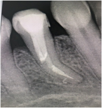
Figure 1: Rentgen picters fractured endodontic instruments in root canal.
The use of CBCT endodontic treatment technologies to diagnose dental clinical practice allows for procedures that are more predictable. Ultrasound is becoming a very useful tool in most stages of endodontic therapy, especially non-surgical and surgical treatments. Operating microscopes have been used for decades many other medical disciplines: ophthalmology, neurosurgery, reconstructive surgery, otorhinolaryngology, and vascular surgery. Its implementation in dentistry in the last fifteen years, especially in endodontics, has revolutionized the way endodontics is practiced worldwide. Precision is important for successful endodontics dental procedures, because operations are performed in small fields under poor lighting. The introduction of the operating microscope revolutionized the practice of endodontics. Incorporating microscopic approach in surgical endodontics, conceptualized by Prof. Kim in the 1990s with Use it is possible to carefully filling of the root canal system and all its branches along the longitudinal axis of the root [6]. Operating surgical microscope greatly increase the image of the structure of the object from 0.2 mm to 0.006 mm or 6 microns, improving the visible vision. The operating microscope is an instrument of great importance for solving various clinical difficulties and situations that arise during endodontic treatment.
Adequate coronal imaging is essential to prevent coronary leakage and to ensure the success of treatment methods, that is, the health of the periradicular periodontium, but according to the literature, it is not uncommon for filling materials to escape from the root of the tooth [7, 9]. The use of nickel-titanium rotary instruments in endodontic practice has gained popularity over the years, however sometimes they break despite their favorable qualities [10-16]. In case of instrument fractures during root canal treatment, the physician is prevented from optimal preparation for obturation of the entire root canal system and negatively affects the long-term prognosis of root canal treatment.
If the instrument is broken during root canal preparation procedures, the approaches chosen are to remove the damaged segment, bypass and seal the fragment in the root canal space, or a true blockage. Many factors must be considered before attempting to remove broken tools [17]. The odds of success must be balanced against potential complications. These factors may include anatomy of the root canal system nature of the material, tools and devices for displacement and removal of individual tools location, size, position and diameter of the destroyed part the experience and ability of the specialist clinician [18].

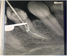
Figure 2: Rentgen picters remove fractured endodontic instruments in root canal.
There is no standardized procedure for successfully removing broken instruments. Previous methods and devices have shown limited success. There is currently no standardized procedure for safe and consistently successful removal. Removing broken fragments with traditional methods is time consuming, risky, and has limited success. Currently, removal of broken instruments is performed using ultrasound, operating microscopes, or micro tube delivery methods. Ultrasonic vibration of broken instrumental segments in combination with irrigation solution is performed under direct visualization and illumination of an operating microscope In the Ultrasonic method, first a direct access created by Gates-Glidden drills, then the ultrasonic tips mounted on the ultrasonic hand piece were used under the operating microscope. Dry ultrasonic tips with a diamond coating were used around the fragment, and then ultrasonic vibrations with ultrasonic tips made of nickel titanium (types 6-8) were used to remove the fragment. In terms of determining success, 74 out of 90 broken instruments were removed or successfully bypassed. This resulted in an 82.2% success rate. The failure rate was 17.7%. The overall success rate was found to be 93.3% ultrasonic hand pieces ultrasonic techniques were found to be more effective in removing instruments [19-22].
A versatile method is the use of fine ultrasonic magnification tips, preferably an operating microscope. If the instrument goes beyond the curvature of the canal or is not visible, the possibilities for removal are reduced, increasing the risk of complications. The first thing to be achieved is direct access to the instrument, which must be removed in order for the instrument to be exposed to 1 mm to 3 mm in its most coronal region using ultrasonic vibrations at that location. This situation can also lead to perforation in the absence of good vision and accurate movement. Several factors will determine whether or not to remove the broken piece. Firstly, its position in the root canal is significant, given that the more apical the fragment, the more difficult it is to remove. In addition, if the instrument goes beyond the curvature of the canal or is not visible, the possibilities are reduced from a few to zero, increasing the risk. If the file breaks during root canal treatment, there are several treatment options available. These solutions may include leaving the fragment where the fracture occurred and including the fragment to form part of the final obturation or removal from the root canal.

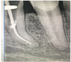
Figure 3: Аfter removal of the instrument, the tooth canal was filled and the tooth was filled.
Removal of a surgical fracture of an instrument from root canals depends on the anatomy of the canal, the location of the fragment in the canal, the length of the separated fragment, the diameter and curvature of the canal itself, as well as the ingress of the instrument fragment into the canal [23]. If individual instruments lie partially around the curvature of the canal and direct access is prepared for the crown of the fractured instrument segments, they can be removed, and with broken instrument segments that are apically located to the curvature of the canal, it is usually impossible. To remove broken instruments, the use of dental microscopes is essential for improved vision. With magnification and microscope illumination, it allows clinicians to observe the most coronal aspects of broken instruments and remove them without any perforation [24]. One of the most difficult situations to address in endodontics is the removal of broken instruments from the canal. In the middle of the canal, 16 out of 21 (76.19%) instruments in straight canals and 9 out of 10 (90%) in curved canals were successfully removed independently from. Fragments located in the crown of one third of the root canal with curved and straight roots were completely removed, however, in the apical third of the canal, 13 of 21 (61.90%) instruments in straight canals and 5 of 10 (50%) in curved canals were removed [25].

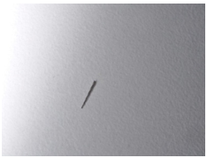
Figure 4: Fractured Endodontic Instruments from Root Canal Systems.
Numerous methods have been described for removing broken instruments from a canal, from using hand files to capture and remove fragments to countless devices built for this purpose [26]. If a decision is made to remove the broken instrument, it must be borne in mind that the procedure can be one of the most difficult to treat.Experiencing an instrument fracture in clinical practice is not uncommon for complicating endodontics, and may include creating inadequate access to the root canal system, anatomical problems and extreme root curvatures, multiple treatments with the same instrument, and the skill set and experience of the treating physician [27]. The use of the dental operating microscope in endodontics, advocated by many professionals, has provided a breakthrough in endodontic treatment [28]. The final decision on the choice of the method should be based on a thorough knowledge of the success rates of each treatment option, balanced'[29]. Analysis of the literature has shown if treat well and there are no signs of apical disease, then the presence of a broken instrument should not reduce the prognosis [30].
Based on this, the long-term study of the results of endodontic therapy is very relevant, which justifies the need for this work.
Purpose
To evaluate the success of using an operating microscope to remove broken instruments from different levels in curved and straight canals.
Patients and Methods
Removal of the broken instrument from the curved canals was performed on 61 teeth using ultrasound under the imaging of an operating microscope or conventional methods. The success of the tool removal methods used were evaluated. The success was determined by the complete removal of the broken fragment of the instrument. All patients underwent a thorough clinical examination according to a generally accepted scheme. After the diagnostic workup was completed, a treatment plan was developed by using a cone beam computed.
The fractured instrument was then bypassed using 6, 8, 10, no K file (Mani inc Japan), and 17% EDTA gel and liquid (Prime Dental).In the present study, the ultrasound technique was successfully applied to the removed broken nickel and titanium segments from narrow and curved root canals under the Dental operating microscope (Carl Zeiss, Germany). The exact location of the damaged file was confirmed under the Dental operating microscope (Carl Zeiss, Germany). To remove broken instruments using the GG drill no. creating stage platform 4 (Mani Inc., Japan). Irrigation for RCTs used 3.5% sodium hypochlorite (Prime dental) and Final rinse 17% EDTA (Prime Dental) followed by 2% CHX (Neelkanth).
An ultrasonic tip # 3 and 4 (pro ultra) was used at power 4. It was placed between the open part of the file and the canal wall and activated in a counterclockwise direction to remove dentin around the separated file.
After loosening the broken instrument from the curved canals, it was caught and removed using special pliers with fine toothed branches. After the broken was removed, we proceeded to the next stage - removal of the residual cement and paste in the coronal part of the root canal with an ultrasonic tip. The sealant was removed from the mid- and apical root canal using ProTaper D1-D3 endo-denture rotary instruments. After ensuring the patency of the root canals (the second canal was detected palatally), the following procedure followed - mechanical and chemical cleaning and the formation of the root canal. Used Crown Down technology and K3 rotary nickel-titanium files. 3% H2O2 and 2.5% sodium hypochlorite were used for short term treatments. A 17% EDTA solution was used to remove the smear applied to the root canal for 1 min. After removal of the instrument, the tooth canal was filled and the tooth was filled.
Results
Postoperative clinical and radiological monitoring was regularly conducted, and criteria for the success were evaluated. In the present study, the ultrasonic technique successful at removing fractured rotary nickel titranium segments from narrow and curved root canals in clinical cases.
Discussion
When removing a broken instrument from a tooth canal The prognosis of endodontic treatment of a tooth depends on several factors, from the equipment to the separation of the instrument, the state of the pulpal or periradicular tissue before treatment, and whether it is possible to remove or bypass the damaged file [30].
A number of factors affect extractions, these factors can be grouped as (1) location, length and type of broken instrument, (2) tooth/canal and (3) doctor's qualifications and available weaponry [31]. A wide range of techniques and devices have been developed to remove the damaged segment of the instrument to make the process easier. These devices can be broadly classified as ultrasonic, microtube devices and pliers/forceps [32]. All methods have similar problems of excessive dentin removal, weakening of the root structure, predisposition to bulge, root perforation or fracture, and possible fragment extrusion. Removing broken instruments outside of the curvatures when direct vision is not possible can be very difficult.
Removal of NiTi instruments is more difficult than removal of stainless steel instruments due to the fact that NiTi instruments are usually split at a shorter fragment length, more apically, in curvature of narrow root canals, with a rota Due to damage from ultrasonic vibration when trying to remove a fragment, NiTi instruments can be further detached or shortened [35]. Many different methods have been used to remove fractured instruments, these methods usually require the use of an operating microscope [36].
Some authors suggest that it is more careful to bypass the broken instrument, especially in those cases when access to the fragment is limited (apical third channel or beyond the curvature of the channel) and its removal can lead to excessive dentin removal with corresponding consequences [37]. A number of factors affect removal. These factors can be broadly grouped as (1) the location, length and type of the broken instrument, (2) the tooth / canal, and (3) the qualifications of the doctor and the weaponry available [38]. Removal of the root canal posts provides access to the endodontic space for thorough cleaning and disinfection. Removal of pillars carries the risk of complications associated with the formation of protrusions, perforations and fractures of the roots of the teeth. The safe removal of metal posts requires knowledge of the appropriate weaponry and technology. The mechanism of ultrasonic vibration impact on the post is associated with the effect of adhesion of the cementitious agent with the subsequent loosening of the post [39,40]. There are three approaches to conservative treatment
- Bypass of the separated instrument,
- Removal of the fractured file,
- Instrumentation and obturation of canal coronally to the fragment
When attempting to remove a fragment due to damage from ultrasonic vibration, NiTi instruments may additionally detach or shorten [41]. As removal of a fractured file is associated with considerable risk,bypassing the instrument should be considered. The removal of files can be expensive in terms of time and equipment and therefore a cost- benefit analysis of the treatment should be considered before selecting a definitive treatment for the patient. Patients should be informed if an instrument fractures during treatment or if a fractured file is discovered during a routine radiographic examination. It is essential legally that the treatment details and the information given to the patient are recorded accurately in the patient’s notes.
Conclusion
Removing broken instruments outside of the curvatures when direct vision is not possible can be very difficult. The clinical procedure of endodontic retreatment under the operating microscope allows to deal with highly complex cases and improve the scope of treatment and its prognosis.
Conflict of Interest and Financial Disclosure
The author declares that he has no conflict of interest and there was no external source of funding for the present study. None of the authors have any relevant financial relationship(s) with a commercial interest.
Consent Statement
Written informed consent was obtained from the patient for publication of this case report and accompanying images.
Funding
The work was not funded.
Ethical Approval
The study was reviewed and approved by the Ethics Committee of the Yerevan State Medical University after M. Heratsi (protocol N16, 5.10.17) and in accordance with those of the World Medical Association and the Helsinki Declaration. Informed consent Patients were informed verbally and in writing about the study and gave written informed consent.
References
- Song M, Kim HC, Lee W, Kim E Analysis of the cause of failure in nonsurgical endodontic treatment by microscopic inspection during endodontic microsurgery. J Endod. 2011;37(11):1516-9
- Siqueira JF J. Aetiology of root canal treatment failure: why well-treated teeth can fail.Int Endod J. 2001;34(1):1-10.
- Kerekes K, Tronstad L Long-term results of endodontic treatment performed with a standardized technique.J Endod. 1979;5(3):83-90.
- Crump MC, Natkin E. Relationship of broken root canal instruments to endodontic case prognosis: a clinical investigation. J Am Dent Assoc. 1970;80(6):1341-7.
- Stabholz A, Friedman S Endodontic retreatment--case selection and technique. Part 2: Treatment planning for retreatment. J Endod. 1988;14(12):607-14.
- Gary B Carr.The Use of the Operating Microscope in Endodontics Dental clinics of North America. 54(2):191-214.
- Sadia Tabassum, Farhan Raza Khan.Failure of endodontic treatment: The usual suspects.Eur J Dent. 2016;10(1):144-147. doi: 10.4103/1305-7456.175682.
- Song M, Kim HC, Lee W, Kim E. Analysis of the cause of failure in nonsurgical endodontic treatment by microscopic inspection during endodontic microsurgery. J Endod. 2011;37:1516-9.
- Siqueira JF., Jr Aetiology of root canal treatment failure: Why well-treated teeth can fail. Int Endod J. 2001;34:1-10
- Parashos P, Gordon I, Messer HH. Factors influencing defects of rotary nickel-titanium endodontic instruments after clinical use. J Endod. 2004;30(10):722-725.
- Simon S, Machtou P, Tomson P, Adams N, Lumley P. Influence of fractured instruments on the success rate of endodontic treatment. Dent Update. 2008;35:172-4.
- Crump MC, Natkin E. Relationship of broken root canal instruments to endodontic case prognosis: A clinical investigation. J Am Dent Assoc. 1970;80:1341-7.
- Castellucci A. Magnification in endodontics: the use of the operating microscope. Pract Proced Aesthet Dent. 2003;15(5):377-384.
- Fors U G H, Berg J O. Endodontic treatment of root canals obstructed by foreign objects. Int Endod J. 1986;19:2-10.
- Madarati A, Qualtrough A J, Watts DC.A microcomputed tomography scanning study of root canal space: Changes after the ultrasonic removal of fractured files. J Endod. 2009;35:125-128.
- Madarati A, Qualtrough A J, Watts D C. Vertical fracture resistance of roots after ultrasonic removal of fractured instruments. Int Endod J. 2010;43:424-429
- Hulsmann M, Schinkel I. Influence of several factors on the success or failure of removal of fractured instruments from the root canal. Endod Dent Traumatol. 1999;15:252-258.
- Madarati A. A., Watts D. C. Qualtrough A. J. E Opinions and attitudes of endodontists and general dental practitioneros in the UK towards the intra‐canal fracture of endodontic instruments. Part 2End Internatioanal Endodontic Journal. 41(12):1079-1087
- Nagai O, Tani N, Kayaba Y, Kodama S, Osada T. Ultrasonic removal of broken instruments in root canals. Int Endod J. 1986;19:298-304.
- Hulsmann M. Removal of fractured instruments using a combined automated/ultrasonic technique. J Endod. 1994;20:144-147.
- Ward JR, Parashos P, Messer HH. Evaluation of an ultrasonic technique to remove fractured rotary nickeltitanium endodontic instruments from root canals: an experimental study. J Endod. 2003;29:756-763.
- Ward JR, Parashos P, Messer HH. Evaluation of an ultrasonic technique to remove fractured rotary nickeltitanium endodontic instruments from root canals: Clinical cases. J Endod. 2003;29:764-767.
- Arun Kulandaivelu Thirumalai, Mahalaxmi Sekar, and Sumitha Mylswamy Retrieval of a separated instrument using Masserann technique. J Conserv Dent. 2008;11(1):42-45.doi: 10.4103/0972-0707.43417.
- Alomairy K H. Evaluating two techniques on removal of fractured rotary nickel-titanium endodontic instruments from root canals; an in vitro study. J Endod. 2009;35:559-562.
- Gencoglua N, Helvacioglu D. Comparison of the different techniques to remove fractured endodontic instruments from root canal systems. Eur J Dent. 2009; J Clin Diagn Res. 2017;11(5):ZC29–ZC35.
- Cujé J, Bargholz C, Hűlsman M.The outcome of retained instrument removal in a specialist practice. Int Endod J. 2010;43:545-554.
- Spili P, Parashos P, Messer H H. The impact of instrument fracture on outcome of endodontic treatment. J Endod. 2005;31:845-850.
- Sunandan Mittal, Tarun Kumar, Jyotika Sharma, and Shifali Mittal An innovative approach in microscopic endodontics. J Conserv Dent. 2014;17(3): 297-298. doi: 10.4103/0972-0707.131812.
- Rosen E, Tsesis I. Evidence-based decision-making in endodontics. Clin Dent. 2017;1:6. https://doi.org/10.1007/s41894-017-0006-0.
- Spili P, Parashos P, Messer HH. The impact of instrument fracture on outcome of endodontic treatment. J Endod. 2005;31(12):845-50.
- McGuigan MB, LoucaC. Duncan H. F Clinical decision-making after endodontic instrument fracture British Dental Journal. 20(214):395–400.
- Sjögren U, Hagglund B, Sunqvist G, Wing K . Factors affecting the long-term results of endodontic treatment. J Endod. 1990;16:498-504.
- Machtou P, Reit C . Non-surgical retreatment. In Bergenholtz G, Hørsted-Bindslev P, Reit C (eds). Textbook of endodontology. 2003;1:300-310.
- Hülsmann M, Schinkel I . Influence of several factors on the success or failure of removal of fractured instruments from the root canal. Endod Dent.
- Ward JR, Parashos P, Messer HH Evaluation of an ultrasonic technique to remove fractured rotary nickel-titanium endodontic instruments from root canals: an experimental study.
- Friedman S, Stabholz A, Tamse A. Endodontic retreatment--case selection and technique. 3. Retreatment techniques.J Endod. 1990;16(11):543-9.
- Peter Spili, Peter Parashos, Harold H Messer.The Impact of Instrument Fracture on Outcome of Endodontic Treatment.Journal of Endodontics. 2005;31(12):845-50.
- Manoel Brito-Júnio Alternative Techniques to Remove Fractured Instrument Fragments from the Apical Third of Root Canals: Report of Two Cases Braz. Dent. 2015;26(1). https://doi.org/10.1590/0103-6440201302446.
- Manoel Brito-Júnior et al .Alternative Techniques to Remove Fractured Instrument Fragments from the Apical Third of Root Canals: Report of Two Cases. Brazilian Dental Journal. 2015;26(1):79-85. ISSN 0103-6440 http://dx.doi.org/10.1590/0103-6440201302446.
- Terauchi Y, O'Leary L, Suda H. Removal of separated files from root canals with a new file-removal system: case reports. Rahimi M, Parashos P. A novel technique for the removal of fractured instruments. J Endod. 2006;32:789-797.
- Nimet Gencoglu, Dilek Helvacioglu. Comparison of the Different Techniques to Remove Fractured Endodontic Instruments from Root Canal Systems.Eur J Dent. 2009;3(2):90-95.
- Nimet Gencoglu, Dilek Helvacioglu.Comparison of the Different Techniques to Remove Fractured Endodontic Instruments from Root Canal Systems.Eur J Dent. 2009;3(2): 90-95.

