
Email id: info@ijclinmedcasereports.com
Temporal Trends and Epidemiological Characteristics of Dermoscopically Evaluated Skin Lesions: A Retrospective Analysis of 1,219 Consecutive Patients
Shkodrani E¹,*, Dorina Ruci³, Cenko F², Cenko A² and Xhaja A¹
¹Clinic of Dermatology, University Hospital Center “Mother Teresa”, Tirana, Albania
²“Lady of Good Counsel” Hospital, Tirana, Albania
³Clinic of Rheumatology, University Hospital Center “Mother Teresa”, Tirana, Albania
Received Date: 21/09/2026; Published Date: 16/11/2026
*Corresponding author: Shkodrani Entela, Clinic of Dermatology, University Hospital Center “Mother Teresa”, Tirana, Albania
Abstract
Background: Dermoscopy is widely used for the evaluation of melanocytic and non-melanocytic skin lesions. Although its diagnostic performance has been extensively investigated, longitudinal data describing the spectrum and temporal distribution of dermoscopically evaluated lesions in Albania, remain limited.
Objective: To characterize the diagnostic distribution, demographic characteristics, and temporal patterns of dermoscopically evaluated skin lesions at a dermatology referral center in Albania over a six-year period.
Methods: This retrospective observational study included 1,219 consecutive patients evaluated at “EB Dermatology Clinic”, Tirana, Albania, between January 2020 and December 2025. Each patient contributed one clinically selected skin lesion, resulting in 1,219 lesions and 1,219 corresponding dermoscopic diagnoses. The six-year period was divided into three consecutive two-year intervals: 2020–2021, 2022–2023, and 2024–2025.
Results: Benign melanocytic and dermal lesions were the most frequent diagnostic category (415/1,219; 34.0%), followed by other dermatological diagnoses (254; 20.8%), basal cell carcinoma (BCC) (173; 14.2%), dysplastic nevi (132; 10.8%), melanoma (88; 7.2%), premalignant melanocytic and keratinocytic lesions (88; 7.2%), and squamous cell carcinoma (SCC) (69; 5.7%). The absolute number of melanoma diagnoses was 36 in 2020–2021, 33 in 2022–2023, and 29 in 2024–2025, while melanoma represented 7.2%, 7.9%, and 9.6% of all evaluated lesions in the respective periods. Most melanoma diagnoses (78/88; 88.6%) occurred in patients older than 25 years. SCC showed a significantly greater male predominance than BCC (p = 0.024).
Conclusion: The diagnostic spectrum of dermoscopically evaluated lesions showed temporal and demographic variation over the six-year study period. The increasing proportion of melanoma among evaluated lesions should not be interpreted as an increase in population-level melanoma incidence because the overall number of patients evaluated declined over time. These findings provide referral-based data from Albania and contribute to addressing the limited evidence on dermoscopic diagnostic patterns in Southeastern Europe.
Keywords: Dermoscopy; Melanoma; Basal cell carcinoma; Squamous cell carcinoma; Skin lesions; Albania; Epidemiology
Introduction
Dermoscopy is a widely used non-invasive technique for the evaluation of melanocytic and non-melanocytic skin lesions. By allowing visualization of subsurface morphological structures that are not readily visible to the naked eye, dermoscopy provides additional diagnostic information during the clinical assessment of suspicious lesions. Evidence from systematic reviews and diagnostic studies has demonstrated that dermoscopic examination can improve diagnostic performance for melanoma and other skin cancers compared with naked-eye examination alone [1,2]. These established findings provide the clinical rationale for the widespread use of dermoscopy; however, diagnostic accuracy was not directly evaluated in the present study. Previous research has focused extensively on the diagnostic performance of dermoscopy, melanoma recognition, and the epidemiology of melanoma and keratinocyte carcinomas. Studies have also reported earlier detection of melanoma and changes in melanoma incidence and mortality in several populations [3-5]. However, important questions remain regarding the spectrum of lesions encountered in routine dermoscopic referral practice and whether the relative distribution of diagnostic categories changes over time within such clinical settings. These patterns may be influenced not only by disease occurrence but also by referral practices, patient selection, healthcare access, clinical awareness, and the characteristics of the population attending a dermatology referral center. This evidence gap is particularly relevant to Southeastern Europe, where longitudinal data describing the distribution of dermoscopically evaluated skin lesions remain limited. Published data specifically characterizing the diagnostic spectrum and temporal patterns of melanocytic, keratinocytic, premalignant, and other dermoscopically evaluated lesions in Albania are scarce. Consequently, it remains unclear whether patterns reported from other European and international populations adequately reflect those encountered in Albanian dermatology referral practice. Population-based studies reporting changes in melanoma incidence and mortality provide important epidemiological context but differ fundamentally from referral-center studies. A clinic-based dataset represents a selected population of patients presenting or being referred for dermatological assessment and therefore cannot be used to estimate population-level incidence or mortality. Accordingly, changes in the frequency or proportion of melanoma within such a dataset should be interpreted as changes in the diagnostic composition of the evaluated clinical population rather than as evidence of changes in melanoma incidence in the general population.
Aim of the Study: The present study aimed to characterize the diagnostic distribution, demographic characteristics, and temporal patterns of dermoscopically evaluated melanocytic and non-melanocytic skin lesions among 1,219 consecutive patients at a dermatology referral center in Tirana, Albania, over a six-year period from January 2020 through December 2025. The study was specifically designed to evaluate patterns within the dermoscopically assessed clinical population and not to estimate population-level incidence of melanoma or other skin cancers.
Materials and Methods
Study Design and Setting: This retrospective observational study included consecutive patients who underwent dermoscopic evaluation of clinically suspicious skin lesions between January 2020 and December 2025 at EB Dermatology Clinic, a dermatology referral clinic in Tirana, Albania. Patients presenting with skin lesions considered to warrant specialist dermoscopic evaluation on clinical grounds were eligible for inclusion. Clinical indications for dermoscopic assessment included suspicious or changing pigmentation or morphology, asymmetry, irregular borders or colors, persistent or progressive lesions, ulceration or bleeding, or other clinical characteristics raising concern for a melanocytic or non-melanocytic neoplasm. A total of 1,219 consecutive patients were included. Each patient contributed one clinically selected lesion to the analysis, resulting in 1,219 dermoscopically evaluated lesions and 1,219 corresponding diagnoses. Each lesion was assigned one specific dermoscopic diagnosis. No patient contributed more than one lesion or diagnosis; therefore, all observations were independent at the patient level. No patients were excluded from the final analysis.
DermoscopicAssessment: Dermoscopic examinations were performed by two dermatologists using the FotoFinder Vexia imaging system. Both dermatologists followed the same predefined diagnostic approach and applied identical dermoscopic assessment/ criteria throughout the study period. Pattern analysis included assessment of lesion symmetry, colors, pigment network, dots and globules, streaks, regression structures, vascular patterns, and other diagnosis-specific dermoscopic features. Established dermoscopic criteria for melanocytic and non-melanocytic lesions were applied consistently during examination [1,2]. Based on the dermoscopic assessment, lesions were classified into the following predefined diagnostic categories:
- benign melanocytic and dermal lesions;
- premalignant melanocytic and keratinocytic lesions;
- melanoma;
- basal cell carcinoma;
- squamous cell carcinoma;
- dysplastic nevi;
- other dermatological diagnoses.
Histopathological examination was performed whenever clinically indicated as part of routine patient management. For each patient, the following variables were recorded: year of diagnosis, age at diagnosis, sex, and dermoscopic diagnosis.
Temporal Grouping: For descriptive temporal comparisons, the six-year study period was divided into three consecutive intervals of equal duration: 2020–2021, 2022–2023, and 2024–2025. Two-year intervals were selected to provide comparable observation periods and to reduce year-to-year variability associated with relatively small numbers within some individual diagnostic categories. The earliest interval, 2020–2021, served as the reference period for temporal comparisons when applicable. Associations between categorical variables were evaluated using the chi-square test, as appropriate. Odds Ratios (ORs) with corresponding 95% Confidence Intervals (CIs) were used for predefined group comparisons where applicable. Statistical significance was defined as a two-sided p value < 0.05. The assumptions underlying categorical comparisons, including adequacy of expected cell frequencies for chisquare testing, were considered when selecting the statistical test. Pearson correlation analysis was not retained in the revised analysis because the temporal dataset comprised only three aggregated two-year intervals, which was considered insufficient for a robust correlation-based assessment of temporal trend.
Ethical Considerations: The study was conducted in accordance with the ethical principles of the Declaration of Helsinki and received approval from the relevant Ethics Committee. All patients provided informed consent permitting the use of their anonymized clinical and dermoscopic data for research purposes. Patient confidentiality was maintained throughout data collection, analysis, and reporting, and no patientidentifiable information was included in the study.
Results
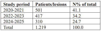
Overall Study Population: A total of 1,219 patients, each contributing one dermoscopicallyevaluated lesion, were included in the analysis. The largest number of patients was evaluated during 2020– 2021, comprising 501 cases (41.1% of the overall study population), followed by 417 cases (34.2%) in 2022– 2023 and 301 cases (24.7%) in 2024–2025, (Table 1).
Table 1: Distribution of patients and dermoscopically evaluated lesions according to study period.
Overall, 691 patients (56.7%) were female and 528 (43.3%) were male, corresponding to a female-to-male ratio of approximately 1.31:1. Benign melanocytic and dermal lesions represented the most frequen diagnostic category, accounting for 415 cases (34.0%). These were followed by other dermatological diagnoses (254; 20.8%), BCC (173; 14.2%), dysplastic nevi (132; 10.8%), melanoma (88; 7.2%), premalignant melanocytic and keratinocytic lesions (88; 7.2%), and SCC (69; 5.7%), (Figure 1).
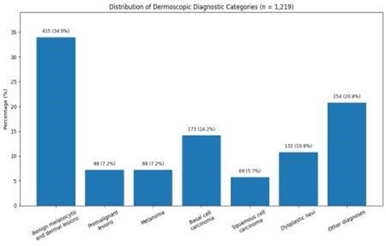
Figure 1: Distribution of dermoscopic diagnostic categories.
Melanoma: A total of 88 melanoma diagnoses were recorded during the six-year study period. The absolute number of melanoma diagnoses was 36 during 2020– 2021, 33 during 2022–2023, and 29 during 2024–2025. When expressed relative to the total number of patients evaluated during each corresponding interval, melanoma represented 7.2% (36/501) in 2020–2021, 7.9% (33/417) in 2022–2023, and 9.6% (29/301) in 2024–2025. Thus, although the absolute number of melanoma diagnoses decreased across the three periods, the relative proportion of melanoma among all dermoscopically evaluated lesions increased. This observation should be considered in the context of the simultaneous decline in the total number of patients evaluated. The mean age of patients diagnosed with melanoma was 46.1 years in 2020–2021, 41.1 years in 2022–2023, and 42.0 years in 2024–2025. During the first period, melanoma occurred equally among males and females (18 males and 18 females). During 2022–2023, 16 cases occurred in males and 17 in females, whereas during 2024–2025, 17 occurred in males and 12 in females. Age-stratified analysis showed that 78 of the 88 melanoma diagnoses (88.6%) occurred in patients older than 25 years, whereas 10 (11.4%) occurred in patients aged 25 years or younger (Table 2, Figure 2).
Table 2: Characteristics of melanoma diagnoses according to study period.
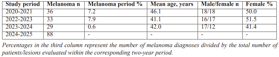
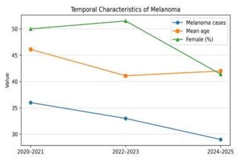
Figure 2: Distribution and demographic characteristics of melanoma diagnoses across the three study periods.
Basal Cell and Squamous Cell Carcinoma: During the study period, 173 basal cell carcinomas (BCCs) and 69 squamous cell carcinomas (SCCs) were diagnosed. The mean age of patients was comparable between the two groups (43.4 years for BCC and 43.5 years for SCC). However, marked differences were observed in sex distribution. SCC showed a pronounced male predominance, with males accounting for 69.6% of all cases, whereas BCC demonstrated a more balanced distribution between sexes, with females representing 46.2% of patients. The difference in sex distribution between SCC and BCC was statistically significant (χ² test, p = 0.024) (Table 3, Figure 3).
Table 3: Comparison between Basal Cell and Squamous Cell Carcinoma.
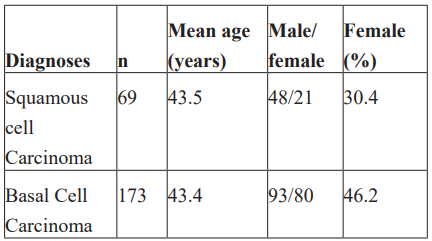
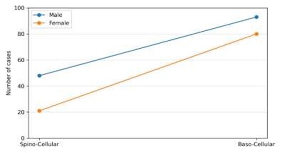
Figure 3: Comparison between Basal Cell and Squamous Cell Carcinoma.
Discussion
The present study highlights a clinically important limitation of conventional outcome assessment after endoscopic dacryocystorhinostomy: an anatomically successful DCR is not necessarily a functionally successful DCR.
In this series of 110 patients, anatomical success was achieved in 87%, confirming the overall effectiveness of eDCR. More importantly, however, 18.9% of anatomically successful procedures remained functionally unsuccessful.
This finding shifts the focus of DCR outcome assessment from a purely surgical question—whether the rhinostomy remains open—to a more clinically relevant question: whether effective tear drainage has actually been restored.
Anatomical success is not synonymous with clinical cure:
Success rates reported after eDCR vary substantially according to the endpoint used.
A systematic review by Leong et al. demonstrated generally favorable results after DCR but also emphasized considerable heterogeneity among studies in surgical technique and definitions of success [1]. Modern series similarly report success rates commonly between approximately 82% and 98%, depending on patient selection and outcome criteria [6].
The 87% anatomical success rate observed in our cohort therefore falls within the expected range of published experience.
However, anatomical success alone overestimates patient-perceived benefit.
Shams et al. specifically evaluated persistent epiphora after anatomically successful DCR and estimated that approximately 5–10% of patients may continue to experience tearing despite a patent lacrimal pathway [7]. Their multicenter study demonstrated that functional epiphora represents a genuine postoperative entity rather than simply undetected ostium closure.
Our observed proportion of functional failure was higher, reaching 18.9% among anatomically successful cases.
Interestingly, Sung et al. reported exactly the same magnitude—18.9% functional failure among anatomically successful eDCR procedures—in their analysis of the effect of age on surgical outcomes [8]. Their study also identified older age and diabetes mellitus as significant correlates of functional failure, strongly supporting the biological and clinical relevance of these factors.
Why can an open DCR remain symptomatic?
Tear drainage is not a passive phenomenon.
Anatomical patency permits tears to reach the nasal cavity, but efficient transport depends on a coordinated lacrimal pump.
Blinking generates complex pressure changes through contraction and relaxation of the orbicularis oculi muscle, while appropriate punctal positioning and eyelid-globe apposition permit tears to enter the canalicular system.
Consequently, a patient can have an entirely patent rhinostomy while remaining symptomatic because the pathway is open but functionally inefficient.
This concept provides a plausible explanation for the ocular adnexal abnormalities observed in our functionally unsuccessful patients.
Age as a determinant of functional failure:
Older age was significantly associated with functional failure in our cohort (P=0.024).
Age-related changes can affect virtually every component of the lacrimal drainage mechanism, including eyelid tone, punctal position, conjunctival anatomy, blink efficiency, and ocular surface homeostasis.
Sung et al. found that although anatomical success did not significantly differ between younger and older groups, functional success was significantly lower among older patients [8]. Older patients with functional failure also exhibited a greater prevalence of eyelid laxity.
Similarly, studies of DCR in elderly populations have suggested that anatomical success may remain high while symptomatic resolution becomes less predictable [11].
These findings suggest that age should not be considered a contraindication to eDCR. Rather, advanced age should trigger a more comprehensive preoperative functional assessment.
Eyelid laxity and conjunctivochalasis: overlooked determinants?
Our observation of frequent eyelid laxity and conjunctivochalasis among patients with functional failure is particularly important.
Eyelid laxity can reduce the efficiency of the lacrimal pump and alter punctal apposition.
Conjunctivochalasis may mechanically interfere with tear flow toward the punctum and can itself produce epiphora through ocular-surface irritation.
Consequently, persistent tearing following DCR may reflect an incorrect assumption that all preoperative epiphora originated exclusively from nasolacrimal duct obstruction.
Preoperative examination should therefore extend beyond lacrimal irrigation.
In elderly patients in particular, careful evaluation should include eyelid position and tone, punctal position, conjunctivochalasis, ocular surface status, and blink dynamics.
Diabetes mellitus and functional outcome:
Diabetes mellitus was significantly associated with functional failure (P=0.008).
This association has also been reported by Sung et al. [8].
Several mechanisms are biologically plausible, including microvascular dysfunction, impaired wound healing, chronic low-grade inflammation, altered epithelial repair, neuropathy, and age-related confounding.
However, because diabetes prevalence increases with age, its independent contribution is difficult to establish without comprehensive multivariable adjustment.
Accordingly, our findings support diabetes as a risk marker, but they should not be interpreted as demonstrating a direct causal mechanism.
Level of obstruction matters:
Another important observation concerns the anatomical level of lacrimal obstruction.
More proximal obstruction was associated with poorer functional prognosis.
This is consistent with the large study by Lee et al., which evaluated 769 external DCR procedures. Although anatomical success reached 98.8%, functional success was only 81.9%. Common canalicular obstruction increased the risk of functional failure by approximately 75%, while canalicular obstruction approximately doubled the risk [9].
These results are physiologically logical.
DCR is exceptionally effective at bypassing distal nasolacrimal duct obstruction, but it cannot fully compensate for impaired tear entry or transport through the canalicular system.
Thus, the more proximal the dysfunction, the less likely creation of a distal bypass alone is to restore normal tear drainage.
Surgical factors versus patient factors:
An intriguing feature of our results is the lack of significant association between functional outcome and several technical variables, including uncinectomy, middle turbinectomy, granulation, membranous obstruction, and intranasal synechiae.
This does not imply that surgical technique is unimportant.
On the contrary, technical failure remains a major cause of anatomical failure.
Recent analysis of revision eDCR has shown that inadequate lacrimal sac exposure, cicatricial closure of the ostium, and synechiae are prominent findings in failed procedures [12].
The distinction is therefore essential:
technical factors may primarily determine whether the rhinostomy remains anatomically patent, whereas patient-related factors may become increasingly important in determining whether an anatomically successful operation is also functionally successful.
The role of silicone intubation:
Silicone intubation remains one of the most debated aspects of eDCR.
A systematic review and meta-analysis of 12 randomized controlled trials encompassing 1,239 procedures reported an overall eDCR success rate of 91.9% and found no statistically significant difference between stented and non-stented procedures [13].
More recent evidence continues to examine this question, with differences according to endpoints and patient selection [14].
Our cohort underwent bicanalicular silicone intubation, but the available analysis did not demonstrate stent-related parameters as independent determinants of poor functional outcome.
This supports the concept that stenting cannot correct dysfunction originating from the eyelid, punctum, canalicular pump, or ocular surface.
A clinically useful classification of DCR failure:
Our findings support separating postoperative failure into two major phenotypes.
Type I — Anatomical failure
This includes:
- ostium closure;
- cicatricial stenosis;
- inadequate sac exposure;
- granulation;
- membranous obstruction;
- synechiae;
- canalicular obstruction.
In this phenotype, revision surgery directed at restoring anatomical patency may be appropriate.
Type II — Functional failure despite anatomical success:
This includes persistent epiphora despite a patent ostium and may be associated with:
- eyelid laxity;
- impaired lacrimal pump;
- punctal malposition;
- conjunctivochalasis;
- proximal canalicular dysfunction;
- ocular surface abnormalities;
- excessive tear production.
In this second phenotype, simply enlarging an already patent rhinostomy may fail to address the actual mechanism of tearing.
This distinction has direct therapeutic consequences.
Toward patient-centered assessment of DCR success:
Traditional surgical studies frequently define success according to endoscopic ostium visualization or lacrimal irrigation.
These remain indispensable measures of technical success, but they should not constitute the sole endpoint.
A more clinically meaningful postoperative evaluation should integrate:
- anatomical ostium patency;
- lacrimal irrigation or functional drainage testing;
- patient-reported epiphora;
- eyelid and punctal examination;
- ocular-surface assessment;
- need for additional lacrimal procedures.
Such a framework would distinguish technical success from patient success.
Our findings therefore argue for a conceptual transition from an ostium-centered model of DCR outcome toward a lacrimal-system-centered model.
Clinical Implications:
The results of this study have several practical implications.
First, elderly patients should undergo systematic assessment for eyelid laxity, punctal abnormalities, and conjunctivochalasis before eDCR.
Second, the anatomical level of obstruction should be clearly characterized because proximal disease may predict poorer functional benefit.
Third, diabetes mellitus should be considered when counseling patients regarding postoperative expectations.
Fourth, persistent epiphora after eDCR should not automatically be interpreted as restenosis.
Before revision surgery, the surgeon should determine whether the primary procedure has failed anatomically or functionally.
This distinction may prevent unnecessary revision of an already patent rhinostomy.
Strengths:
A strength of this study is the consecutive nature of the series, comprising 110 endoscopic DCR procedures performed over a 10-year period at a single tertiary referral centre with a uniform surgical protocol.
Another important strength is the deliberate distinction between anatomical and functional outcomes.
Rather than considering ostium patency alone, our analysis explores why some technically successful procedures remain clinically unsuccessful.
The evaluation of demographic, systemic, intranasal, lacrimal, and ocular adnexal factors further provides a multidimensional assessment of surgical outcome.
Limitations:
Several limitations should be acknowledged.
First, the retrospective design introduces potential selection and information biases.
Second, standardized quantitative patient-reported epiphora scores were not available in the source dataset.
Third, detailed quantitative measurements of postoperative ostium size and dynamic lacrimal pump function were unavailable.
Fourth, several associations may be confounded by age, particularly diabetes mellitus and eyelid laxity.
Fifth, the available dataset does not provide all adjusted odds ratios and 95% confidence intervals required to construct a formal predictive model.
Finally, the sample size of 110 patients limits statistical power, particularly for the subgroup of anatomically successful procedures in which functional failure was assessed; non-significant associations should therefore be interpreted as inconclusive rather than as evidence of absence of effect, and the reported proportions carry correspondingly wide confidence intervals.
Future prospective studies should combine standardized anatomical assessment with validated epiphora scores, objective functional drainage testing, detailed eyelid examination, and multivariable predictive modeling.
Conclusion
Endoscopic dacryocystorhinostomy provides favorable anatomical outcomes, with an overall anatomical success rate of 87% in this series of 110 patients.
However, the most important finding of this study is that 18.9% of anatomically successful procedures remained functionally unsuccessful.
Advanced age, diabetes mellitus, proximal lacrimal obstruction, eyelid laxity, and conjunctivochalasis emerged as clinically relevant characteristics associated with or potentially contributing to poor functional outcome.
These results demonstrate that ostium patency alone is an incomplete measure of DCR success.
Persistent epiphora following an anatomically successful procedure should prompt evaluation of the entire lacrimal functional unit—including the puncta, canaliculi, eyelids, lacrimal pump, conjunctiva, and ocular surface—before revision surgery is considered.
Ultimately, the goal of DCR should not merely be to create an open rhinostomy.
The goal should be to restore effective tear drainage and relieve the patient's symptoms.
References
- Leong SC, Macewen CJ, White PS. A systematic review of outcomes after dacryocystorhinostomy in adults. Am J Rhinol Allergy, 2010; 24: 81–90.
- Wormald PJ. Powered endoscopic dacryocystorhinostomy. Laryngoscope, 2002; 112: 69–72.
- Tsirbas A, Wormald PJ. Mechanical endonasal dacryocystorhinostomy with mucosal flaps. Br J Ophthalmol, 2003; 87: 43–47.
- Cokkeser Y, Evereklioglu C, Er H. Comparative external versus endoscopic dacryocystorhinostomy: results in 115 patients. Otolaryngol Head Neck Surg, 2000; 123: 488–491.
- Woog JJ. The incidence of symptomatic acquired lacrimal outflow obstruction among residents of Olmsted County, Minnesota, 1976–2000. Trans Am Ophthalmol Soc, 2007; 105: 649–666.
- Onerci M, Orhan M, Ogretmenoglu O, Irkec M. Long-term results and reasons for failure of intranasal endoscopic dacryocystorhinostomy. Acta Otolaryngol, 2000; 120: 319–322.
- Shams PN, Chen PG, Wormald PJ, et al. Management of functional epiphora in patients with an anatomically patent dacryocystorhinostomy. JAMA Ophthalmol, 2014; 132: 1127–1132.
- Sung JY, Lee YH, Kim KN, Kang TS, Lee SB. Surgical outcomes of endoscopic dacryocystorhinostomy: analysis of age effect. Sci Rep, 2019; 9: 19861.
- Lee MJ, Khwarg SI, Kim IH, Choi JH, Choi YJ, Kim N. Surgical outcomes of external dacryocystorhinostomy and risk factors for functional failure: a 10-year experience. Eye (Lond), 2017; 31: 691–697.
- Ali MJ, Psaltis AJ, Wormald PJ. Dacryocystorhinostomy ostium: parameters to evaluate and DCR ostium scoring. Clin Ophthalmol,
- Tooley AA, Klingler KN, Bartley GB, et al. Dacryocystorhinostomy for acquired nasolacrimal duct stenosis in the elderly (≥80 years of age). Ophthalmology, 2017; 124: 263–267.
- Arora R, et al. Endoscopic dacryocystorhinostomy: reasons for failure and possible solutions. Eur Arch Otorhinolaryngol, 2024/2025.
- Kang MG, Shim WS, Shin DK, Kim JY, Lee JE, Jung HJ. A systematic review of benefit of silicone intubation in endoscopic dacryocystorhinostomy. Clin Exp Otorhinolaryngol, 2018; 11: 81–88.
- Dar A, Jalal AA, Ibrahim M, et al. Safety and efficacy of silicone intubation compared to standard techniques in endoscopic dacryocystorhinostomy: a systematic review and meta-analysis. Int Ophthalmol, 2026; 46: 291.
- Xie C, Zhang L, Liu Y, Ma H, Li S. Comparing the success rate of dacryocystorhinostomy with and without silicone intubation: a trial sequential analysis of randomized controlled trials. Scientific Reports, 2017.
- Baek JS, Jeong SH, Lee JH, Choi HS, Kim SJ, Jang JW. Cause and management of patients with failed endonasal dacryocystorhinostomy. Clin Exp Otorhinolaryngol. 2017; 10(1): 85-90. doi: 10.21053/ceo.2016.00192.
- Fayers T, Laverde T, Tay E, Olver JM. Lacrimal surgery success after external dacryocystorhinostomy: functional and anatomical results. Ophthalmic Plastic and Reconstructive Surgery, 2009.
- Marcet MM, Kuk AK, Phelps PO. Evidence-based review of surgical practices in endoscopic endonasal dacryocystorhinostomy for primary acquired nasolacrimal duct obstruction and other indications. Curr Opin Ophthalmol, 2014; 25(5): 443-448. doi: 10.1097/ICU.0000000000000084.
- Vinciguerra A, Nonis A, Resti AG, et al. Long-term outcomes of endoscopic dacryocystorhinostomy: failure rate and risk factors. Laryngoscope, 2021; 131(1): E1-E6.
- Hurwitz JJ, Merkur S, DeAngelis D. Outcome of lacrimal surgery in older patients. Can J Ophthalmol, 2000; 35: 18–22.