
Email id: info@ijclinmedcasereports.com
Glaucoma Study in Northern Sudan: Patient’s Demographics, Classification, Response to Interventions and Disease Severity
Isra Mohammed O Sid Ahmed*, Marouf Mohammed Marouf, Nosiba Mohammed Khoajali Ballah, Jammaa Ahmed and Yaseen Siddig Yaseen Ahmed
Department of Ophthalmology, Al-Jawareh hospital, Sudan
Received Date: 30/06/2026; Published Date: 04/09/2026
*Corresponding author: Isra Mohmmed O Sid Ahmed, Department of Ophthalmology, Al-Jawareh hospital, Sudan
Abstract
Background: glaucoma is a major cause of vision impairment and blindness worldwide. Africa has a unique clinical profile of glaucoma presentation and disease progression. In Sudan, early diagnosis and treatment of glaucoma is a challenge especially after the eruption of war and scarcity of eye care facilities.
Purpose: to describe glaucoma patients ‘demographics, clinical profile and response to medical treatment and surgical interventions.
Method: cross-sectional, retrospective and descriptive study. The data was retrieved from the medical records of a tertiary hospital.
Result: 338 patients diagnosed with glaucoma were enrolled in the study. the mean age was 59.4 ± 13.0 years (median 60 years, IQR 50–70; range 18–90 years). 209 patients (62.0%) were female and 128 (38.0%) were male. Only 81 patients were sub-classified in the medical records to glaucoma types. The mean baseline intraocular pressure (IOP) was 20.98 ± 10.87 mmHg in the right eye and 19.95 ± 10.91 mmHg in the left eye. Elevated baseline IOP (≥21 mmHg) was observed in 123 of 324 right eyes (38.0%), 99 of 321 left eyes (30.8%), and in either eye in 164 of 332 patients.
Only 81 patients were classified in the medical records into glaucoma sub-types. Primary open-angle glaucoma was identified in 18 patients (22.2%), pseudoexfoliative glaucoma in 16 (19.8%), neovascular glaucoma in 13 (16%), angle-closure suspect in 9 (11.1%), primary angle-closure glaucoma in 7 (8.6%), angle-closure glaucoma in 6 (7.4%), glaucoma suspect in 5 (6.2%), and uveitic glaucoma in 3 patients (3.7%). Visual impairment was graded according to the best-corrected visual acuity (BCVA) of the worse eye. Mean logMAR BCVA was 1.39 ± 1.12 for the worse eye. Of the 315 patients with available data, 74 (23.5%) had normal or near-normal vision (≥6/12), 42 (13.3%) had mild visual impairment (6/12–6/18), 41 (13.0%) had moderate visual impairment (6/24–6/60), and 3 (1.0%) had severe visual impairment (<6/60–CF), whereas 155 patients (49.2%) had profound visual loss or blindness (HM/PL/NPL). Among 152 patients with available data, 9 (5.9%) had mild disease structural damage (CDR ≤0.5), 44 (28.9%) had moderate disease progression (CDR 0.6–0.8), and 99 (65.1%) had severe disease progression (CDR ≥0.9).
Overall, 302 patients (89.3%) received medical treatment including topical IOP-lowering therapy. Carbonic anhydrase inhibitors were the most frequently prescribed medications, used by 241 patients (79.8%). A total of 189 patients underwent surgical intervention. Trabeculectomy was the most common procedure, performed in 77 patients (40.7%), followed by phacoemulsification in 75 (39.7%), YAG laser peripheral iridotomy in 37 (19.6%), combined phacotrabeculectomy in 34 (18.0%),), transscleral micropulse cyclophotocoagulation in 11 (5.8%).
Paired pre- and post-treatment IOP measurements: In the right eye, mean IOP decreased from 22.82 ± 11.47 mmHg at baseline to 16.93 ± 10.64 mmHg at the last follow-up, corresponding to a mean reduction of 5.89 ± 11.57 mmHg. In the left eye, mean IOP decreased from 20.52 ± 10.20 mmHg to 14.36 ± 6.57 mmHg, with a mean reduction of 6.16 ± 10.36 mmHg. These reductions were statistically significant in both eyes (Wilcoxon signed-rank test, p < 0.001). At the last follow-up, 134 of 169 right eyes (79.3%) and 144 of 168 left eyes (85.7%) achieved an IOP below 21 mmHg, whereas 118 (69.8%) and 127 (75.6%), respectively, achieved an IOP below 18 mmHg.
Conclusion: glaucoma is more prevalent among older population. Although more prevalent among females but the male’s mode of disease is found to be more severe. 49% of the patients presented with blindness. IOP which is the only known modifiable risk factor has shown considerable response to variable options of medical and surgical interventions.
Abbreviations: CAI - Carbonic Anhydrase Inhibitor; IOP - Intra-Ocular Pressure; POAG - Primary Open Angle Glaucoma
Introduction
Globally, glaucoma is the second cause of irreversible blindness after cataract. Glaucoma is defined as the ocular disease entity that is characterized by the progressive damage of ganglia cells of the retina. The main risk factor known is increased intra-ocular pressure, although the retinal ganglia cell loss can occur in glaucoma without an increase in intra-ocular pressure. Other risk factors are: aging, decreased central corneal thickness, myopia, family history and diabetes mellitus [1].
The clinical profile of glaucoma among African patients is unique in the disease severity in younger age. Evidence revealed blindness risk from glaucoma among Africans is 6-8 times more than patients from other ethnic groups [1].
Pathophysiology of glaucoma:
Increased prevalence of glaucoma especially of the open-angle type among African patients is an outcome of multiple factors. Environmental and genetic factors predispose to open- angle glaucoma in African patients [2].
Glaucoma is classified according to etiology into secondary or primary. Each variant is classified further according to pathophysiology into open or closed-angle glaucoma. Secondary glaucoma result from an insult to the eye, either caused by trauma, inflammation (uveitis), neovascular or as a part of pseudoexfoliation syndrome [3].
Medical and surgical intervention basically aim at control of disease progression through lowering intra-ocular pressure.
Glaucoma is a public health burden. More than 60 million people are diagnosed with glaucoma 8 million people lost their vision due to glaucoma. It is presumed that by 2040, 111 million will be affected with glaucoma. The prevalence of glaucoma in Sub-Saharan Africa is 4% among adult population above 40 years old compared to the 1% global prevalence [4].
In addition to being a public health concern, glaucoma is a disease with an economic impact as it is considered a major cause of visual impairment and hence profoundly affecting the patient’s productivity [5].
Materials and Methods
This a cross-sectional descriptive study. The study was conducted at Al-Jawareh hospital, Dongola –Sudan. Al- Jawareh hospital is a tertiary care hospital established 2 years ago. Study population was selected through systematic data sampling.
Inclusion and exclusion criteria:
The participants included in the study are patients diagnosed with glaucoma during the year 2025 above 18 years old, not known to have any other cause of optic neuropathy.
Parameters of the study:
Demographic data collected was sex, age occupation and residency. Concurrent ocular diseases were reported. Diabetes and hypertension were also noted. Visual acuity with correction was documented from the first visit. IOP of the first and last visits were documented.
Medical treatment received by the patients as well as surgical procedures were also noted. Detailed description of medical eye drops for each patient was given. Surgical interventions undergone for glaucoma management in the medical records were noted. IOP before and after intervention were written down to explore the patient’s response to treatment.
The severity of the disease was expressed as the cup disc ratio for each patient.
Statistical methods:
This analysis was conducted on a clinic-based sample of 338 patients presenting to a glaucoma service. Continuous variables are reported as Mean ± Standard Deviation (SD) and median with Inter-Quartile Range (IQR); categorical variables as frequency and column percentage. Visual acuity was converted from Snellen notation to the logarithm of the minimum angle of resolution (logMAR) using the standard mapping. Disease severity was graded by the Cup-to-Disc Ratio (CDR) of the worse-affected eye: mild (≤ 0.5), moderate (0.6–0.8), and severe (≥ 0.9). For paired pre-/post-treatment Intra-Ocular Pressure (IOP) comparisons, the Wilcoxon signed-rank test was applied to the subset of patients with both baseline and follow-up measurements. Denominators reflect the number of records with non-missing data on each variable. Analyses and figures were produced in Python 3 (pandas, SciPy, Matplotlib).
Results
Three hundred and thirty-eight patient records were reviewed in this study. Age data were available for 316 participants, with a mean age of 59.4 ± 13.0 years (median 60 years, IQR 50–70; range 18–90 years). Age and sex:
The largest age group was 51–65 years, accounting for 141 patients (44.6%) (Figure 1). Sex distribution for the study population: 209 (62.0%) were females and 128 (38.0%) were males.
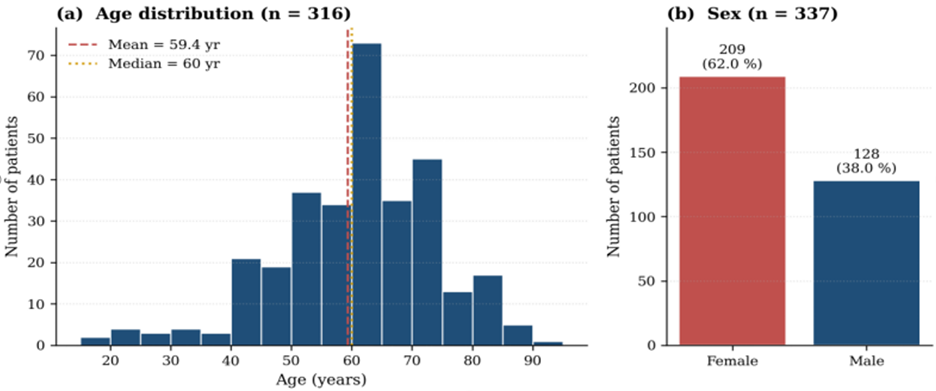
Figure 1: Demographic profile of study participants.
Systemic co-morbidities:
Regarding systemic comorbidities, a history of diabetes mellitus was present in 74 of 149 participants (49.7%), while hypertension was reported in 50 of 118 participants (42.4%).
Ocular co-morbidities:
Among ocular comorbidities, cataract was the most common finding, affecting 92 patients (27.2%). Diabetic retinopathy was present in 14 patients (4.1%), age-related macular degeneration in 6 (1.8%), and central retinal vein occlusion in 5 patients (1.5%)
Visual acuity:
Visual impairment was graded according to the best-corrected visual acuity (BCVA) of the worse eye. Mean logMAR BCVA was 1.39 ± 1.12 for the worse eye. Of the 315 patients with available data, 74 (23.5%) had normal or near-normal vision (≥6/12), 42 (13.3%) had mild visual impairment (6/12–6/18), 41 (13.0%) had moderate visual impairment (6/24–6/60), and 3 (1.0%) had severe visual impairment (<6/60–CF), whereas 155 patients (49.2%) had profound visual loss or blindness (HM/PL/NPL).
Glaucoma classification:
Only 81 patients were classified in the medica records as glaucoma sub-types. Primary open-angle glaucoma was identified in 18 patients (22.2%), pseudoexfoliative glaucoma in 16 (19.8%), neovascular glaucoma in 13 (16%), angle-closure suspect in 9 (11.1%), primary angle-closure glaucoma in 7 (8.6%), angle-closure glaucoma in 6 (7.4%), glaucoma suspect in 5 (6.2%), and uveitic glaucoma in 3 patients (3.7%).
Intra-ocular pressure IOP (the first visit):
The mean baseline intraocular pressure (IOP) was 20.98 ± 10.87 mmHg in the right eye and 19.95 ± 10.91 mmHg in the left eye. Elevated baseline IOP (≥21 mmHg) was observed in 123 of 324 right eyes (38.0%), 99 of 321 left eyes (30.8%), and in either eye in 164 of 332 patients (49.4%).
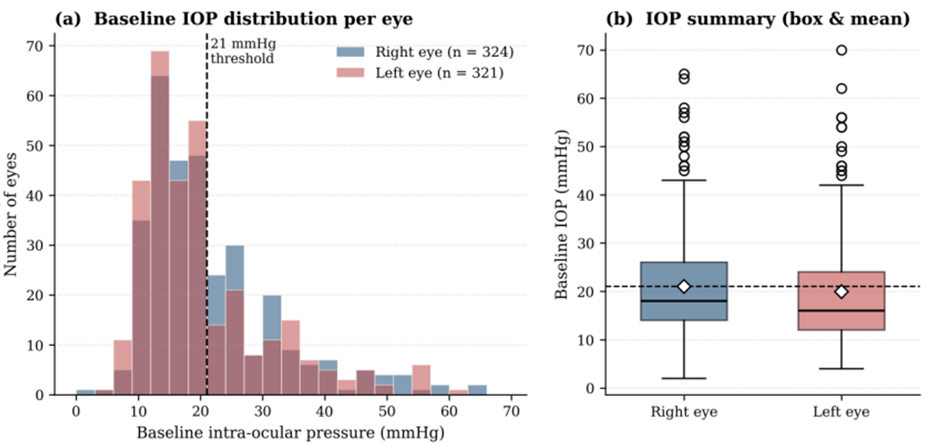
Figure 2
Pre-versus post treatment IOP:
Paired pre- and post-treatment IOP measurements were available for 169 right eyes and 168 left eyes. In the right eye, mean IOP decreased from 22.82 ± 11.47 mmHg at baseline to 16.93 ± 10.64 mmHg at the last follow-up, corresponding to a mean reduction of 5.89 ± 11.57 mmHg. In the left eye, mean IOP decreased from 20.52 ± 10.20 mmHg to 14.36 ± 6.57 mmHg, with a mean reduction of 6.16 ± 10.36 mmHg. These reductions were statistically significant in both eyes (Wilcoxon signed-rank test, p < 0.001). At the last follow-up, 134 of 169 right eyes (79.3%) and 144 of 168 left eyes (85.7%) achieved an IOP below 21 mmHg, whereas 118 (69.8%) and 127 (75.6%), respectively, achieved an IOP below 18 mmHg.
Cup/disc ratio (CDR):
Disease severity was graded by the cup-to-disc ratio (CDR) of the worse-affected eye: mild (≤ 0.5), moderate (0.6–0.8), and severe (≥ 0.9)
Disease severity was assessed according to the cup-to-disc ratio (CDR) of the worse eye. Among 152 patients with available data, 9 (5.9%) had mild disease (CDR ≤0.5), 44 (28.9%) had moderate disease (CDR 0.6–0.8), and 99 (65.1%) had severe disease (CDR ≥0.9)
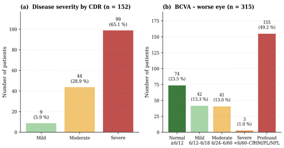
Figure 3: Disease severity and visual function profile.
Medical treatment:
Overall, 302 patients (89.3%) received medical treatment including topical IOP-lowering therapy and topical steroid. Carbonic anhydrase inhibitors were the most frequently prescribed medications, used by 241 patients (79.8%), followed by beta-blockers in 237 (78.5%), alpha-2 agonists in 129 (42.7%), prostaglandin analogues in 127 (42.1%), and topical corticosteroids in 3 patients (1.0%)
Combined medication:
Combination therapy was common, with 253 patients (83.8%) receiving more than one class of IOP-lowering medication. Among the treated patients, 108 (35.8%) were receiving two medications, 106 (35.1%) three medications, and 39 (12.9%) four medications, whereas 47 patients (15.6%) were controlled on a single drug class.
Surgical intervention:
A total of 189 patients underwent surgical intervention. Trabeculectomy was the most common procedure, performed in 77 patients (40.7%), followed by phacoemulsification in 75 (39.7%), YAG laser peripheral iridotomy in 37 (19.6%), combined phacotrabeculectomy in 34 (18.0%), bleb needling in 16 (8.5%), transscleral micropulse cyclophotocoagulation in 11 (5.8%), and intravitreal anti-VEGF injection in 7 patients (3.7%)
Response of IOP to intervention:
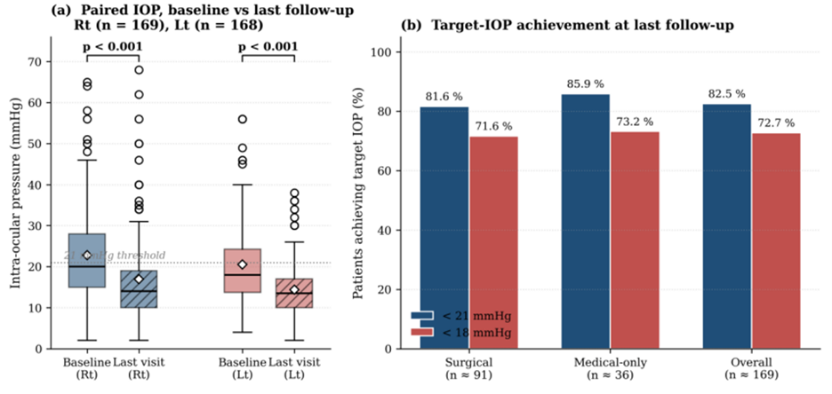
Figure 4: Response to intervention – IOP reduction and target achievement.
Postoperative complications:
were documented in 20 patients (5.9%) among those with available surgical follow-up data relation between disease severity and age, sex and diabetes:
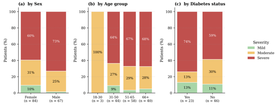
Figure 5: Disease severity (worse eye) stratified by selected patient characteristics.
Discussion
Demographics:
Age:
In our study most of the patients age ranged from 51-65 years. The same fact was proved by Kyei and colleagues. They even found glaucoma prevalence among population above 40% in 4 times more among sub-Saharan population than another ethnic group [5].
The pathophysiology of glaucoma explained the increased prevalence of the disease among adults above 50 years.
Incidence of glaucoma is increased among aging population. Aging is a known risk factor as neurological damage at the cellular level through oxidative stress. Ocular tissues involved in this process are the trabecular meshwork and schlemm’s canal. The disturbance of those structures would decrease the aqueous fluid outflow.
Laboratory studies have shown that retinal ganglia cells and axons are more vulnerable to IOP changes among older population. Structural changes associated with aging will increase the morbidity of the disease [6].
Sex:
Male gender predilection for glaucoma was noted by studies from South Korea, Ghana and Nigeria. In our study, female noticeably out rated males (62% and 38% respectively).
In our study, females considerably outnumbered males affected with glaucoma. The study by Vajaranant and colleagues revealed that female hormones might be a risk factor for glaucoma as females with early menopause are more susceptible to open angle glaucoma. Also, normal tension glaucoma is more prevalent among females. Previous studies found that women are at higher risk of developing angle closure glaucoma [7].
Intraocular pressure:
IOP is not only the most important risk factor of glaucoma, it is also the main modifiable factor which is targeted by medication and surgical intervention to slow or stop the disease’s progression [8].
In our study, 164 of 332 patients presented with IOP greater than 21. Only ten patients presented with IOP above 50 mm Hg.
Any patient presenting with IOP above 21mmHg is considered glaucomatous if this IOP is associated with the characteristic optic nerve head changes [9].
Glaucoma classification:
Primary open angle glaucoma is the most prevalent classification of glaucoma.
1 in 100 of general population might be affected with open angle glaucoma, POAG is usually asymptomatic in the early stages. On the other hand, angle closure glaucoma presents earlier with on and off visual symptoms and severe pain associated with the acute attacks [10].
Optic nerve head changes and glaucoma progression:
Optic nerve head cupping and pallor can be found in cases of compressive optic neuropathies, but the structural changes of the optic nerve head seen in glaucoma patients are unique and not usually seen in other etiologies of the optic nerve neuropathies. The variations of the normal optic nerve C/D ratio add to the challenge. Nevertheless, the longitudinal direction of CDR enlargement is found to be characteristic for glaucoma structural changes of the optic nerve head [11].
In the study by Kokou and colleagues, a vertical increase of CDR f 1.79% occurs annually is the study population, associated with neuro-retinal rim thinning and RNFL thinning of 1.64% and 0.93% respectively [12].
Increased diameter of the nerve cup optic associated with glaucoma indicating the loss in the retinal ganglion cells. The cup-disc ratio (CDR) is a known diameter of the disease progression and the resulting structural damage reflecting glaucoma severity.
Although recently there is a running debate regarding the specificity of CDR as an indicator for glaucoma diagnosis or prognosis, CDR is still a recognized popular glaucoma progression parameter. Therefore, OCT machines manufactured today usually include an in-built software for estimating CDR and comparing it with the saved data of normal population [13].
Glaucoma medical treatment:
Treatment options for glaucoma are chosen based on the classification of glaucoma and the baseline IOP. The most effective anti-glaucoma drugs nowadays are Prostaglandin F2alpha analogues. Topical beta-blockers can reduce the IOP by 20-27%. Carbonic Anhydrase Inhibitors (CAI) are available in both systemic and topical forms. Adrenergic agonists lower IOP by reducing aqueous production. combination therapy showed better efficiency and less side effects than prescribed multiple eye drops. 35% of our participants received two drugs-therapy, the percentage received three drugs. 13% required 4 drugs to control IOP in some point of the course of the disease. Studies proved that beta-blockers when added to a prostaglandine agonist, CAI, Adrenergic agonist or pilocarpine will provide better IOP lowering effect compared to the effect of each drug separately prescribed [14].
Prostaglandin analogue is considered the first line of medical treatment of glaucoma. In our study only 42.1% were using prostaglandin analogue while 79.3% were on CAI. According to Sarfo and colleagues the choice of medication for glaucoma in Sub-Saharan Africa depends on its price and availability. Sudan was in a state of war in the time of the study, unavailability of certain drugs might limit medication choice [15,16].
Disease severity, associated factors:
The African population showed a severe form of the disease compared with the Caucasians. They have a higher IOP on presentation and a more rapid progression of glaucoma [10,17].
Patients with diabetes: diabetic patients with uncontrolled fasting blood sugar are more susceptible to elevated IOP. Glaucoma is more prevalent among diabetic patients compared to non-diabetic patients. Studies emphasize on oxidative stress and cell apoptosis as common physiopathological processes shared between diabetes and glaucoma. Glial cells dysfunction is also a degenerative process shared by both diabetes and glaucoma which eventually leads to severe form of glaucoma progression among diabetic patients. Additionally, diabetes might cause structural changes of trabecular meshwork leading to elevation of IOP [18,19].
In our study, 74% of glaucomatous patients have a severe form of the disease compared to 59% of the non-diabetic cases.
In the Canary Islands study by Gonzalez-Hernandez and colleagues, 203,115 diabetic patients were followed for 15 years, the study revealed a higher glaucoma incidence over time, increasing with advanced age. They also concluded that vascular diabetic changes would add up to optic nerve structural changes in glaucomatous patients. The researchers concluded that diabetic patients are candidate for glaucoma screening especially with advanced age [20]. Aging process is associated with increased glaucoma severity and structural optic nerve head changes expressed as CDR changes, although in our study the age groups from (31-50 years), (51-65 years) and (>66 years) showed very approximate percentages of glaucoma severity which were: 64%, 67% and 68% respectively. Cases below 30 years showed only moderate CDR progression. These findings support the evidence that glaucoma among Africans progresses before 40 years into the severe form of the disease.
Glaucoma progression relation to the patient’s sex: in this study 73% of the male patients presents with the severe form of the disease compared to 60% of the females. As we discussed above the female patients outnumbered the male patient in our participants but they showed a milder form of the disease [21].
Sex hormone might have a protective role in glaucoma progression, estrogen in pre-menopausal females protects the optic nerve head in glaucoma slowing structural damage of the neural cells. On the other hands, males and post-menopausal females develop more structural damage with the same levels of IOP changes [22].
Conclusion
Glaucoma causes profound functional and structural damage with almost half of the patients ending blind. Lowering IOP which is the aim of different glaucoma therapies was achieved considerably with medical treatment and surgical interventions. Male gender, aging and diabetes are associated with more optic nerve damage among glaucoma patients.
Limitations:
The data was retrospectively retrieved from medical records so some parameters were missing.
The participants represent the Northern state, more diversity is required for more generalization of Sudanese population.
References
- Asiamah R, Kyei S, Owusu G, Agyiri PE. Prevalence of glaucoma in Africa: A systematic review and Bayesian meta-analysis. PloS one, 2025; 20(8): e0330567. https://doi.org/10.1371/journal.pone.0330567.
- Siegfried CJ, Shui YB. Racial Disparities in Glaucoma: From Epidemiology to Pathophysiology. Missouri medicine, 2022; 119(1): 49–54.
- Dietze J, Blair K, Zeppieri M, et al. Glaucoma. In: StatPearls [Internet]. Treasure Island (FL): StatPearls Publishing, 2026.
- Ezinne NE, Ojukwu CS, Ekemiri KK, Akano OF, Ekure E, Osuagwu UL. Prevalence and clinical profile of glaucoma patients in rural Nigeria-A hospital based study. PloS one, 2021; 16(12): e0260965. https://doi.org/10.1371/journal.pone.0260965.
- Kyei S, Obeng PA, Kwarteng MA, Assiamah F. Epidemiology and clinical presentation of glaucoma in a referral facility in Ghana: Any lessons for public health intervention? PloS one, 2021; 16(1): e0245486. https://doi.org/10.1371/journal.pone.0245486.
- Zhang Y, Huang S, Xie B, Zhong Y. Aging, Cellular Senescence, and Glaucoma. Aging and disease, 2024; 15(2): 546–564. https://doi.org/10.14336/AD.2023.0630-1.
- Vajaranant TS, Nayak S, Wilensky JT, Joslin CE. Gender and glaucoma: what we know and what we need to know. Current opinion in ophthalmology, 2010; 21(2): 91–99. https://doi.org/10.1097/ICU.0b013e3283360b7e.
- Fu DJ, Ademisoye E, Shih V, et al. Survival of medical treatment success in primary open-angle glaucoma and ocular hypertension. British Journal of Ophthalmology, 2024; 108: 1701-1707.
- Aboushahma, Yassen. Clinical Presentation of Newly Diagnosed Glaucoma Patients. Misurata Medical Sciences Journal, 2015; 2: 41-43. doi: 10.36602/mmsj/2015/02/03.
- Olawoye O, Kizor-Akaraiwe N, Pons J, Sarimiye T, Washaya J, Hughes S, et al. STAGE Research Group Clinical Characteristics and Stage at Presentation of Glaucoma Patients in Sub-Saharan Africa. Journal of glaucoma, 2022; 31(9): 717–723. https://doi.org/10.1097/IJG.0000000000002068.
- Lee EJ, Han JC, Kang M, et al. Longitudinal changes in optic disc cupping from the baseline in chiasmal lesion optic neuropathy and glaucoma. Sci Rep,2024; 14: 8889. https://doi.org/10.1038/s41598-024-59419-3.
- Kokou Vonor, Tchilabalo Tchodjoou, Yaovi Tété, Yawa Nagbé, Roger Ahlonko Kuaovi-Koko, Koffi Didier Ayéna, et al. Progression of Primary Open Angle Glaucoma on Optical Coherence Tomography of the Optic Nerve Head Open Journal of Ophthalmology, 2022. DOI: 10.4236/ojoph.2022.124031.
- McCalla A, Wang M, Eslami M, Kazeminasab S, Luo Y, Rana H, et al. Association Between Cup-to-Disc Ratio and Structural and Functional Damage Parameters in Glaucoma: Insights from Multiparametric Modeling. Translational vision science & technology, 2025; 14(4): 17. https://doi.org/10.1167/tvst.14.4.17.
- Crawley L, Zamir SM, Cordeiro MF, Guo L. Clinical options for the reduction of elevated intraocular pressure. Ophthalmology and eye diseases, 2012; 4: 43–64. https://doi.org/10.4137/OED.S4909.
- Fu DJ, Ademisoye E, Shih V, et al. Survival of medical treatment success in primary open-angle glaucoma and ocular hypertension British Journal of Ophthalmology, 2024; 108: 1701-1707.
- Crawley L, Zamir SM, Cordeiro MF, Guo L. Clinical options for the reduction of elevated intraocular pressure. Ophthalmology and eye diseases, 2012; 4: 43–64. https://doi.org/10.4137/OED.S4909.
- Sarfo JO, Mordi P, Aggrey EK, Quaicoe ASP, Attafuah PYA. Glaucoma prevalence and treatment in sub-Saharan Africa's elderly population: a scoping review. BMC geriatrics, 2025; 25(1): 255. https://doi.org/10.1186/s12877-025-05901-0.
- Song BJ, Aiello LP, Pasquale LR. Presence and Risk Factors for Glaucoma in Patients with Diabetes. Current diabetes reports, 2016; 16(12): 124. https://doi.org/10.1007/s11892-016-0815-6.
- Crawley L, Zamir SM, Cordeiro MF, Guo L. Clinical options for the reduction of elevated intraocular pressure. Ophthalmology and eye diseases, 2012; 4: 43–64. https://doi.org/10.4137/OED.S4909.
- Gonzalez-Hernandez M, Gonzalez-Hernandez D, Betancor-Caro N, Guedes-Guedes I, Guldager MK, Gonzalez de la Rosa M. Glaucoma Incidence and Progression in Diabetics: The Canary Islands Study Using the Laguna ONhE Application. Journal of clinical medicine, 2022; 11(24): 7294. https://doi.org/10.3390/jcm11247294.
- Jammal AA, Berchuck SI, Thompson AC, Costa VP, Medeiros FA. The Effect of Age on Increasing Susceptibility to Retinal Nerve Fiber Layer Loss in Glaucoma. Investigative ophthalmology & visual science, 2020; 61(13): 8. https://doi.org/10.1167/iovs.61.13.8.
- Maria Rizk, Alice Grise-Dulac, Damien Gatinel. Glaucoma in women: What do we know so far - A systematic review,AJO International, 2024; 1(1): 100013. https://doi.org/10.1016/j.ajoint.2024.100013.