Impactful Research Articles on Dieulafoy’s Lesions: Bibliometric Insights from the 50 Most Cited Studies
Tochukwu Ikpeze*, Mohamad Hijazi, Bassel Bitar, Fernando Mateo, Ankitha Vinaparti, Kenan Rahima and David Hess
Department of Internal Medicine, TriHealth, Cincinnati, OH, USA
Received Date: 31/08/2024; Published Date: 28/10/2024
*Corresponding author: Tochukwu Ikpeze, MD, MBA, Department of Internal Medicine, TriHealth, Cincinnati, OH, USA
Introduction
Dieulafoy’s lesions are complex, rare, and potentially life-threatening vascular malformations that cause gastrointestinal bleeding [1]. A Dieulafoy’s lesion is characterized by the protrusion of a normal blood vessel with a widened diameter, which protrudes into the mucosa [1,2]. Roughly 6.5% of all non-variceal, upper gastrointestinal bleeds are caused by Dieulafoy’s lesions [1,2]. Treatment currently consists of endoscopic manipulation using thermal or heat tropes, regional injection-epinephrine, or mechanical banding and hemoclips [1-3]. Identifying the most impactful articles addressing Dieulafoy’s lesions can be both beneficial and valuable to patient care and ongoing research endeavors.
Methods
The study design is a bibliometric analysis. In June of 2024, we used ISI Web of Science (v5.11, Thomas Reuter, Philadelphia, Pennsylvania, USA) to search for the following key phrases: “Dieulafoy’s Lesion “, “Dieulafoy’s disease” or “Dieulafoy’s ulcer”. Search areas included general surgery, gastroenterology, surgical endoscopy, radiology, oncology, and nuclear medicine and imaging. Articles were searched from 1900 to 2024.
The articles were ranked based on number of citations. The results were then evaluated to determine articles most clinically relevant to the management of Dieulafoy’s lesions. The top 50 articles that met the search criteria were further characterized on the basis of: title, author, citation density, journal of publication, year (and decade) of publication, institution, and country of origin.
Results
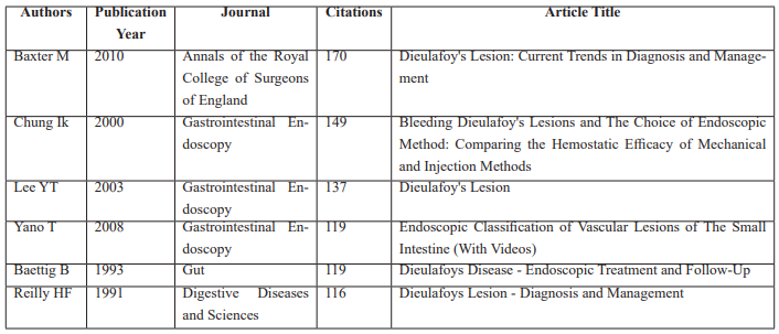
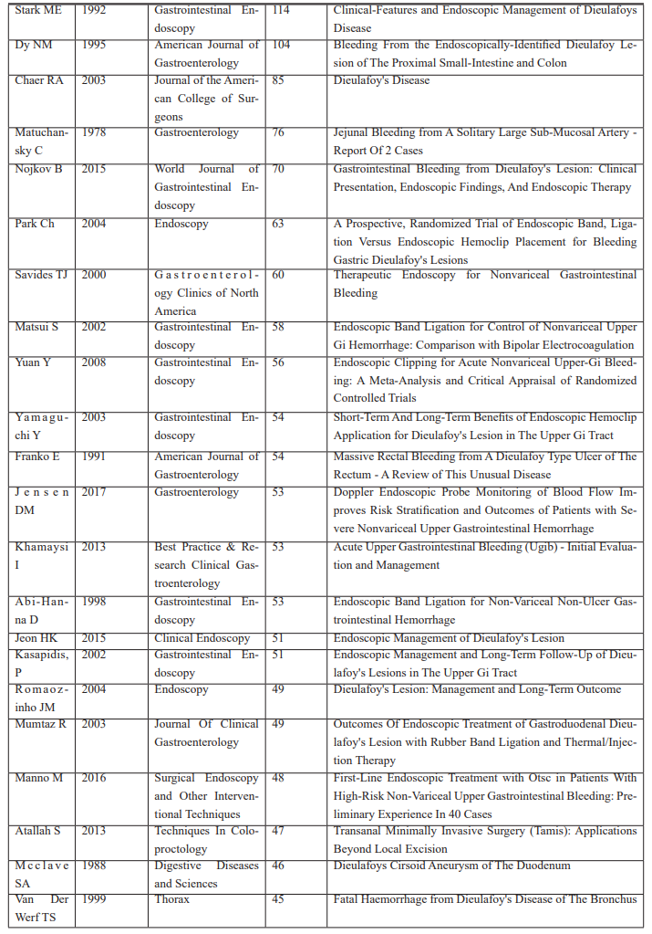
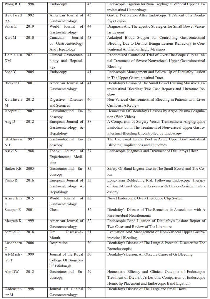
A total of 540 articles matched the search criteria. The most influential 50 articles ranged from 29 to 170 in number of citations. The articles were published between 1978 and 2021, and all articles were published in English. The top cited article was the 2010 work by Baxter et al. discussing the current trends in diagnosis and management of Dieulafoy’s lesions. The second most cited article was published in 2000 by Chung et al. and discussed endoscopic methods for bleeding Dieulafoy’s lesions. Third on the list was the article by Lee et al. discussing the clinical characteristics of Dieulafoy’s lesions (Table 1).
Table 1: Most Influential Articles.
Twenty publications (40%) originated from the United States, 6 (12%) from Japan, 4 (8%) from United Kingdom and South Korea, and 2 (4%) each from Portugal, Italy, and Switzerland.
Table 2: Countries of origin.

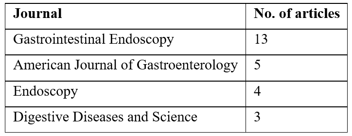
Table 3: Top journals of publication.
Most articles published on Dieulafoy’s Lesions were in Gastrointestinal Endoscopy (13). The second most common journal destination was the American Journal of Gastroenterology (5), followed by Endoscopy (4) and Digestive Diseases and Science with three (3) articles.
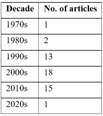
The 2000s was the most active decade of publication (18 papers) followed by 2010s with fifteen (15) articles published in that decade. This was followed by the 1990s with thirteen (13) articles and the 1980s with two (2) articles. The 1970s and 2020s were the least active with 1 published article per decade.
Table 4: Decades of publication.
A total of 48 institutions contributed to the top 50 articles. Mayo clinic and University of California (Los Angeles) contributed the most with two articles each (Table 6).
Table 5: Top institutions of publication.

There were two top published authors: Franko E with two articles and Jensen DM with two articles as well. The remaining authors published an article each.
Table 6: Top cited authors.

Discussion
Dieulafoy’s lesions are rare, upper gastrointestinal anomalies that are primarily managed by a multi-disciplinary team primarily composed of gastroenterologists, intervention radiologists, and vascular surgeons [4]. Historically, Dieulafoy’s lesions was treated with either gastrectomy or gastronomy [1,4]. However, endoscopic modalities have replaced the surgical approaches, which include mechanical banding with hemoclips, sclerotherapy with regional epinephrine or norepinephrine regional injection, and use of heat, thermal or plasma coagulation [1,4,5]. Understanding the top cited articles may serve as a vehicle to drive advances in Dieulafoy’s research.
The most cited article was the 2010 work by Baxter M, which discusses current trends in the Diagnosis and Management of Dieulafoy’s lesion [6]. The article was published in the Annals of the Royal College of Surgeons of England and cited 170 times. Using the Medline database, the authors identified 45 relevant articles, which were analyzed for the review. They found that 80% of all Dieulafoy’s lesions were caused by peptic ulcers, esophageal and duodenal erosions [6]. Moreover, if left undiagnosed or untreated, may cause a mortality rate of up to 80% as well. While he reported no consensus on the treatment of Dieulafoy’s at the time, therapeutic endoscopy was utilized up to 90% of the time, with angiography proposed as a viable alternative in the event of treatment failure. Lastly, the authors credited the reduction of mortality from 80% to roughly 9% to the advancements in endoscopy [6].
The second most cited article was published in 2000 by Chung et al. in the journal Gastrointestinal Endoscopy and discussed the choice of endoscopic methods on treating Dieulafoys lesions [7]. A total of 24 patients were randomized into either the mechanical endoscopic method using hemoclips and banding, or the endoscopic injection therapy. The authors found that less therapeutic endoscopic sessions were needed to achieve permanent hemostasis for the mechanical therapy group compared to the injection therapy group (1.17 vs 1.67) [7]. Moreover, a higher percentage of initial hemostasis was achieved in the mechanical therapy group compared to the injection therapy cohort. (91% vs 75%). Based on their results, the authors recommended endoscopic mechanical therapy for the treatment of Dieulafoy’s lesions when compared to other endoscopic approaches as it improves initial hemostasis, requires less endoscopic sessions, and has a lower rate of recurrent bleeding [7].
The third most cited article was the 2003 review article by Lee TY et al titled Dieulafoy’s Lesion, which was also published in the journal Gastrointestinal Endoscopy.8 In this article, the authors detail important characteristics of Dieulafoy’s lesion found on histologic examination. In the slides shown, they point out persistent artery tracking through the gastric submucosa, which ultimately becomes exposed, erodes, and causes bleeding [8]. They also call to attention different findings reported by other pathologists to help explain the lesion, which include pressure erosion of the ectatic vessel through the overlying epithelium, and abnormally fixed vessel in the muscularis mucosa. Ultimately, the authors subscribed to the findings that dysplastic changes leading to subintimal fibrosis, loss of elastic fibers near the necrotic arterial wall, and thinning of arterial fibers were terminal histologic finds that led to the Dieulafoy’s pathology [8].
The most recent highly cited paper on the list is by Jensen DM et al. published in 2021. This was a randomized controlled trial where 53 patients were placed into either standard endoscopic hemostasis using hemoclips [28] or large over-the-scope clips (OTSC) [25] for treatment of severe Dieulafoy’s bleeding [9]. Both treatment groups had similar baseline risk factors [9]. The authors found that the OTSC group had significantly less rebleeding (4% vs 28.6%), complications (0% vs 14.3%), and transfusions when compared to the standard endoscopic treatment group [9].
The oldest highly cited paper was the 1978 article by Matuchansky et al. and details the report of two isolated cases of massive intestinal bleeding from solidary submucosal arterial abnormality [10]. Both bleeding arteries were discovered in the jejunal submucosa with the aid of abdominal angiography. Histopathological examinations revealed characteristics like that of previously reported Dieulafoy’s lesions [10]. The authors’ proposed to call their findings “Dieulafoy-like erosion”. Treatments were not discussed in this article.
Most centers where the top cited articles originated from were in the United States. Several other countries such as Japan, South Korea, United Kingdom, and Italy were also represented in the top 50 cited list as well. The Gastrointestinal Endoscopy journal accounted for 26% of all publications on the list. The most active decade of publication was the 2000s. Two authors: Jensen DM and Franko E were the top cited authors.
We acknowledged some limitations to our study. First, given the dynamic natura of citations, the results from an earlier search (June 2024) may have changed if conducted at present. Nevertheless, a drastic or dramatic change would be unlikely. Another notable limitation is the publication frequency of a journal. For example, some journals may be published quarterly, while others are monthly or biweekly. Consequently, they may appear more often in the top cited list. Lastly, excluding non-English publications may have limited or altered the search results.
To our knowledge, this is the first study that evaluates the most clinically impactful, top cited research articles about Dieulafoy lesions. Most articles originated in the top-cited list originated from the United States and published in the 2000s. The most frequently cited journals were Gastrointestinal Endoscopy and American Journal of Gastroenterology. Understanding the rarity of these vascular abnormalities, historical findings, and current trends will help advance research in Dieulafoy’s lesions. Moreover, rapid advancements in endoscopic treatment will undoubtedly impact the incidence, prevalence, complications, and mortality of Dieulafoy’s lesions. As a result, it would be worthwhile to revisit the inquiry regarding the top cited Dieulafoy lesion articles in the future as this article describes the current state of the most impactful articles.
References
- Malik TF, Anjum F. Dieulafoys Lesion Causing Gastrointestinal Bleeding. [Updated 2023 Apr 27]. In: StatPearls [Internet]. Treasure Island (FL): StatPearls Publishing, 2024.
- Joarder AI, Faruque MS, Nur-E-Elahi M, et al. Dieulafoy's lesion: an overview. Mymensingh Med J, 2014; 23(1): 186-194.
- Fockens P, Tytgat GN. Dieulafoy's disease. Gastrointest Endosc Clin N Am, 1996; 6(4): 739-752.
- Levy AR, Broad S, Loomis Iii JR, Thomas JA. Diagnosis and Treatment of a Recurrent Bleeding Dieulafoy's Lesion: A Case Report. Cureus, 2022; 14(11): e32051. doi:10.7759/cureus.32051.
- Norton ID, Petersen BT, Sorbi D, Balm RK, Alexander GL, Gostout CJ. Management and long-term prognosis of Dieulafoy lesion. Gastrointest Endosc, 1999; 50(6): 762-767. doi:10.1016/s0016-5107(99)70155-0.
- Baxter M, Aly EH. Dieulafoy's lesion: current trends in diagnosis and management. Ann R Coll Surg Engl, 2010; 92(7): 548-554. doi:10.1308/003588410X12699663905311.
- Chung IK, Kim EJ, Lee MS, et al. Bleeding Dieulafoy's lesions and the choice of endoscopic method: comparing the hemostatic efficacy of mechanical and injection methods. Gastrointest Endosc. 2000;52(6):721-724. doi:10.1067/mge.2000.108040.
- Lee YT, Walmsley RS, Leong RW, Sung JJ. Dieulafoy's lesion. Gastrointest Endosc, 2003; 58(2): 236-243. doi:10.1067/mge.2003.328
- Jensen DM, Kovacs T, Ghassemi KA, Kaneshiro M, Gornbein J. Randomized Controlled Trial of Over-the-Scope Clip as Initial Treatment of Severe Nonvariceal Upper Gastrointestinal Bleeding. Clin Gastroenterol Hepatol, 2021; 19(11): 2315-2323.e2. doi:10.1016/j.cgh.2020.08.046
- Matuchansky C, Babin P, Abadie JC, Payen J, Gasquet C, Barbier J. Jejunal bleeding from a solitary large submucosal artery. Report of two cases. Gastroenterology, 1978; 75(1): 110-113.
- Ikpeze T, Mesfin A. The top 50 cited articles on chordomas. J Spine Surg, 2018; 4(1): 37-44. doi:10.21037/jss.2018.03.13

