Clinical Prediction and Characterisation of Digital Pressure Magnitude During Vertical Oscillatory Pressure on Patients with Chronic Low Back Pain
Taofik O afolabi1,*, Chidozie E Mbada2, Aanuoluwapo D Afolabi1, Esther Ajibaye1 and Olagoke Adaramola1
1Physiotherapy Department, Faculty of Medical Rehabilitation, University of Medical Sciences, Ondo, Nigeria.
2Medical Rehabilitation Department, Obafemi Awolowo University, Ile-Ife, Nigeria
Received Date: 30/05/2024; Published Date: 04/10/2024
*Corresponding author: Taofik O Afolabi, PhD, Department of Physiotherapy, University of Medical Sciences, Ondo, Nigeria
Abstract
Aim: This study predicted and characterized the optimal dosage for Vertical Oscillatory Pressure in terms of the Digital Pressure Magnitude (DPM), number of oscillations and frequency of treatment, somatotype, and pain intensity.
Method: Twenty- four (24) Participants with Low Back Pain were purposively recruited for dose optimization which involved the measurement of Digital Pressure Magnitude (DPM) with a Digital Pressure Sensor Machine (DPSM), consideration of a number of oscillations and frequency of treatment during VOP. Participants’ physical and clinical characteristics (pain intensity, height and weight, somatotype) were used to predict and characterize DPM.
Result: A linear regression model was used to predict the digital pressure magnitude based on pain intensity number of oscillations, frequency, height, weight, somatotype and pain intensity. It was found that pain intensity (B = 0.558, p = 0.181), height (B = -2.857, p = 0.935), weight (B= 0.612, p = 0.102), somatotype (B = -251, p = 0.624). PI (β= 0.288, p= 0.181), somatotype (β = -112, p=0.624), weight (β= 0.379, p=0.102), height (β=-017, p=0.935) were not significant predictors of DPM. Also, the second quartile (50th Percentile) was taken as the average quartile. At Q2, based on the age of the participants, the average DPM applied for each range: 20-40 years (20.0 Newton), 41-60 years (17.0 Newton) and greater than 60 years (19.0 Newton); Based on somatotype: Average DPM applied were: endomorph (18.0 Newton), ectomorph (22.0 Newton), mesomorph (14.5 Newton) and based on pain intensity: at low-intensity pain the force applied was 18.0 Newton, at high intensity pain, the force applied was 20.0 Newton.
Conclusion: Pain intensity, weight, height, and somatotype cannot predict digital pressure magnitude. However, the minimum and maximum force applied can be characterized based on pain intensity, somatotype, and age of the participants.
Keywords: Digital-Pressure; Vertical-Oscillatory-Pressure; Low-Back pain
Introduction
The magnitude of applied manual force is defined as the amount of force applied by the practitioner on a body [1]. The Force–Velocity Relationship explains how the force of fully activated muscle varies with velocity [2]. Applying oscillatory Posterior anterior mobilization techniques or Vertical Oscillatory Pressure, the maximum magnitude of applied force is usually reported as the mean of the force peaks that occur during a specified period [3,4]. Studies by [5], Harm et. al., (2010), and [6] all quantified mobilization force in terms of magnitude of force applied, frequency of oscillation, and duration of treatment.
Force magnitudes have been measured for mobilization techniques applied to the lumbar spine and, to a lesser extent, the thoracic and cervical spines [5]. Mobilizations are quantified by measurement of both the applied force and the displacement (movement) that occurs as a result of the applied force ([3], Harm et. al, 2010).
The magnitude of a mobilization or how hard the Physiotherapist pushes on the spine is usually reported as the magnitude of force ([5], Harm et al, 2010). However, the sensations felt by a patient during mobilization will be affected by the concentration of the applied force (namely, the pressure). Studies have noted that pressure is defined by force, the surface area where the force is applied can affect the pressure measured and likely the sensation of pressure felt by the patient (Maitland et al., 2005, [7]). For example, a person receiving mobilization will feel a different sensation if the force is applied over a smaller compared with a larger surface area, such as when a Physiotherapist mobilizes with a thumb grip vs a pisiform grip. In previous research and clinical trials on mobilization techniques, researchers have usually reported the force applied without measuring the surface area upon which it is applied (Matyas et al., 1985)
In some previous studies, Force applied during mobilization has been measured in connection with different parameters such as frequency of oscillation [5,8,9] also explained that the magnitude of force applied can be categorized in terms of the frequency of oscillation and the force amplitude ([4], Petty et al., 2001). The frequency of the oscillating force during mobilization is another potential source of variation between Manual Therapists or when Physiotherapists repeat techniques on subsequent occasions, which may be subject to the Physiotherapist’s skill level ([7], Maitland et al., (2001)] recommended applying mobilizations at a rate ranging between 1 oscillation every 2 seconds and 2 to 3 oscillations per second, depending on patient factors ([5], Cormdie et al., 2004, Lee et al., 2005). And research indicated that Physiotherapists apply digital pressure forces at a rate of 1 to 1.5 Hz (ie,1-1.5 oscillations per second) regardless of the grade of mobilization [6]. Moreover, [7] indicated that oscillatory movement can be applied on the spinous during mobilization about twice or thrice for about 15-30 seconds depending on the acuteness of the pain. [7,10] also noted that oscillatory movement is dependent on the somatotype of the patient. According to [7], a good and effective oscillation can be characterized by smoothness of the movement, amplitude, velocity, and rhythm. Another factor is the amplitude of force. Amplitude of Force amplitude is the difference between the minimum and maximum forces applied during mobilization; that is, the difference between the force recorded at the trough and the force recorded at the peak of an applied oscillatory force on a force-time curve. [7] noted that patient anthropometric makeup could interfere with force applied during VOP, however, other factors such as pain intensity, number of oscillations and frequency of treatment were not considered as determinants to predict the magnitude of force applied. Also, the average force applied has not been expressed in terms of these parameters such as pain intensity and somatotype.
In view of the above, this study predicted the digital pressure force applied on the body tissue with pain intensity and somatotype. Also, expressed the average force that can be applied in terms of pain intensity, somatotype, and age of the participant.
Methods
Twenty- four (24) Participants with Low Back Pain were purposively recruited for dose optimisation of dose which involved the measurement of Digital Pressure Magnitude (DPM) with Digital Pressure Sensor Machine (DPSM), consideration of some oscillations and frequency of treatment during VOP. Participants’ physical characteristics (pain intensity, height and weight, somatotype) were determined and used to predict and characterize DPM [11].
The somatotype was calculated and stratified. Quantification of the somatotype for participants Ten anthropometric dimensions were needed to calculate the anthropometric somatotype: Height, Weight, stretch physique, body mass, four skinfolds (triceps, subscapular, supraspinal, medial calf), two bone breadths (bi-epicondylar humerus and femur), and two limb girths (arm flexed and tensed, calf). The following descriptions are adapted from). All anthropometric parameters were taken with their standard instruments and participants were stratified into somatotypes (endomorph, ectomorph, and mesomorph), anthropometry calculation and stratification were done using the Heath Cater method.
Digital Pressure Magnitude (DPM) with Digital Pressure Sensor Machine (DPSM) was applied using the number of oscillations, and frequency of treatment during VOP application on patients with Low-Back pain based on somatotype and age. Twenty- four (24) Participants with LBP were recruited to participate in the study. Participants’ physical characteristics (pain intensity, height, and weight, somatotype) were used to predict and characterize DPM.
Results
Prediction of digital pressure magnitude
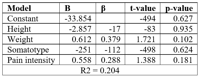
Linear regression model was used to predict the digital pressure magnitude based on pain intensity number of oscillations, frequency, height, weight, somatotype and pain intensity. It was found that pain intensity (B = 0.558, p = 0.181), height (B = -2.857, p = 0.935), weight (B= 0.612, p = 0.102), somatotype (B = -251, p = 0.624). And this equation was derived to predict Digital Pressure Magnitude: from the table above this equation can be derived to predict Digital Pressure Magnitude (DPM): - 33.85 – 2.85 (Height) + 0.612 (Weight) – 2.51(Somatotype) + 0.58 (Pain intensity).
Characterization of Digital Pressure Magnitude (DPM)
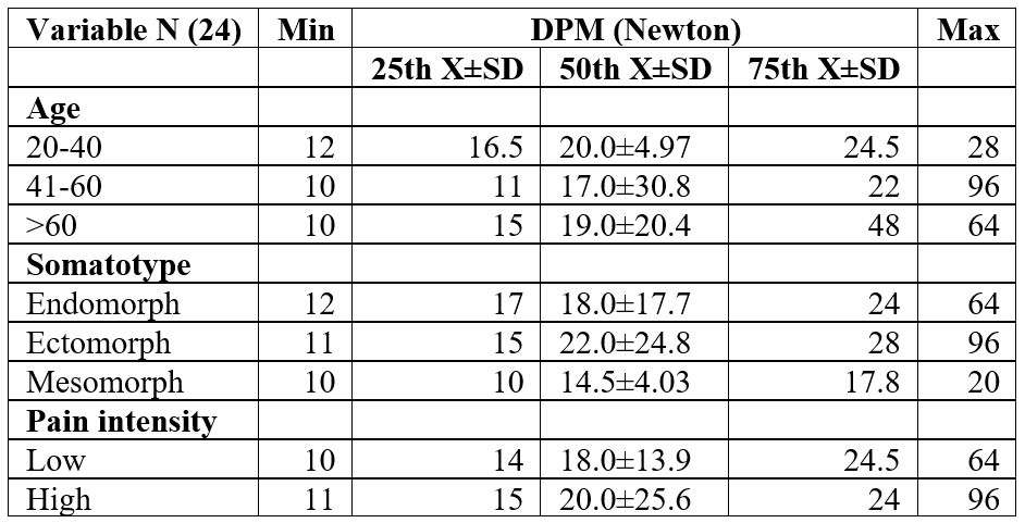
Using the percentile cut-point, Digital Pressure Force was characterized based on some characteristics such as age, somatotype, and pain intensity. This table showed the percentile cut at first (Q1=25th), second (Q2=50th), and third quartile (Q3=75th). The second quartile (50th Percentile) was taken as the average quartile. At Q2, based on the age of the participant, the average DPM applied for each characterized by age range: 20-40 years (20.0 Newton), 41-60 years (17.0 Newton), and greater than 60 years (19.0 Newton); Based on somatotype: Average DPM applied were: endomorph (18.0 Newton), ectomorph (22.0 Newton), mesomorph (14.5 Newton) and based on pain intensity: at low-intensity pain the force applied was 18.0 Newton, at high-intensity pain, the force applied was 20.0 Newton.
Table 1: Regression model to predict for digital pressure magnitude (Newton) using number of oscillations, height, weight, somatotype and pain intensity.

Table 2: Digital pressure magnitude characteristics of age, somatotype, pain intensity and percentile data of the participants.

Discussion
This research predicted and characterized the optimal dosage for VOP in terms of the Digital Pressure Magnitude (DPM), number of oscillations and frequency of treatment, somatotype, and pain intensity.
The result of this study showed that somatotype, pain intensity, weight, and height of the participant could not significantly predict the digital pressure magnitude (Table 2 This is partly supported by a study by [12,13] who explained that clinical characteristics could predict digital pressure magnitude unlike physical characteristic which could not. The findings of this study reaffirmed the conclusion of [10,12,14] that the effectiveness of digital pressure magnitude before the oscillatory phase of digital pressure may be attributed to the various clinical skills and physique of the Physiotherapists applying digital Pressure. Also, [5] supported the outcome of this study but highlighted other patient characteristics such as pain intensity, patient irritability, pain severity, and nature of patient symptoms which could influence digital pressure magnitude ([15], Magaray et al., 1984). However, [16] contradicted the outcome of this study which stated that Some Physical and clinical characteristics may predict the magnitude of digital pressure.
The prescription of treatment technique is known to be a dosage mechanism based on certain factors such as quantity and duration (Arsonson and Hardman, 1992, [17]). Dosage was developed for the safety of the process and monitoring of prognosis in a treatment [17]. Designing the correct dosage regimen is important for achieving the desired therapeutic efficacy and avoiding undesired effects. Several processes and techniques including exercise are in dosage (Maxwell, 2016). Research as indicated the application of dosage mechanisms in manual therapy (Harm et al., 2010; Snoddgrass et al., 2016), these aforementioned studies corroborated with this study on the development of average force considering different patient characteristics such as pain intensity, patient’s weight, patient’s height, and somatotype for effective guidance during digital pressure application [18-25].
Conclusion
Pain intensity, weight, height, and somatotype cannot predict digital pressure magnitude. However, the minimum and maximum force applied can be characterized based on pain intensity, somatotype, and age of the participants.
References
- Cuthbert SC, Goodheart GJ Jr. On the reliability and validity of manual muscle testing: a literature review. Chiropr Osteopat, 2007; 15: 4. doi: 10.1186/1746-1340-15-4.
- Duane, Knudson. Fundamentals of Biomechanics, 2007; Page 67-75.
- Chiradejnant A, Latimer J, Maher CG. Forces applied during manual therapy to patients with low back pain. Journal of Manipulative and Physiological Therapeutics, 2002; 25(6): 362-369. doi: 067/mmt.2002.126131.
- Harms MC, Bader DL. Variability of forces applied by experienced therapists during spinal mobilization. Clinical Biomechanics, 1997; 2: 393-399.
- Harms MC, Innes SM, Bader DL. Forces measured during spinal manipulative procedures in two age groups. Rheumatology (Oxford), 1999; 38: 267-274.
- Snodgrass SJ, Rivett DA, Sterling M, Vicenzino B. Dose optimization for spinal treatment effectiveness: a randomized controlled trial investigating the effects of high and low mobilization forces in patients with neck pain. J Orthop Sports Phys Ther, 2014; 44(3): 141-52.
- Nwuga VCB. Case Histories in Manual Treatment of Back Pain. 2nd Edition, William Publishers, 2007; page 199–211.
- Pentelka L, Hebron C, Shapleski R, Goldshtein I. The effect of increasing sets (within one treatment session) and different set durations (between treatment sessions) of lumbar spine posteroanterior mobilisations on pressure pain thresholds. Manual Therapy, 2012; 17(6): 526-530.

