Effectiveness of Ureteroscopy in clearance of Ureteric Calculi: An Observational Study
Abijith B Krishna1,* and Nilanjan Roy22
1Institute of Serology, ASCE, India
2Department of General Surgery, AFMC, India
Received Date: 17/02/2024; Published Date: 28/06/2024
*Corresponding author: Abijith B Krishna, ASCE, Institute of Serology, Kolkata, West Bengal, India
Abstract
Background: Urolithiasis, which has been a well-known entity for many centuries, is the formation of calculi in the urinary system, i.e., from the renal pelvis to ureter, urinary bladder or in the urethra. There is a shift in treatment in the recent years from open surgery to endourologic management due to advancement of technology, experience in procedures and miniaturisation of endourologic instruments. This study aims to evaluate the effectiveness of ureteroscopy in clearance of ureteric calculi.
Methods: A prospective observational study was conducted in patients who presented with ureteric stones to a tertiary care centre in India during the time period from January 2019 to June 2020. The patients of age group 18 to 60 years with ureteric calculi undergoing ureteroscopic removal of stones were included in the study. The patients with prior ureteric perforations, anatomic ureteric aberrations were excluded from the study. The flexible ureteroscopic lithotripsy is performed by the written informed consent obtained from all the patients. Stone clearance was assessed per-operatively and postoperatively. Complications and its management were also documented.
Results: The patients consisted of 79 males and 19 females with the male to female ratio being 4.16. The total number of calculi in this cohort was 106 stones with 11 patients having multiple calculi. The maximum representation was proximal ureteric region (29.2%), followed by distal ureteric (25.4%) and Vesico-Ureteric Junction (VUJ) region (24.5%). Right sided stones were commoner as compared to left side, with comparable distribution of calculi in both the sides. Stone size of 5 to 10 mm (58.5%) were in majority, followed by those sized 10-15mm and size less than 5 mm in diameter. The mean +SD for the calculi was 8.79+3.54 and 9.25+3.59 respectively for the right and left sided calculi. Average detection of X-rays for calculi in various regions is 65.1% with a range of 71.4% for renal calculi to lowest of 58% for proximal ureteric calculi. The average detection value of urolithiasis by ultrasonography was 84.9%, highest for mid-ureteric calculi at 100% and minimum of 65.4% for VUJ calculi. The overall efficiency in clearing urolithiasis by size was 97.9%. There was failure in clearance of stones in one patient each with distal ureteric urolithiasis and multiple calculi. The requirement for DJ stenting was felt the most in patients with multiple calculi who were stented universally for clearance of gravel, particles and small stones.
Conclusion: The flexible ureteroscopy with laser lithotripsy is recommended as a first line procedure for removal of ureteric and renal stones till size of 20 mm.
Keyword: Ureteric calculi; Ureteroscopy; Lithotripsy; Urolithiasis
Introduction
The introduction of minimally invasive surgery has brought a revolution in management of various surgical illnesses. Historically, urologists have always been at the forefront of minimally invasive surgery having mastered the procedures for lower urinary tract over the 20th century. Since the last two decades, introduction of ureteroscopes including flexible ureteroscopes and percutaneous techniques has extended the minimally invasive approach to upper urinary tracts as well. This technology permits urologists to extend their endoscopic expertise as high as the pyelo-calyceal system, not just for the stone disease, but also for a myriad of benign and malignant processes.
Urolithiasis is one of the commonest diseases of the urinary tract which is known to afflict mankind since time immemorial. Urinary calculi have been identified in the mummies of ancient Egypt and they were probably frequent, as can be deduced from references to bloody urine in old papyri. It is also mentioned in the medical manuscripts of various civilisations such as Greek, Indian, Babylonian and Egyptian civilisations. It is estimated to affect approximately 11% of men and 7% of women during their lifetime [1]. Urinary stones produce acute unilateral flank pain radiating to the groin, which is often accompanied by nausea, vomiting, and other urinary symptoms [2]. More than 1 million Patients presenting with suspected urolithiasis to emergency departments (ED) each year in the United States exceed the figure of one million [3]. A much greater yearly number is reported from the Indian subcontinent.
Treatment of stones with surgery had been described almost two thousand years back [4]. Surgeons in ancient India also described operative methods for bladder stones in 600 BC. Till 19th century, surgery did not take any large steps forward due to lack of effective means of controlling pain and the serious adverse effects of postoperative infection. With the advent of anaesthesia and development of new kinds of instruments, the surgical procedures multiplied and surgeons began to specialise in the field of Urology. The procedure for lithotripsy was developed to avoid high Renal Tubular Acidosis rate for open operations for bladder stones. It was first performed in Paris on a live patient by a surgeon called Civiale in the year 1824 [5]. He utilised a stiff metal tubular device which was passed blindly through the urethra to crush the bladder stones. The open surgery for urolithiasis remained the preferred mode of management till the 1980s. These open procedures produced good results as in 80 to 90% of all surgeries it was possible to remove all macroscopic stones. However, there was a high rate of accompanied adverse events such as post-operative pneumonia, infection, pain and bodily disfigurement.
In 1976, first percutaneous method was performed for removal of kidney stones [6]. The role of open surgery diminished with introduction of methods for percutaneous access to the kidney. It made possible for renal and ureteral stones to be removed either directly or through a variety of methods (ultrasound, electrohydraulic lithotripsy and mechanical crushing). It had better success rates than open surgery at 98 percent of all cases, but being an open invasive procedure, it was susceptible to complications such as post-operative bleeding and urosepsis. Historically till the introduction of ureteroscopic methods, larger renal stones requiring surgical methods were managed with percutaneous nephrolithotomy (Percutaneous Nephrolithotomy), Shockwave Lithotripsy (SWL), or a combination of both [7]. Open surgery has been used relatively sparingly for the treatment of stones, with only selective indications, such as patients presenting with complex collecting systems, having excessive morbid obesity, and those with extremely poor function of the affected renal unit [8].
The management of urolithiasis has undergone a drastic change since the early 1980s with the advent of various minimally invasive treatment modalities, technical advancements in endoscopic procedures and equipment, and also an increase in surgical skills in using them [9]. The miniaturisation of diameter of rigid, semi rigid and flexible ureteroscopes, increased scope flexibility, improvement of accessories, holmium laser technology as well as recent advancements in urological laparoscopic surgery has almost eliminated open stone surgery in the favour of minimally invasive stone removal procedures. Retrograde ureteroscopy for the management of urolithiasis is a sought-after modality being both patient and surgeon friendly. The success rate of retrograde ureteroscopy have been gradually improving over the years. The reasons behind the improved success rates are significantly better understanding of endoscopic anatomy of the ureter and kidneys, the availability of refined state-of-the-art ureteroscopes along with an array of useful gadgets, more advanced methods of intra-ureteral lithotripsy and better recognition and management of complications. In case of distal ureteral calculi there is hardly any doubt that retrograde ureteroscopy has a definite edge over Extracorporeal Shockwave Lithotripsy as it is more efficacious. But when it comes to proximal ureteric calculi the indications for ureteroscopy are less well defined.
This study was done to assess the effectiveness of ureteroscopic methods in the clearance of ureteric calculi. It also assesses the effectiveness of ureteroscopy for clearance of stones as per size and site. The complications of URSL and the need for DJ stenting after URSL were also assessed.
Methods
A prospective observational study was conducted in the department of General Surgery at Armed Forces Medical College and Command Hospital (Southern Command), Pune, a tertiary care, referral and teaching hospital for a period of one and a half years from January 2019 to June 2020. Both the out-patients and in-patients diagnosed with ureteric calculi undergoing ureteroscopic removal of stones in this center were recruited in the study after informed consent.
Patients of age group18 years to 60 years with ureteric calculi undergoing ureteroscopic removal of stones were included in the study. The patients who are already on DJ stent, who are undergoing a repeat URSL, those with prior ureteric perforation or anatomical ureteric aberrations (Duplicate ureter/ primary obstructive megaureter) or those who are on chronic use of analgesics and steroids were excluded from the study. Any patient who has undergone prior Extracorporeal Shockwave Lithotripsy before being taken up for URSL were also excluded.
The patients presenting with new ureteric calculi, diagnosed on imaging X Ray KUB and NCCT who undergo ureteroscopic removal of stones were screened for eligibility. They were informed about the trial and a written informed consent was obtained (consent form attached in annexure) before inclusion in the study.
The detailed demographic data and clinical features of patients including name, age, sex, occupation, socioeconomic status, general physical examination, systemic examination was collected in a pre-designed format. The flexible ureteroscopic lithotripsy procedure was performed under local anesthesia by the treating urologists. No pre-operative antibiotics were given. The patient was placed in lithotomy position, perineal region was prepared and cleaned with 10% povidone iodine and draped. The Flexible Ureteroscopy protocol was standardized. The ureteric stone was positioned in the excretory phase. The access to the ureter was performed by using an 8F flexible ureteroscope (Karl Storz SE & Co.) through the cystoscope under fluoroscopy. A Flexi-Tip Dual Lumen Ureteral Access Catheter was used for insertion of a second 0.038-inch guidewire. Subsequently, ureteral access sheath (12/14F) was used to insert the ureteroscope into the ureter. Disintegration of ureteric stones was performed using a 200-micron holmium laser fiber at an energy level of 0.5-0.8 J and at a rate of 10-20 Hz. Fragments measuring 2-3 mm or lower were extracted; larger stone fragments being further lased upon to reduce size. Plain NS was used as irrigating fluid at a perfusion pressure of was <40 cm H2O. The irrigating fluid volume used was <2,000 ml for all patients during the surgery. Stone clearance was assessed per operatively by direct visual uretero-pyeloscopy. It was also done post-operatively by means of radiograph, ultrasonography or CT scan depending on the surgeon's choice. Evidence of impaction and requirement for retropulsion for stones for management were noted. DJ stenting was done in cases of high stone load, proximal impacted stones, residual stone being present or when excessive graveluria was observed. The secondary outcome of post-operative pain was assessed further with the VAS scale (ranging from 1-10) on days 2 and 14. Episodes of hematuria were noted and any other complications observed were investigated as per requirement. The patients were further followed up at 6 months. Any residual calculus, impacted fragments, pain or persistent hematuria were documented.
All the data was analysed using the SPSS Ver. 22 software. The student’s t test used for analysing the quantitative variables with normal distribution. The Chi (χ2) square test was used where distribution was skewed and for categorical variables.
Results
A total of 98 patients were recruited in this study to assess the effectiveness of ureteroscopy in treating ureteric calculi. Among them 79 were males and the rest were females (Figure 1). The age of patients ranged between 21 to 60 years with majority being in the group 31-40 years in both genders. The χ2 value noted was 1.7588 with p – value of 0.6239 which was statistically not significant. The mean age was 39.9 years with SD of 8.73 years (Table 1). The size of the calculi is shown in Table 2. The maximum number of ureteric calculi were found in proximal ureteric region (29.2%), followed by distal ureteric (25.4%) and vesico-ureteric junction (VUJ) region (24.5%). On comparing for both sides, no significant difference was noted. The χ2 value noted was 1.1242 with p – value of 0.89 which was statistically insignificant (Figure 2).
The renal calculi were found to be smallest in number in our cohort with a total of seven calculi (7.7%), was usually found in association as a part of multiple calculi disease in a single affected individual and the usual size was less than 5 mm.
The next least common distribution site was mid ureteric region which accounted for only fifteen calculi (14.1%).
The majority commonest sized calculi found in our cohort comprised of size range from 5.1 mm to 10mm at almost 58.5% of the cohort (Table 2). The next commonest size was 10.1 – 15 mm calculi at 23.6%. No calculi of sizes more than 20 mm were found in the cohort (Figure 3). The distribution between the left and right sided calculi was again insignificant. The mean +SD for the calculi was 8.79+3.54 and 9.25+3.59 respectively for the right and left sided calculi. On comparing the means again no significant difference was noted (p-value 0.05).
On taking the CT scan finding as the gold standard method for detection of urinary calculi, the average detection of X-rays for calculi in various regions is 65.1% with a range of 71.4% for renal calculi to lowest of 58% for proximal ureteral calculi (Table 3). The similar average value for the ultrasonography detection of urolithiasis was 84.9%. The range of detection with USG was highest for mid-ureteric calculi at 100% and minimum of 65.4% for VUJ calculi (Figure 4). The distribution was non-significant with χ2 values of 1.685 corresponding to the p-value of 0.98.
A vast majority of calculi (80.2%) were associated with hydro-uretero-nephrosis (Figure 5). With the resolution and removal of stones after Flexible Ureteroscopy, majority of these resolved spontaneously. Multiple and impacted calculi were noted in 11 patients each.
The overall efficiency in clearing urolithiasis by size was approximately ⁓98%.(Table 4) All the stones of size 5-10mm were cleared by 6 months. There was failure to clear stones in single instance for both the sizes of 10-15 mm and 15-20 mm. in both these cases, there was persistence of single renal stone of size 4 mm or more which was taken as failure (Figure 6).
The overall efficiency in clearing urolithiasis by site again was approximately ⁓98% (Figure 7). There was failure in clearance of stones in one patient each with distal ureteric urolithiasis who developed renal stone > 4mm on follow-up and in a single patient with multiple calculi (Table 5).
The requirement for DJ stenting was felt the most in patients with multiple calculi who were stented universally for clearance of gravel, particles and small stones. The next commonest requirement was in patients of proximal ureteric calculi (⁓52%) followed by patients with distal ureteric calculi (⁓45%) (Figure 8). The least requirement was for patients with VUJ calculi. The complications of the procedures documented (Figure 9) included significant pain,hematuria ,graveluria and persistent calculus (Table 7).
Table 1: Age distribution of the patients.
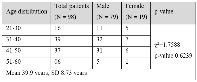
Table 2: Size of Calculi.

Table 3: Efficacy of radiological modalities in detection of urolithiasis.
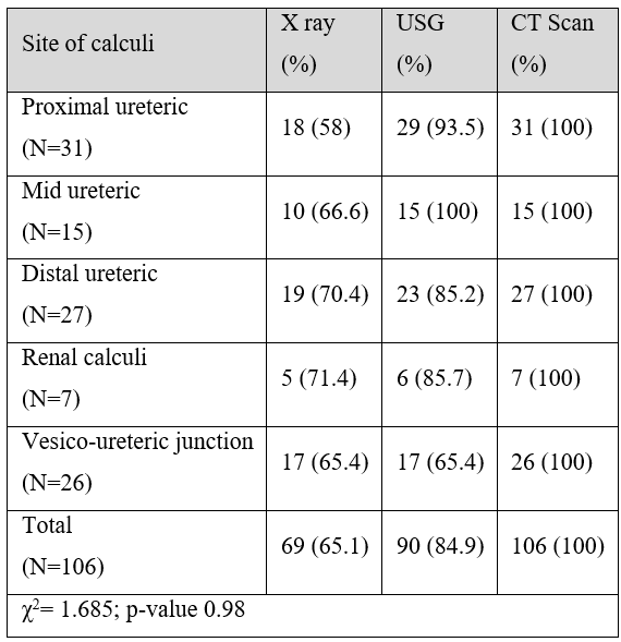
Table 4: Clearance of stone by ureteroscopy.
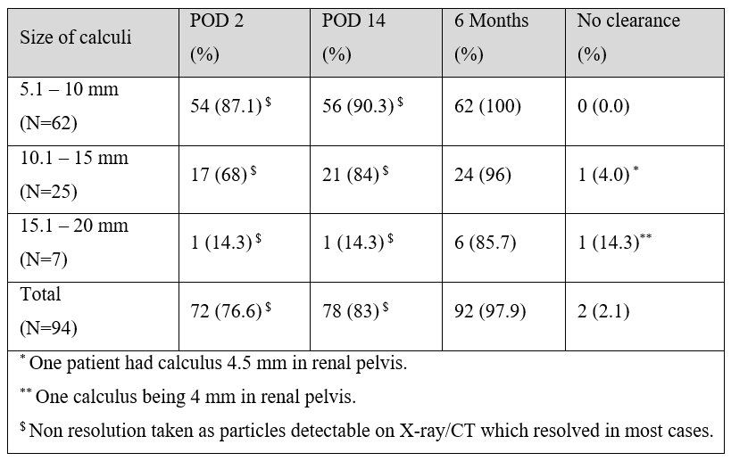
Table 5: Clearance of stone by ureteroscopy by site.
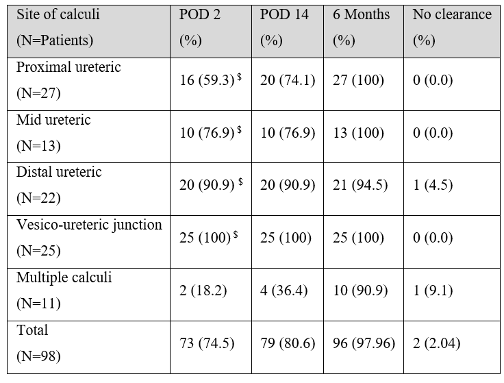
Table 6: Requirement for DJ stenting.
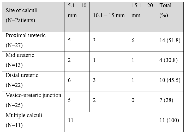
Table 7: Post-op complications post ureteroscopy.
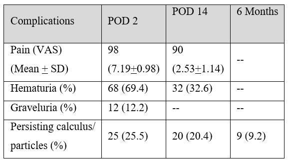

Figure 1: Gender distribution in percentages.

Figure 2: Distribution of calculi according to location.

Figure 3: Size comparison of calculi.

Figure 4: Comparison of other modalities with CT scan for detection of urinary calculi.

Figure 5: Complications due to calculi.

Figure 6: Stone clearance depending on size.
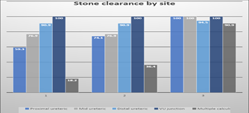
Figure 7: Stone clearance depending on site.
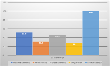
Figure 8: Requirement for DJ stenting.
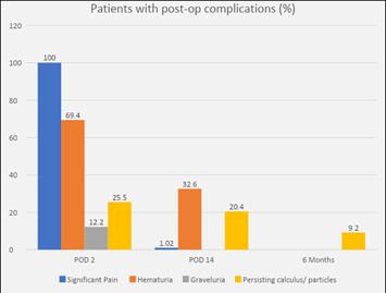
Figure 9: Patients with post op complications.
Discussion
Urolithiasis is one of the commonly encountered diseases in urology practice. I have a prevalence of 2 to 3 % in general population [10]. Flexible ureteroscopy is rapidly becoming the treatment of choice in ureteric and renal calculi due to lower complication rates and high efficacy.Recently a novel robotic catheter system has also been developed for performing retrograde ureterorenoscopy. Desai et al. had performed remote robotic flexible ureterorenoscopy bilaterally in five pigs using a 14F robotic catheter system [11]. The potential advantages of robotic flexible URS are multiple including an increased range of motion, instrument stability, and improved ergonomics. Still relevant comparative studies are warranted in addition to refinement of this technology.
The advantage of flexible ureteroscopy is that, it can also be employed as day care procedure and patient can be discharged on same day [12]. It is most effective at vesico-ureteric junction stones and becomes progressively difficult as the stone becomes larger and more proximal in location. The factors determining the effectiveness is clearance of stones, lesser post-operative complications and reduced invasiveness of procedure. In addition, cost factor and availability of facilities also determines the treatment modality used. This prospective study was done to determine the effectiveness of ureteroscopy in clearing ureteric calculi, study its complications and also assess the requirement of DJ stenting.
In our study a total of 98 patients were treated for ureteric calculi by Flexible Ureteroscopy and wherever required DJ stenting was also performed during the procedure. The patients consisted of 79 males and 19 females with the male to female ratio being 4.16. The likely reason for this disparity in the representation of both genders is that the study was being conducted in an army service hospital. Additionally, males are also more prone to urolithiasis due to their work profile and likely to solicit medical attention for the problem. Similar findings have been noted in other studies in the tropical Asian region [13], even though the overall ratio has already fallen below 1 in India [13]. The age of patients in the study ranged between 21 to 60 years with overwhelming majority being in the group 31-50 years (77.5%) in both genders. This was also comparable to other recent studies in Indian subcontinent by Kale et al [14] and Silva et al [15]. Similar results have also been noted in other countries in the Asian region [13]. The mean age noted for this cohort was 39.9 years with a SD of 8.73 years. The overall age distribution for both the genders was comparable.
The total number of calculi in this cohort was 106 stones with 11 patients having multiple calculi. The maximum representation was proximal ureteric region (29.2%), followed by distal ureteric (25.4%) and vesico-ureteric junction (VUJ) region (24.5%) followed by mid-ureteric region and least common were renal calculi (usually a part of complex multiple calculi in single individual). Right sided stones were commoner as compared to left side, even though the distribution of calculi were comparable in both the sides. On comparing the size of stones, the incidence of stone size of 5 to 10 mm (58.5%) were in majority. The next commonest size noted was 10-15mm followed by smaller stones less than 5 mm in diameter. No stone more than 20 mm was reported in the present study. The distribution between the left and right sided calculi based on size was again insignificant. The mean +SD for the calculi was 8.79+3.54 and 9.25+3.59 respectively for the right and left sided calculi. On comparing the means no significant difference was noted (p-value 0.05). A vast majority of calculi (80.2%) were associated with hydro-uretero-nephrosis. With the resolution and removal of stones after Flexible Ureteroscopy , majority of these resolved spontaneously. Multiple and impacted calculi were noted in 11 patients each.
On taking the CT scan finding as gold standard method for detection of urinary calculi, the average detection of X-rays for calculi in various regions is 65.1% with a range of 71.4% for renal calculi to lowest of 58% for proximal ureteric calculi. The average detection value of urolithiasis by ultrasonography was 84.9%. The range of detection with USG was highest for mid-ureteric calculi at 100% and minimum of 65.4% for VUJ calculi. The distribution was non-significant with χ2 values of 1.685 corresponding to the p-value of 0.98.
Calculus clearance rate is the most important factor in effectiveness of removal of stones by enabling the patient to have a symptom free life. It is defined as no residual stone or residual stone less than 4mm in the last imaging examination (X-ray, ultrasonography, or CT) without clinical symptoms on follow-up 1 ~ 3 months after the operation [16]. The overall efficiency in clearing urolithiasis by size was 97.9%. In present cohort, all the stones of size 5-10mm were cleared by 6 months. Rayamajhi et al [17] and Tripathy et al [18] have also reported stone clearance rates > 90% in stones less than 10 mm. It was in contrast to the study done by Preminger et al who reported clearance rates of only 80% with ureteroscopy. However, these rates were noted with semi-rigid and early flexible ureteroscopes; which have now dramatically improved with improving technology of visualisation, flexible material and better grasping devices and laser lithotripsy. There was failure to clear stones in single instance for individuals with stones sizes each of 10-15 mm and 15-20 mm. In both these cases, there was persistence of single renal stone of size 4 mm or more on follow up at 6 months which was taken as failure of clearance [16]. The study by Darakh et al who performed semirigid ureteroscopy for stones >15mm reported a success rate of only 66.67%. [12]. In our series it was noted to be 85.7%. Chen et al [16] in their meta-study comparing Flexible Ureteroscopy and Percutaneous Nephrolithotomy also concluded that stone clearance rate decreases with the increasing size of ureteric calculi similar to findings in the present index study. The final clearance rates noted in the study by Li et al [19] was 87.8% and 94.5% with Flexible Ureteroscopy alone and Flexible Ureteroscopy with metallic ureteric stent respectively.
The overall efficiency in clearing urolithiasis by site again was 97.96%. There was failure in clearance of stones in one patient each with distal ureteric urolithiasis who developed renal stone > 4mm on follow-up and in a single patient with multiple calculi. According to the American Urological Association /EAU ureteral stones guideline panel the stone free rate for ureteroscopy (URS) in the treatment of upper ureteric calculi is around 81%. In our study there was complete clearance of stones with Flexible Ureteroscopy and laser lithotripsy in proximal ureteric stones. The study by ElGanainyl et al in 2009 [20], reported a stone clearance rates of 91% with semirigid ureteroscope for stone sized 9-20 mm. It will be prudent to say that our study proves the clearance rates to be better with better flexible ureteroscopes.
The requirement for DJ stenting was felt the most in patients with multiple calculi who were stented universally for clearance of gravel, particles and small stones. The next commonest requirement was in patients of proximal ureteric calculi (⁓52%) followed by patients with distal ureteric calculi (⁓45%). The least requirement was for patients with VUJ calculi. All patients in the cohort reported substantial discomfort or pain (VAS >6) on Day 2 post procedure, however, this proportion decreased to insignificant levels by two weeks (1.02%). Hematuria was noted in 69% patients on day 2, which decreased to 32.6% by two weeks and was reported nil on long term follow-up at 6 months. Graveluria was reported only in 12.2% cases on POD 2, nil thereafter. A proportion of 9.2 % patients had persistent calculi on follow-up, however only in two patients (2.04%) it was significant (>4mm) requiring further follow-up.
Conclusion
Flexible ureteroscopy is rapidly becoming the treatment of choice in ureteric and renal calculi due to lower complication rates and high efficacy with an added advantage of being employed as day care procedure. Present study had a total of 98 patients treated for ureteric calculi by Flexible Ureteroscopy.DJ stenting wherever required was also performed during the procedure. The patients consisted of 79 males and 19 females with the male to female ratio being 4.16. The total number of calculi in this cohort was 106 stones with 11 patients having multiple calculi. The maximum representation was proximal ureteric region (29.2%), followed by distal ureteric (25.4%) and Vesico-Ureteric Junction (VUJ) region (24.5%). Right sided stones were commoner as compared to left side, with comparable distribution of calculi in both the sides. Stone size of 5 to 10 mm (58.5%) were in majority, followed by those sized 10-15mm and size less than 5 mm in diameter. The mean +SD for the calculi was 8.79+3.54 and 9.25+3.59 respectively for the right and left sided calculi. Average detection of X-rays for calculi in various regions is 65.1% with a range of 71.4% for renal calculi to lowest of 58% for proximal ureteric calculi. The average detection value of urolithiasis by ultrasonography was 84.9%, highest for mid-ureteric calculi at 100% and minimum of 65.4% for VUJ calculi. The overall efficiency in clearing urolithiasis by size was 97.9%. In present cohort, all the stones of size 5-10mm were cleared by 6 months. There was failure to clear stones in single instance for individuals with stones sizes each of 10-15 mm and 15-20 mm. In both these cases, there was persistence of single renal stone of size 4 mm or more on follow up at 6 months which was taken as failure of clearance. There was failure in clearance of stones in one patient each with distal ureteric urolithiasis who developed renal stone > 4mm on follow-up and in a single patient with multiple calculi. The requirement for DJ stenting was felt the most in patients with multiple calculi who were stented universally for clearance of gravel, particles and small stones. The next commonest requirement was in patients of proximal ureteric calculi (⁓52%) followed by patients with distal ureteric calculi (⁓45%). At two weeks post procedure, no patients had significant pain or graveluria. 32,6% had some hematuria and 20.4% had evidence of small calculi.The flexible ureteroscopy (Flexible Ureteroscopy ) with laser lithotripsy is recommended as a first line procedure for removal of ureteric and renal stones till size of 20 mm.
Acknowledgements: We acknowledge Dr. Ambily Sahadevan ADMO/CH/GRC for the technical assistance.
Funding: None
Conflict of interest: None
Ethical approval: Ethical approval obtained
References
- Scales CD, Smith AC, Hanley JM, et al. Project Urologic Diseases of America Project. Prevalence of kidney stones in the United States. Eur Urol, 2012; 62: 160-165.
- Teichman JMH. Clinical practice. Acute renal colic from ureteral calculus. N Engl J Med, 2004; 350: 684-693.
- Fwu C-W, Eggers PW, Kimmel PL, et al. Emergency department visits, use of imaging, and drugs for urolithiasis have increased in the United States. Kidney Int, 2013; 83: 479-486.
- Drach GW. Surgical overview of urolithiasis J Urol, 1989; 141: 711.
- Butt AJ. Treatment of urinary lithiasis Springfield, Illinois Charles С Thomas, Publishers, 1960.
- Bernstrom I, Johanson В Percutaneous pyelolithotomy a new extraction technique Scand J Urol Nephrol, 1977; 10: 257.
- Wiesenthal JD, Ghiculete D, D’A Honey RJ, Pace KT. A comparison of treatment modalities for renal calculi between 100 and 300mm2: Are shockwave lithotripsy, ureteroscopy, and percutaneous nephrolithotomy equivalent? J Endourol, 2011; 25: 481-485.
- Preminger GM, Assimos DG, Lingeman JE, et al. Chapter 1: AUA guideline on management of staghorn calculi: Diagnosis and treatment recommendations. J Urol, 2005; 173: 1991–2000.
- Breda A, Ogunyemi O, Leppert JT, et al. Flexible ureteroscopy and laser lithotripsy for single intrarenal stones 2 cm or greater—is this the new frontier? J Urol, 2008; 179: 981–984.
- Menon M, Parulkar BC, Drach GW. Urinary lithiasis: etiology, diagnosis and medical management. In: Walsh PC, et al., eds. Campbell's Urology. 7th ed. Philadelphia: Saunders,1998; 2661-2733.
- Desai MM, Aron M, Gill IS. Flexible robotic retrograde renoscopy: description of novel robotic device and preliminary laboratory experience. Urology, 2008; 72: 42–46.
- Darakh PP, Ramesh RB, Doshi C. Comparative study of different modalities of treatment for large upper ureteric calculi. International Surgery Journal, 2019; 6(5): 1534-1539.
- Liu Y, Chen Y, Liao B, Luo D, Wang K, Li H, et al. Epidemiology of urolithiasis in Asia. Asian journal of urology, 2018; 5(4): 205-214.
- Kale SS, Ghole VS, Pawar NJ, Jagtap DV. Inter-annual variability of urolithiasis epidemic from semi-arid part of Deccan Volcanic Province, India: climatic and hydrogeochemical perspectives. Int J Environ Health Res, 2014; 24: 278-89.
- Silva GR, Maciel LC. Epidemiology of urolithiasis consultations in the hilade valley. Rev Col Bras Cir, 2016; 43: 410-415.
- Chen Y, Wen Y, Yu Q, Duan X, Wu W, Zeng G. Percutaneous nephrolithotomy versus flexible ureteroscopic lithotripsy in the treatment of upper urinary tract stones: a meta-analysis comparing clinical efficacy and safety. BMC urology, 2020; 20(1): 1-2.
- Rayamajhi BB, Khadka A, Thapa N. A Prospective Study to assess the effectiveness of extracorporeal shock wave lithotripsy versus ureteroscopy for proximal ureteral calculi between sizes 5 to 10 mm. MJSBH, 2020; 19(2): 65-69.
- Tripathi SP, Jain DK, Dinesh M, Pathak P. Comparative study of ureteroscopy versus extracorporeal shock wave lithotripsy in management of upper ureteral calculi. IJMHS, 2018; 8(8): 88-94.
- Li T, Sun X, Li X, He Y. Flexible ureteroscopy lithotripsy combined with metallic ureteral stents for the treatment of patients with upper urinary tract calculi. Experimental and Therapeutic Medicine, 2020; 20(4): 3330-3335.
- ElGanainy E, Diaa AH, Elgammal MA, Abd- Elsayed AA, Shalaby M. Experience with impacted upper ureteral Stones; should we abandon using semi rigid ureteroscopes and pneumatic lithoclast? Int Arch Med, 2009; 2: 13.

