The Impact of Petroleum Polluted Environment on Female Reproductive Function in Ogoni Rivers State, Nigeria
Blessing L Dum-Awara1, Solomon M Uvoh2,*, Onokpite Emmanuel3 and Benson C Ephraim-Emmanuel4
¹,²Department of Human Physiology, Faculty of Basic Medical Sciences, College of Health Sciences University of port Harcourt, Rivers State Nigeria
³Department of Anaesthesia and Intensive Care, Faculty of Clinical Sciences, College of Medicine, Delta State University Abraka, Delta State Nigeria
4Environmental Health, Africa centre of Excellence for public Health and Toxicological Research, University of Port Harcourt, Nigeria
Received Date: 06/09/2023; Published Date: 02/02/2024
*Corresponding author: Dr. Solomon M Uvoh, Department of Human Physiology, Faculty of Basic Medical Sciences, College of Health Sciences University of port Harcourt, Rivers State Nigeria
Abstract
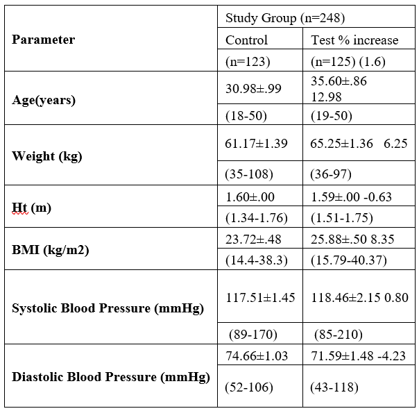
Infertility is one of the major global problems experienced among women of child bearing age on daily basis, hence necessitating research for the study of reproductive function among female residents of petroleum host communities. The number of female subjects in petroleum impacted environment studied was one hundred and twenty-five compared with one hundred and twenty-three from non-petroleum impacted environment. Methods used for the determination of serum hormonal profile was Tiets and layman method while auscultatory method was employed for blood pressure determination and the weight divided by the square of its height for body mass index. The result from this study indicates a percentage increase in the anthropometric parameters among the test subjects group compared with the control. However, there was a decrease in height percentage (-0.63%) among the test group. The duration of menstrual bleeding and loss of pregnancy in the test group was significantly higher (4.53days and 0.9) compared with control (4.00days and 0.18) group. The parity (2.73), menstrual cycle (28.29), and age at menarche (14.98) was significantly higher among control subjects compared with the test (14.23) group and a non-significant p-value observed. The test group hormonal profile indicates a significant level of estrogen and progesterone decrease with an increase in luteinizing and follicle stimulating hormone observed among the control compared with test group. Furthermore, there was a positive correlation between pregnancy loss and serum vanadium including serum lead though weak. This study has clearly shown the adverse effect of petroleum polluted environment on endocrine and general physiology of females within child bearing age resulting in developmental disorders and reproductive malfunction and pregnancy loss among women residents in petroleum polluted environment.
Keywords: Hormones; Estrogen; Progesterone; Menarche; Females; Petroleum
Introduction
The continuity of life from its onset of origin to present day is possible only because of the normal functioning of the reproductive organs associated with the endocrine glands. Hormones are chemical substances built in the endocrine glands within the body. These chemicals control most major bodily functions, from simple primary needs eg hunger to complex systems like reproduction, emotions and mood. Hormones are vital for natural developmental role such as growth, development and Metabolism i.e., how your body gets energy from food consumed, Sexual function, Reproduction and Mood.
Reproductive hormones are the chemicals produced and released to regulate reproduction while the interaction of these hormones controls the reproductive cycle, the female reproductive system is primarily regulated by five major hormones including estrogens, progesterone, gonadotropin releasing hormone, FSH and LH. These hormones play crucial role in different stages of development in female reproductive system [1,2].
Estradiol and progesterone are produced in ovaries of non-pregnant females. Sex Hormones such as estradiol and progesterone in females and testosterone in males are produced in the gonads under the influence of follicle stimulating hormone and luteinizing hormone.
FSH, Luteinizing hormone and Progesterone
Follicle-Stimulating Hormone (FSH) is a gonadotropin, a glycoprotein polypeptide hormone synthesized and extracted by the gonadotropin cells of the Anterior Pituitary Gland (APG), which regulates the development, growth, pubertal maturation, and reproductive processes consisting of two polypeptide units. The alpha subunits of the glycoprotein’s of FSH consist of 96ᾳ and has a beta subunit of 111 amino acids (FSH β), which confers its specific biological action, and is answerable for interaction with the FSH receptor [3]. In females, FSH initiates follicular growth, specifically affecting granulosa cells, but with the concomitant rise in inhibin B, FSH levels reduce in the late follicular phase. It induces the growth and recruitment of immature ovarian follicles in ovaries. In addition, it is also shown that gonadotropin surge-attenuating factor produced by small follicles in the first half of the follicular phase also apply negative feedback on pulsatile LH secretion amplitude, thus allowing a more favourable environment for follicular growth and preventing premature luteinisation, [4]. When a woman draws closer to pre menopause, the quantity of small antral follicles recruited in a cycle diminishes and consequently insufficient Inhibin B is produced to fully lower FSH and the serum level of follicle stimulating hormone begins to rise, at last the FSH level becomes so high that down regulation of FSH receptors occurs and by post menopause any remaining small secondary follicles has no FSH nor LH receptors [5].The increase in serum estradiol cause a decrease in FSH production by restraining GnRh production in the hypothalamus [6]. FSH regulates the development, growth, pubertal maturation and reproductive process in the body, [7]. In both genders, FSH stimulates the differentiation of primordial germ cells. In males, FSH induces Sertoli cells to secrete androgen-binding proteins (ABPs) regulated by inhibin's negative feedback mechanism on the anterior pituitary gland. Specifically, activation of Sertoli cells by FSH sustains spermatogenesis and stimulates inhibin B secretion. FSH helps control the menstrual cycle and eggs growth. FSH reached its highest levels just before an egg is released by the ovary (Medline plus, 2020).FSH is synthesized by gonadotropin cells for development and pubertal maturation processes.
Progesterone is among steroid hormones called progestogens [8]. Progesterone form corpus luteum, a temporary endocrine gland that is produced after ovulation. Progesterone prepares endometrial linings during pregnancy after ovulation. It triggers the lining to accept a fertilized egg. It also prohibits the uterus muscular contraction which will reject an egg. This change sparks menstruation and if the body does conceive, progesterone will continue to increase for vasculatures that will feed the growing foetus. Once the placenta develops, progesterone continues to increase throughout the pregnancy so that the body does not produce more eggs. It prepares the breasts for milk production. (The Hormone Health Network, 2021). Decreased progesterone result in irregular periods, difficulty conceiving, pregnancy complications while estrogen helps develop and maintain the sex organs. It helps to stimulate the eggs follicular growth, maintains the thickness trusted and promotes lubrication.
Luteinizing hormone is a heterodimeric glycoprotein produced by gonadotropic cells in the APG and is regulated by GnRh from the hypothalamus [9]. Each monomeric unit is a glycoprotein molecule consisting of one alpha and one beta subunit making it the full functional protein. An acute rise of LH triggers ovulation and growth of the corpus luteum [10]. The protein dimer contains 2 glycopeptides subunits labelled alpha- and beta- subunits that are non-covalently associated [3]. LH has a beta subunit of 120 amino acids (LHB) that confers its specific biologic action and is answerable for the occurrence with the LH receptor. It supports theca cells in the ovaries that provide androgens and hormonal precursors for estradiol production. In females LH surge triggers ovulation, which initiate the turning of the residual follicle into a corpus luteum that in turn produces progesterone to prepare the endometrium for a possible implantation. LH is necessary to maintain luteal function for the second two weeks of the female periodic cycle. LH levels are normally low during childhood but increases in women after menopause. During the reproductive years, typical levels are between 1–20 IU/L.
According to Rehman (2018) [11], ovarian cycle consists of alternating follicular and luteal phases. Menstrual phase is by interaction of luteinizing hormone and follicle-stimulating hormone [1]. FSH and LH raised levels is of ovarian malfunction [12]. FSH feedback mechanism will be lost and LH rise-Turners syndrome in women. Around day 14, LH levels rise causes ovarian follicle to rupture and release a mature oocyte (egg) from ovary, a process called ovulation [13]. Estrogen level is high at this phase while the Progesterone and estrogen cause uterus linings to thicken more, for possible fertilization and if the egg is not fertilized there will be corpus luteum degeneration with no progesterone production while the estrogen level decreases, the top layers break down and menstrual bleeding occurs- called hypergonadotrophic- hypogonadism, due to ovarian failure.
Estrogen
Estrogen is the primary female sex hormone needed for development, regulation of female reproductive system and sex characteristics with influence on fertility and infertility in mammals. They are members of steroid hormone family produced principally by the gonads and placenta, but in other numerous tissues such as breast, bone, skin, vasculature, adipose tissues, mesenchymal cells, and numerous sites in the brain. They have negative and positive control effects on the hypothalamic pituitary axis [14]. It was established that estrogens acted on target organs such as uterus, hypothalamus, pituitary, bone, mammary tissue, and liver, including having local actions within the gonads [15].
Estrogen enhances and maintains the mucous lining of the uterus (Wikipedia, 2021). This hormone also helps prevent flow of milk after weaning. An imbalance of estrogen leads to irregular or no menstruation, light/heavy bleeding during menstruation, more severe premenstrual or menopausal symptoms, mood changes sleeping disorders, low sexual desire, vaginal dryness and vaginal atrophy, feelings of anxiety, infertility, painful intercourse etc. Estrogen increase serotonin, a chemical that boosts mood and serotonin decline that contributes to mood swings or depression. (Medical news, 2021). Low estrogens’ levels can affect sexual functions and your risk for obesity, osteoporosis, and cardiovascular diseases.
Regulation of female reproductive hormones
Reproduction is regulated by hormones released by the hypothalamus and the pituitary gland. In both sexes, the hypothalamus releases GnRH and the GnRH secretion is pulsatile which acts on gonadotropes to stimulate, synthesis and release LH and FSH (Pawson et al., 2005). The pituitary gland produces two different hormones, follicle-stimulating hormone (FSH) and luteinizing hormone (LH).

Figure 1: Regulation of hormones.
Materials and Methods
Questionnaires and Consent forms were given to the subjects after sensitization to get their history and consent from volunteered subjects. Sterile swab, needles, spirit, toniquet, gloves, face mask etc were among the materials used during the study.
Collection of blood sample
5ml peripheral apparently healthy women of child bearing age blood samples were collected from the cubital vein at different location after sanitization from each subject and then introduced into a plain sample bottle. Samples were centrifuge at 5000rpm for 5min. It was stored in freezer and then transported in an ice cube cooler for laboratory analysis.
Determination Methods for Hormones
The Tiets methods was used to determine progesterone (ng/ml) and estrogen (pg/ml) while the follicle stimulating and luteinizing hormones were determined using the Layman’s method in M/u/ml.
Ethical approval
This research was duly approved by the University of Port Harcourt Research Ethical Committee in accordance with Helsinki declaration with approval number UPH/CEREMAD/REC/MM67/019, the Rivers and Cross Rivers State ministry of Health Research Ethics Committee with the certificate number CRCMOH/REC/2020/113 obtained.
Study Population- The study population comprises of two hundred and forty-eight apparently healthy women of age 18-40 years drawn from both Rivers and Cross river state of Nigeria.
Inclusion criteria: Female resident between 18-40 years of age only who must have lived in the petroleum oil impacted environment for at least ten years.
Exclusion criteria: Male subjects from the study population.
Females above forty years of age and females outside the study population environment were not allowed to participate in the study.
Results
Table 1: Anthropometric Parameters of The Study Population (Range in parenthesis).

Table 2: Reproductive Characteristics of The Study Population.

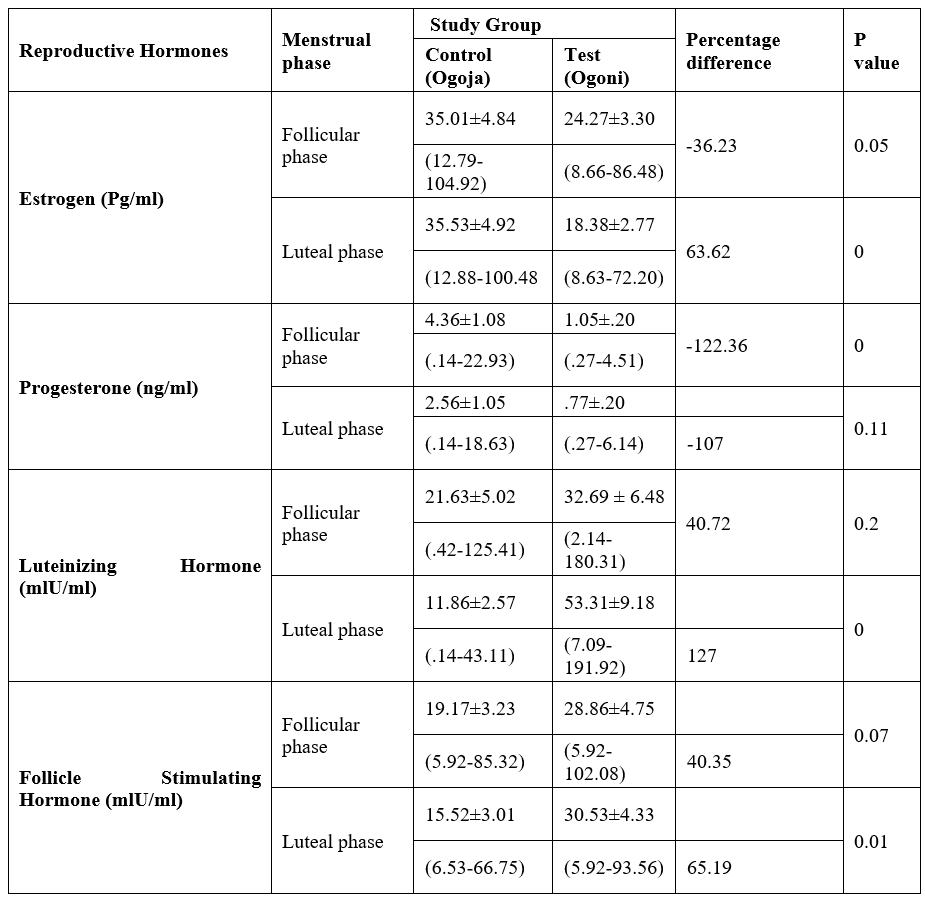
Table 3: Female Reproductive Hormones of Studied Population.

Table 4: Correlation between heavy metals and reproductive function parameters.


Figure 2: Shows a weak positive correlation between pregnancy loss and serum vanadium with a linear R²=0.33.

Figure 3: Shows a weak positive correlation between pregnancy loss and serum lead with a linear R²=0.23.
Discussion
The findings from this study revealed shorter menstrual cycle length among women resident in petroleum impacted areas. The study observed heavy and longer bleeding duration among the women subjects as well as high level of pregnancy lost due to their consistent exposure to petroleum polluted environment as area of residence.
Female reproductive Hormones
The female reproductive hormones in this study were analysed based on their menstrual cycle (follicular and luteal) phase. The result showed a lower mean value of oestrogen follicular phase when compared with the control but significantly on a borderline while the luteal phase was significantly lower than the control. Progesterone was significantly lower at follicular phase when compared with the control but statistically not significant at luteal phase though the mean value was low when compared with the control. The mean value of Luteinizing Hormone at follicular phase for the group is higher than the control but not significant while the luteal phase was significantly higher in the test group compared with the control. For follicle stimulating hormone, the mean values for both follicular and luteal phase were higher than the control group but was not statistically significant.
According to Mahesh et al., (2012) [16] anti-androgens in adult animals increase the serum levels of androgens, luteinizing hormone and estrogen. Some of the reported adverse effects on both farm animals and human on the endocrine system and general physiology have resulted in reproductive malfunction and developmental disorder [17]. This study is in agreement with the study of Uboh et al, [18,19] who evaluated that Exposure to gasoline fumes results in a significant decrease in serum estradiol and progesterone levels in female rats but not in agreement with the gonadotropic hormones who no significant effect on the serum gonadotropin level because this study showed significant increase in the serum level of gonadotropic hormones. According to Mathias et al, (2020) [20] Serum levels of FSH, LH, estradiol and progesterone were significantly lower among fuel attendants. This is in agreement with the sex hormones in this study but opposite for the gonadotropic hormones because the mean values for follicle stimulating hormone and luteinizing hormone were highly significantly higher compared with the control. This finding is also in agreement with [21] that says the results of reproductive hormonal profile assay showed that the serum levels of estradiol was consistently low in most respondents with menstrual irregularities regardless of the duration of exposure to petroleum but not in agreement with other reproductive hormones (LH, FSH and progesterone) having fluctuation because this study shows a significant increase in the level of the gonadotropin hormones (LH and FSH) of the exposed group and reduction in progesterone level when compared with the control. This is to say that petroleum causes alteration in women reproductive hormonal level which may have adverse effect on reproduction, and this is of serious health concern. This could be as a result of low functioning pituitary gland or alteration in the ovary due to petroleum impaction for estrogen. This study is in agreement with the study of [22], who established that Oil-Related Environmental Contaminants (OREC) can affect reproductive processes by directly influencing ovarian cell proliferation, apoptosis, viability, hormone release, and response to gonadotropins. This study is also in congruent with [23,24] who observed an increase in the age at menarche among female residents in non-gas flaring communities compared with gas flaring female resident subjects in Bayelsa state of Nigeria.
Conclusion
Petroleum causes alteration in women reproductive hormonal levels which may have adverse effect on reproduction. The decreased in estrogen and progesterone to be précised that is adequately needed for foetal development must have been responsible for the pregnancy loss while the decrease in the menstrual cycle length could be a cause for concern with regards to infertility among women residents in petroleum host communities.
Conflict of Interest: The authors declare that there is no any form of conflict of interest is this study.
References
- Cahoreau C, Klett D, Combarnous Y. "Structure-function relationships of glycoprotein hormones and their subunits' ancestors". Frontiers in Endocrinology, 2015; 6: 26.
- Clarke H, Dhillo WS, Jayasena CN. Comprehensive Review on Kisspeptin and Its Role in Reproductive Disorders. Endocrinol Metab (Seoul), 2015; 30(2): 124-141.
- Jiang X, Dias JA, He X. "Structural biology of glycoprotein hormones and their receptors: insights to signalling". Molecular and Cellular Endocrinology, 2014; 382(1): 424–451.
- Fowler BA. Mechanisms of kidney cell injury from metals. Environ Health Perspect, 1993; 100: 57–63.
- Vihko KK. Gonadotropins and ovarian gonadotropin receptors during the perimenopausal transition period. Maturitas, 1996; 23 Suppl (Supplement): S19-22.
- Dickerson LM, Shrader SP, Diaz VA. "Chapter 8: Contraception". In Wells BG, DiPiro JT, Talbert RL, Yee GC, Matzke GR (Eds.). Pharmacotherapy: a pathophysiologic approach. McGraw-Hill Medical, 2008; 1313–1328.
- Ulloa-Aguirre A, Reiter E, Crépieux P. "FSH Receptor Signaling: Complexity of Interactions and Signal Diversity". Endocrinology, 2018; 159(8): 3020–3035.
- King SR. Neurosteroids and the Nervous System. Springer Science and Business Media, 2012; 44–46.
- Stamatiades George A, Kaiser Ursula B. "Gonadotropin regulation by pulsatile GnRH: Signaling and gene expression". Molecular and Cellular Endocrinology. Signaling Pathways Regulating Pituitary Functions, 2018; 463: 131–141.
- Nosek Thomas M."Section 5/5ch9/s5ch9_5". Essentials of Human Physiology,
- Rehman K, Fatima F, Waheed I, Akash MSH. Prevalence of exposure of heavy metals and their impact on health consequences. J Cell Biochem, 2018; 119(1): 157-184.
- Bowen R."Gonadotropins: Luteinizing and Follicle Stimulating Hormones". Colorado State University, 2004.
- Johnson LR. Essential Medical Physiology. Academic Press, 2003; p. 770.
- Diczfalusy E, Fraser IS. The discovery of reproductive steroid hormones and recognition of their physiological roles. In: Fraser IS, Jansen RPS, Lobo RA, 1998.
- Hall JM, Couse JF, Korach KS. The multifaceted mechanisms of estradiol and estrogen receptor signaling. J Biol Chem, 2001; 276: 36869–36872.
- Mahesh VB. "Hirsutism, virilism, polycystic ovarian disease, and the steroid-gonadotropin-feedback system: a career retrospective". American Journal of Physiology. Endocrinology and Metabolism, 2012; 302(1): E4–E18.
- Indarto D, Izawa M. Steroid Hormones and Endocrine disruptors: Recent advancesin receptor - mediated actions. Yonago Acta Medica, 2001; 44: 1-6.
- Uboh FE, Ebong PE, Eka OU, Eyong EU, Akpanabiatu MI. Effect of inhalation exposure to kerosene and petrol fumes on some anaemia-diagnostic indices in rats. Global J. Environmental Science, 2005; 3(1): 59-63.
- Uboh FE, Akpanabiatu MI, Ndem JI, Alozie Y, Ebong PE. Comparative nephrotoxic effect associated with exposure to diesel and gasoline vapours in rats, J. Toxicol Environ Health Sci., 2009; 1: 68-74.
- Mathias A Emokpae, Fidelis O Oyakhire. Levels of some reproductive hormones, cadmium and lead among fuel pump attendants in Benin City, Nigeria, Afr. J. Med. Health Sci, 2020; 19(6): pp. 70-77.
- Christopher E, Ekpenyong K, Davies ND. Effects of Gasoline Inhalation on Menstrual Characteristics and the Hormonal Profile of Female Petrol Pump Workers, Journal of Environmental Protection, 2013; 4: 65-73.
- Alexander S, Attila K, Attila K, Andrej B, Jan K, Abdulkarem A, et al. Mechanisms of the direct effects of oil-related contaminants on ovarian cells, Journal of Environmental Science and Pollution Research, 2020; 27(9): 1-9.
- Solomon MU, Kiridi Emily EG, Alagha BE, Onokpite E. Onset of menarche among adolescent girls in gas and non-gas flaring environment in bayelsa state, Nigeria.Sourth Asian Research Journal of Medical Science, 2022; 4(3): 13-18.
- Bowen R. "Luteinizing and Follicle Stimulating Hormones", 2019.

