Creatine Phosphokinase Serum Levels in Acute Closed Isolated Femur Fractures
Dorcas Chomba*, Edward Gakuya and John Atinga
Orthopedic and Trauma Consultant, Kenya Orthopedic Association, Kenya
Received Date: 28/02/2023; Published Date: 15/05/2023
*Corresponding author: Dr. Dorcas Chomba, Orthopedic and Trauma Consultant, Kenya Orthopedic Association, Kenya
Abstract
Background: Soft-tissue injuries in closed fractures are less obvious than in open fractures, but still have enormous importance. The evaluation of soft tissue injury in closed fractures can be much more difficult than open fractures and the severity is easily underestimated. Of note is that closed fractures are much more common than open fractures, i.e., of all fractures recorded it is estimated only 3% are open fractures. Understanding soft tissue status is important as the effective treatment of any fracture depends upon good soft tissue management. The current methods of soft tissue assessment include clinical examination or use of imaging. Specifically, MRI and U/S which are expensive or not readily available. This study aimed at establishing the relationship between the patterns of closed fracture femur as defined in the AO classification and severity of soft tissue injury by measuring the creatine phosphokinase levels in serum.
Objective: To measure the serum creatine levels in patients with closed fracture femur as a biochemical marker of degree of muscle injury and correlate this to AO fracture pattern classification.
Study Design: Cross-sectional multi-center hospital-based study – KNH and Kikuyu Mission Hospital.
Methodology: Consecutive sampling of patients presenting in KNH with isolated closed fracture femur was done. Their blood samples were collected within 48hrs following injury. The blood samples were transported to Lancet at specified conditions optimum for CK analysis. The CK analysis was done using the COBAS INTEGRA 400/800 whose test principle measures the rate of production of β-Nicotinamide Adenine Dinucleotide. This is directly proportional to CK activity. The levels of CK were correlated to AO fracture pattern classification of femur shaft fractures.
Data Processing: The collected data was coded and analyzed using Microsoft Excel, SPSS 20. and summarized as percentages and means (95% confidence intervals).
Results: Males 51 (77%) were more than the female 15 (23%). There was a correlation between mechanism of injury and CK levels but no correlation between CK levels and fracture pattern. None of the patients had clinical signs of compartment syndrome or ABI less than 0.9. Females had a generally lower amount of CK compared to the males. Those who sustained pathological fractures had CK levels within normal.
Conclusion: There is a correlation between amount of energy involved during injury and CK levels but no correlation was noted between fracture pattern and CK.
Introduction
Creatine kinase is one of the most useful markers of muscle wellbeing [1]. The level of creatine phosphokinase in serum has been used to assess among other iatrogenic muscle injury in arthroplasty, myocardial infarction and muscular dystrophies [2].
The current definition of a fracture is a soft tissue injury with a break in the bone [13]. In closed injuries, the degree of injury and ischemic tissue may not be clinically apparent and this can make diagnosing extent of soft tissue injury difficult [7].
The current classifications of soft tissue injury are mostly subjective hence likely to have intra-observer and inter-observer variability. Many modern imaging techniques including MRI and ultrasound permit qualitative assessment of closed soft-tissue injuries. Clinically useful biochemically based quantitative assessment of soft tissue damage in relation to fracture pattern has not been explored.
Muscle injury can be as a result of direct force to the muscle, kinetic cellular disruption, hypoxia at zone of injury and resultant inflammatory cascade, leading to rhabdomyolysis. This results in increased leak of intracellular proteins including creatine phosphokinase. From previous studies, it has been observed that CK starts to increase within 12hours of onset of muscle injury, reaches its peak in 24-72 hours then declines gradually over 72-120 hours as long as muscle injury is not continuing [15,16,2].
By and large quantifying the degree of soft tissue injury through biochemical marker is superior to imaging techniques.
However, there’s no study correlating femur fracture pattern, AO classification and CK levels as a marker of soft tissue injury.
This study aimed to correlate the degree of muscle injury sustained in closed diaphyseal femur fracture pattern based on AO classification to levels of CK.
Study question: Is there appreciable correlation of CK serum levels and AO classification of fracture pattern
Methodology
Study design: Multi-center hospital based cross-sectional study
Study site: This study was conducted at the A&E department, orthopedic ward and orthopedic clinic in Kenyatta National Hospital and Kikuyu Mission Hospital.
Study population: 66 patients aged between 18– 5 years presenting at the two hospitals with isolated acute closed fracture femur and met the inclusion criteria were included in the study. Evaluated and recruited within 48 hours of injury. X-ray images were reviewed and pattern of fractures classified according to AO criteria. 4mls of venous blood sample blood sample collected and analyzed. The CK analysis was done using the COBAS INTEGRA 400/800.
Inclusion criteria: Isolated closed fracture femur
Exclusion criteria: Recent sports injury, known muscle disease, cardiac disease and trauma, neurodegenerative diseases, renal failure, hypoxic brain disease, any patient requiring CPR, or recently operated on.
Results
Data was analyzed using excel office for windows 2010 and SPSS 20.0
Total patients, gender distribution, mean age, AO classification.
Table 1: Data Demographics- Gender Distribution.
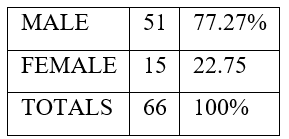
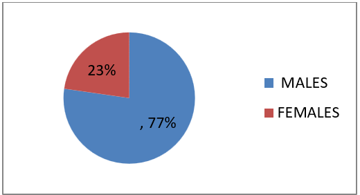
Figure 1: 66 patients gender distribution.

Figure 2: AO fracture classification pattern as per gender distribution.
Table 2: CK levels plotted against estimated amount of energy involved during injury A-low, B-moderate, C-high.

Table 3: CK Level Means Calculation.
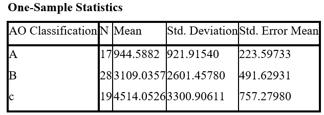
Table 4: CK level means calculation.

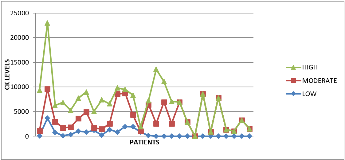
X COORDINATES (serum CK levels)
Figure 3: CK levels plotted against (moi) graded energy involved during injury.
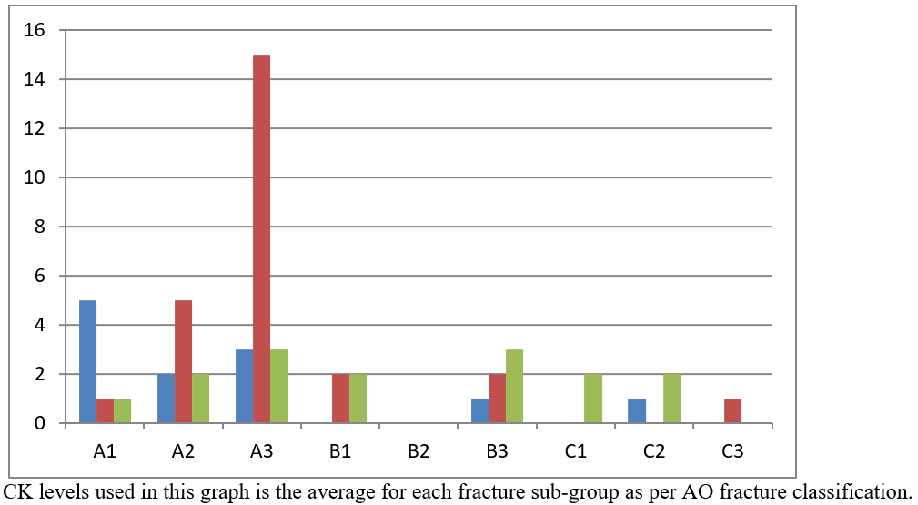
Figure 4: Graded mechanism of injury plotted against fracture pattern.
Table 5: Correlation between ck levels and fracture pattern.
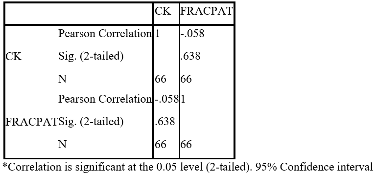
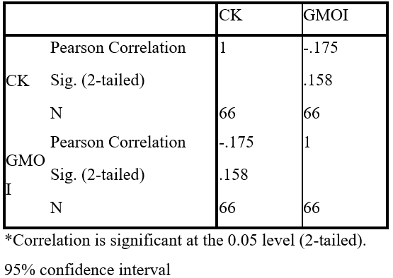
Table 6: Correlation between ck levels and graded energy involved during injury.
Results
A total of 66 patients who had fracture femur were recruited and blood samples taken for CK levels. Of the 66, 15 were females and 51 were males. Females -22.7%, Males -77.27% (Table 1).
Females had a generally lower level of CK levels compared to their male counterparts even with similar fracture patterns. Patients who were more muscular had a higher CK level compared to less muscular patients (this was observed by the investigators although there were no objective measurements done). Mechanism of injury associated with direct hit or crush to the muscles e.g., caved in by soil/collapsed wall or direct bumper hit to the limb were generally associated with higher levels of CK, Figure 4. The amount of energy was a subjective measurement obtained during history taking with a detailed review of the mechanism of injury. One patient with a pathological fracture broke his femur while turning in bed, this was graded as low energy, while those who were in high-speed accidents, falling walls were categorized as high energy (Figure 3). Amount of energy involved during injury appears to have a better correlation with CK levels compared to the fracture pattern.
However, the Pearson’s correlation calculated for the correlations between CK levels and fracture pattern yielded a non-significant level of more than 0.05. Although noted that the mean differences calculated for the CK levels for the three main groups of fractures was found to have a difference that was significant, 0.01.
Discussion
In closed injuries, the degree of injury and ischemic tissue may not be apparent and this is a challenge in making soft tissue injury diagnosis and therapeutic decisions [8]. The purpose of this study was to evaluate whether there is a correlation between CK serum levels and fracture pattern as classified by AO classification of fractures. CK serum levels was used as a marker of muscle injury [6,7]. AO classification was used as each pattern of fracture in the classification represents the expected amount of energy involved in the injury [13]. Isolated femur fracture was used following the argument that fractures of the femoral shaft are markers of high-energy injuries and the femur is well enveloped by muscles [10-12].
Normal expected levels of serum CK in general population varies between 35–175 U/L [5]. In this study the laboratory range for CK provided by the laboratory was 35 - 184U/L. Elevated levels clearly represent a strong disruption of striated muscle tissue with concomitant release of intracellular muscle components into the circulation [6,7].
It is well known that there are still marked sex differences in human beings in CK serum levels at rest, with lower values in females than in males [22]. In this study it was also noted that the CK levels even when elevated was still lower in females with similar fracture patterns compared to the male patients. One of the explanations for this could be due to the muscle bulk in females being lower than in males [23]. The second reason could be related to the amount of energy involved during injury; female patients tend to be involved in lower energy accidents compared to the male patients [25]. However, it is notable that the majority of female patients in this study were >50 hence probability of insufficiency fractures was higher which would also explain the low energy fractures hence, low CK serum levels and the fracture pattern sustained [26].
It is well known that the pattern of fracture sustained on the bone is mostly related to the amount of energy involved. Fractures of the femoral shaft are markers of high-energy injuries and since the femur is well enveloped by muscles; some of the energy is absorbed by the soft tissue envelope [10-12]. However, in this study the levels of CK did not match very well with the fracture pattern but there was a definite correlation between the CK levels and amount of energy involved during injury. Patients who sustained fractures through high energy mechanism or direct force/crush injury type to the thigh/femur had higher levels of CK regardless of the fracture pattern sustained (Figure 3). The authors thought this could be explained by assuming that in those fractures of lower grading as per AO that had higher levels of CK than expected, the reason could be because most of the force involved was absorbed by the muscle envelope therefore, less of the energy reached the bone to cause a higher-grade fracture pattern. As already defined earlier, a fracture, is a soft tissue injury with a break in the bone [13]. The other reasons could be of soft tissue injury such as direct impact, implosion, and displacement during transportation, degree of fracture displacement, inflammatory process, and ischemia due to blood supply interruption or edema. These reasons which the investigator did not have control of could explain the reason for the mismatch between CK levels and fracture pattern. It is important to note that none of the patients had signs of compartment syndrome clinically or vascular injury as checked by measuring the ABI. The other factor to consider that may explain the mismatch was the fact that all these patients received intravenous fluids as part of their initial resuscitation measures. The effect of intravenous fluid on the levels of CK could have been through dilution or increased clearance through the kidneys [27]. Also, to be considered is whether all the CK released from the injured muscles actually leaked into circulation.
Patients who sustained pathological fractures had CK levels within normal range. The mechanism of injury in these patients was indirect rotational forces which seemed to be transmitted through bone and muscle but was not sufficient to cause significant muscle injury and only caused a break in the bone at an area of weakness [28].
Conclusion
1. There was a clearer correlation between amount of energy in injury and CK levels as compared to CK levels and fracture pattern [29,30].
2. Females had lower CK level elevation in general even after trauma and this could be related to the muscle bulk or mechanism of injury.
3. There was no correlation between the serum CK levels and pattern of fractures as per the AO classification.
Recommendations: a follow up study that would include larger numbers, capacity to assess for muscle bulk more objectively, an objective way to measure the number of forces absorbed by the soft tissue envelope, long term follow up of the patients to assess for bone healing and functional outcome and development of myositis ossificans.
References
- Fu FH, You CY, Kong ZW. Acute changes in selected serum enzyme and metabolite concetrations in 12yr to 14yr old athletes after an all-out 100m swimming sprint. Percept Motor Skills, 2002; 95: 1171-1178.
- Musil D, Stehlík J, Verner M. A comparison of operative invasiveness in minimally invasive anterolateral hip replacement (MIS-AL) and standard hip procedure, using biochemical markers]. Acta Chir Orthop Traumatol Cech, 2008; 75(1): 16-20.
- Totsuka S, Nakaji K, Suzuki K, Sugawara and Sato K. “Break point of serum creatine kinase release after endurance exercise,”Journal of Applied Physiology, 2002; 93(4): pp. 1280–1286.
- Brancaccio P, Maffulli N Limongelli FM, “Creatine kinase monitoring in sport medicine,”British Medical Bulletin, 2007; 81-82: pp. 209–230.
- Gagliano D, Corona G, Giuffrida, et al. “Low-intensity body building exercise induced rhabdomyolysis: a case report,”Cases Journal, 2009; 2(1): article 7.
- Efstratiadis G, Voulgaridou A, Nikiforou D, Kyventidis A, Kourkouni E, Vergoulas G. “Rhabdomyolysis updated,” Hippokratia, 2007; 11(3): pp. 129–137.
- Huerta-Alardín AL, Varon J, Marik PE. “Bench-to-bedside review: rhabdomyolysis—an overview for clinicians,”Critical Care, 2005; 9(2): pp. 158–169.
- Levin SL. Condit DP Combined injuries—soft tissue management. Clin Orthop Relat Res; 1996; 327: 172–181.
- Tscherne H, Oestern HJ. A new classification of soft-tissue damage in open and closed fractures. 1982; 85(3): 111–115.
- Salminen ST, Pihlajamaki HK, Bostman ON. Population based epidemiologic and morphologic study of femoral shaft fractures. Clin Orthop Relat Res, 2000; 372: 241-249.
- Bruce H Ziran, Natalie L Talboo, Navid M Ziran Operative Techniques in Orthopaedic Surgery1st Edition,Chapter 10 pg 569 published by LWW(Lipincot Williams Wilkins).
- AO foundation- Diaphyseal fracture principles by Piet de Boer.
- Müller ME, Nazarian S, Koch P, et al. The Comprehensive Classification of Long Bone Fractures. Berlin Heidelberg New York: Springer-Verlag, 1990.
- Ward MM. “Factors predictive of acute renal failure in rhabdomyolysis,” Archives of Internal Medicine, 1988; 148(7): pp. 1553–1557.
- Roth D, Alarcon FJ, Fernandez JA, Preston RA, Bourgoignie JJ. “Acute rhabdomyolysis associated with cocaine intoxication,” The New England Journal of Medicine, 1988; 319(11): pp. 673–677.
- Rice EK, Isbel NM, Becker GJ, Atkins RC, McMahon LP. “Heroin overdose and myoglobinuric acute renal failure,” Clinical Nephrology, 2000; 54(6): pp. 449–454.
- Strecker W, Gebhard F, Rager J, et al. “Early biochemical characterization of soft-tissue trauma and fracture trauma”, 1999; 47(2): 358-364.
- Bergin PF, Doppelt JD, Kephart CJ, et al “comparison of minimally invasive direct anterior versus posterior total hip arthroplasty based on inflamation and muscle damage markers”. 2011; 93(15): 1392-1398. doi: 10.2106/JBJS.J.00557
- Mjaaland KE, Kivle K, Svenningsen S, et al “Comparison of markers for muscle damage, inflammation and pain using minimally invasive direct anterior versus direct lateral approach in total hip arthroplasty”. J Orthop Res, 2015; 33(9): 1305-1310. Doi:10.1002/jor.22911.
- Bernd Fink, Alexander M, Martin SS, et al “Comparison of minimally invasive posterior approach and the standard posterior approach for total hip arthroplasty” Journal of Orthopaedic Surgery and Research, 2010; 5: 46.
- Morgan DB, Carver ME, Payne RB. “Plasma creatine and urea: urea:creatine ratio in patients with raised plasma urea. Br.Med J, 1977; 2(6092): 929-932. PMID 912370.
- Bayer PM, Gabi F, Gergely T, et al. Isoenzymes of Creatine kinase in the perinatal period. J Clin Chem Biochem, 15: 349-352.
- Fallon KE, Sivyer G, Dare A. The biochemistry of runners in a 1600km ultra-marathon. Br J Sports Med, 1999; 33: 264-269.
- Bhandari M, Tornetta P, Sprague S, et al. Predictors of reoperation following operative management of fractures of the tibia shaft. J Orthop Trauma, 2003; 17(5): pp.3-361.
- Ayman El-Menyar, Hany El-Hennawy, Rifat Latifi, et al Traumatic Injury Among Females: does gender matter? Journal of Trauma Management and Outcomes 2014; 8: 8.
- Aasis Unnanuntana, Brian P, Eve Donelly, et al. The Assessment of Fracture Risk. J.Bone Joint Surg Am, 2010; 2(3): pp 743-753.
- Enas Samir, Sherry N. Rizk, Samar A. Abdou. Influence of intraoperative fluid administration on creatine kinase and its effect on kidney function after laparascopic nephrectomy. Egyptian Journal of Anaesthesia, 2012; 28(3): 211-215.
- Sari S, Harri P, Veikko A, et al. Speciifc features associated with femoral shaft fractures caused by low energy trauma. J Trauma, 1997; 43: 117-122.
- Michael JH, mCcarthy, Simon Gatehouse, Monica Steel, Ben Goss, Richard Williams. The influence of the energy of trauma, the timing of decompression, and the impact of grade of SCI on outcome.
- Robert Perlau. Shortcut to Orthopaedics: What's Common and What's Important for Canadian students and primary care physicians, 2015; 48.

