Estimation of the Skin to Erector Spinae Fascial Plane Space Depth in Children Using Computer Tomography
Sabashnee Govender*, Adrian Bosenberg and Albert-Neels Van Schoor
Department of Anatomy, School of Medicine, Sefako Makgatho Health Sciences University, South Africa
Department of Anatomy, Section of Clinical Anatomy, School of Medicine, Faculty of Health Sciences, University of Pretoria, South Africa
Department Anaesthesiology and Pain Management, University Washington and Seattle Children’s Hospital, Seattle, United States of America.
Received Date: 18/01/2022; Published Date: 02/02/2022
*Corresponding author: Dr Sabashnee Govender, PhD, Department of Anatomy, School of Medicine, Sefako Makgatho Health Sciences University, PO Box 232, Ga-Rankuwa 024, South Africa
Abstract
Background: Nerve blocks may present technical challenges in children of different ages due to growth and developing anatomy. The erector spinae plane block is a versatile technique that can be used as an alternative to paravertebral and epidural blocks to provide postoperative analgesia for truncal surgery. However, there is limited data reporting on the skin to structure distance should this block be performed in children without the use of ultrasound guidance. This study aimed to determine the skin to erector spinae fascial plane space depth using data obtained from one hundred and fifty computer tomography scans to assist in performing an erector spinae plane block in children up to twelve years of age.
Methods: Measurements vital to performing an erector spinae plane block were taken from computer tomography scans at two vertebral levels to represent thoracic and abdominal spread, respectively. Statistical analyses were performed to determine whether there was a relationship between age, gender and the measurement in different groups. Groups were divided as follows: group 1 (0 – 2 months), group 2 (2 months – 2 years) and group 3 (2 – 12 years).
Results: Results revealed no correlation with sex, age and measurements in group 1 or 2. While a weak to moderate correlation was found between age and measurements in group 3.
Conclusion: The erector spinae plane block is a novel interfascial block that can be performed in various age groups. Although the transverse process acts as an anatomical landmark, it is not as evident in young children. Therefore, the optimal success of the block depends on direct visualization using ultrasound. However, should ultrasound guidance not be available, predicted measurements for various age groups may facilitate correct needle placement and potentially reduce the risk of complications.
Keywords: Erector spinae plane block; Regional anaesthesia; Depth estimation; Paediatrics
Introduction
Nerve blocks may present technical challenges in children of different ages due to growth and developing anatomy. These include thinner muscle layers, sliding fascial planes and loose connective tissue (Aksu and Gürkan 2018).[1] Moreover, variations with age and body habitus such as height, weight and gender, require technical adjustments to achieve success and avoid potential complications. Although ultrasound guidance increases the success rate of regional blocks, it is not always available - particularly in the developing world (Masir et al. 2006) [2]. Landmark-based or loss of resistance techniques or nerve stimulation is relied upon when performing nerve blocks in low resource institutions.
The erector spinae plane block is a novel interfascial block that is performed in a tissue plane deep to the erector spinae muscle (Govender, Mohr, Neels Van Schoor, et al. 2020; Roy et al. 2020).[3,4] The erector spinae plane block is a versatile technique that can provide postoperative analgesia for truncal surgery (Aksu and Gürkan 2020; Govender, Mohr, Bosenberg, et al. 2020) [5,6]. Erector spinae block has been used as an alternative to paravertebral and epidural blocks because it targets the same spinal nerves but away from neuraxial structures (De la Cuadra-Fontaine et al. 2018; Forero et al. 2016; Govender, Mohr, Bosenberg, et al. 2020; Kaushal et al. 2020; Peng et al. 2019; Vidal et al. 2018) [6-11] reducing the risk of spinal cord injuries (Govender, Mohr, Bosenberg, et al. 2020) [6]. Knowledge of the skin to erector spinae fascial plane space depth potentially increases the safety and success rate when performing the erector spinae plane block with or without ultrasound guidance. To the best of our knowledge, this is the only study that investigates the needle depth to the transverse process, the bony landmark used when performing an erector spinae plane block, at different vertebral levels (T5 and T8) in different age groups.
The primary objective of this study was to determine to erector spinae fascial plane space depth using data obtained from computer tomography (CT) scans to assist in performing an erector spinae plane block in children up to twelve years of age at two vertebral levels (T5 and T8). The secondary aims were to determine the mean distance from the spinous process to the needle entry site (over the lateral tip of the transverse process) at the two vertebral levels. In addition, to determine whether there is a relationship between age, gender and the skin to erector spinae fascial plane depth in different age groups.
Materials and Methods
This study was approved by the PhD and Research Ethics Committee, University of Pretoria, South Africa (94/2019). Permission was also obtained from the Head of the Department of Radiology and CEO of Steve Biko Academic Hospital to retrospectively source CT scans from patient archives. Written consent was obtained from the patients (or their parents/guardians) upon capturing the scans. All records obtained were kept confidential, as not to reveal the identity of patients.
After conducting power analysis for multiple linear regression analyses to determine the sample size. One hundred and fifty CT scans were selected from the database of radiographic images over fifteen years (2005-2019). The patients were scheduled for a variety of thoracic or abdominal procedures. Demographic information such as age and gender were recorded. Scans were grouped according to group 1 (0 – 2 months), group 2 (2 months – 2 years) and group 3 (2 – 12 years). Scans with abnormal vertebral column development (e.g. kyphosis and scoliosis), visceromegaly or space-occupying lesions, as diagnosed by the consulting radiologist, were excluded.
The scans were then analysed using RadiAnt, a Digital Imaging and Communication in Medicine (DICOM) viewer. Using the on-screen measuring function, calibrated for each image, measurements were made bilaterally at vertebral levels T5 and T8 (to represent thoracic and abdominal spread, respectively) in a transverse section. Measurements included: A – the distance from the spinous process to the transverse process; B – the depth from the skin to the lateral tip of the transverse process (representing the level erector spinal fascial plane space); C – the depth from the skin to the most superficial point of the erector spinae muscle; D – the depth from the skin to the most superficial point of the rhomboid muscle; E – the depth from the skin to the most superficial point of the trapezius muscle (Figure 1).
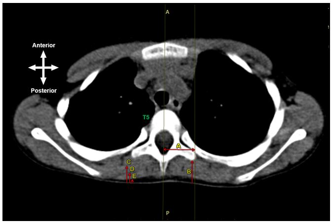
Figure 1: A CT scan of a transverse section through the thorax at vertebral level T5. Measurements that were taken include; A – the distance from the spinous process to the transverse process; B – the depth from the skin to the tip of the transverse process (i.e., erector spinae fascial plane); C – the depth from the skin to the most superficial point of the erector spinae muscle; D – the depth from the skin to the most superficial point of the rhomboid muscle; E – the depth from the skin to the most superficial point of the trapezius muscle. Key: A – anterior, P – posterior, T5 – vertebral level T5.
Statistical analysis
All measurements were inputted into a Microsoft Excel spreadsheet. Further statistical analysis of the measurements and the subsequent comparisons of those measurements with the available demographic profile was performed using Statistic Data Analysis (STATA), version 16. In order to ensure the validity and accuracy of the results obtained, intra- and inter-observer reliability checks were conducted. The primary investigator repeated 25% of the initial measurements, while an independent researcher repeated 20% of the initial measurements.
After testing for normality, comparisons were made between left and right sides using a paired t-test to test for statistical significance. Additionally, the Bonferroni correction method was adopted to adjust the p-values to reduce the chance of obtaining false-positive results (type I errors) when multiple paired tests are performed on a single set of data. Measurements that were not statistically significant, were pooled together to create a mean before continuing with the statistical analysis.
Linear regression models were then performed to establish whether a linear relationship/correlation existed between the dependent variables – the measurement – and the independent variables – age and gender. Although these tests were run for all measurements, this article concentrates on the skin to erector spinae fascial plane space depth and the lateral distance from the spinous process to the needle entry site.
Results
Upon intra- and inter-observer analysis, a student t-test was performed to compare the two sets of data (measurements taken by the primary investigator versus measurements taken by the secondary investigator) in order to ensure that the measurements obtained, were valid. The statistical results revealed a p-value greater than 0.05 for both the intra- and inter- reliability checks, which indicated that there was no statistically significant difference between the data sets. The initially obtained data measurements were thus considered to be correct.
Paired t-tests were performed to test for statistical significance between right versus left side measurements. Normality was further confirmed as the mean for each measurement was twice the standard deviation. Overall, there were a total of 9 comparisons per age group. After adopting the Bonferroni correction method, the new p-value was 0.0056. Addendum A summarises the results of the paired t-test for the three age groups.
From the total sample size of group 1, 22 scans belonged to females while the remaining 23 belonged to males. Based on the p-values, a significant difference was noted between the right- and left sides for T5 skin to erector spinae fascial plane space, as well as the right- and left sides of T8 skin to erector spinae muscle in the neonatal group (even though measurements were assessed for outliers). Statistically significant measurements (only the T5 skin to erector spinae fascial plane space) were then plotted on a bar graph reflecting the mean and standard error (Figure 2).
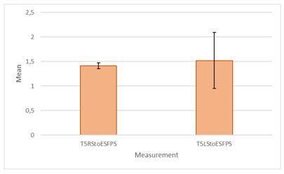
Figure 2: Bar graph showing the results from the paired t-test for the statistically significant measurements. The error bar represents the standard error in relation to the mean. Key: T5RStoESFPS – at vertebral level T5 right side, skin to the erector spinae fascial plane space and TLRStoESFPS – at vertebral level T5 left side, skin to the erector spinae fascial plane space.
As we can see from figure 2, the measurements from the skin to the erector spinae fascial plane at vertebral level T5, was greater on the left side than on the right side. The error bar represents the standard error of the mean. The standard error is used to measure the accurateness with which a sample distribution represents a population by using the standard deviation. Error bars indicate the spread of data around the mean or how accurately the mean of the measurements represents the data set (ie the variability). Furthermore, standard error bars can be used to estimate whether or not a difference is truly significant depending on the overlapping of the bars – or lack thereof. If standard error bars overlap as indicated in figure 2, the difference is less likely to be statistically significant, whereas a slight overlapping of bars, indicates that there is a probability that the difference is statistically significant. Overall, there is statistical significance between the measurements. However, the actual difference between the right- and left sides is small. Additionally, the small error bar (on the right-hand side) indicates the concentration of the data around the mean, making the results more reliable.
No significant difference was noted between any of the measurements for groups 2 (28 females and 21 males) and 3 (31 females and 26 males). Subsequently, a comparative analysis was performed between individual groups to determine if there was a significant difference between individual measurements and the group (1, 2 or 3) before pooling the data. Results revealed a significant difference (p-value < 0.05) between; spinous process to transverse process, skin to erector spinae fascial plane space and age groups. Due to the statistical difference between groups, the data was not pooled, and further statistical testing was performed on the groups individually.
Measurements (from each age group) that was not statistically significant was pooled to create averages for each measurement with a new standard deviation (Addendum B). Regression analysis was then performed to evaluate the correlation between the measurements – the dependent variable – and fixed factors such as sex and age – the independent variables. From the results for age group 1, a weak correlation – adjusted R2-value ≤ 0.3 – was found between the measurements and sex. Likewise, a weak correlation – adjusted R2-value ≤ 0.3 – was found between the measurements and age. In age group 2, a weak correlation was found between the measurements and sex or age (adjusted R2-value of ≤ 0.1). While in age group 3 a weak correlation was found between T5 skin to erector spinae fascial space and age (adjusted R2-value of 0.38), and a moderate correlation was found between T8 skin to erector spinae fascial space and age (adjusted R2-value of 0.45), T8 spinous process to transverse process (adjusted R2-value of 0.42). Measurements with a moderate correlation (≥ 0.40) were then further plotted on a scatter plot to display the relationship of the correlation (Figure 3).
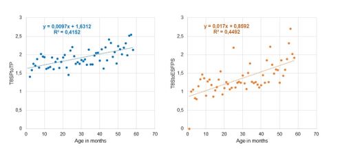
Figure 3: Scatter plot displaying the correlation between T8 skin to erector spinae fascial plane space in cm to age in months (right image, in orange), T8 spinous process to transverse process in cm to age in months (left image, in blue).
From figure 3, the adjusted R2-values indicate how much of the attribution is caused by age. Therefore, the skin to the erector spinae fascial plane space at vertebral level T8, 45% of the variations can be explained by age or is caused by age. While for T8 spinous process to transverse process, 42% of the variation can be explained by age or is caused by age.
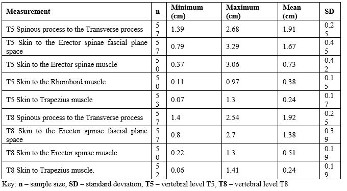
The mean distance from the spinous process to the transverse process in group 1 at vertebral level T5 was 1.28 cm with a standard deviation of 0.20, while the distance at vertebral level T8 was 1.25 cm with a standard deviation of 1.95. In group 2, the mean distance was 1.59cm (standard deviation of 0.30) and 1.59cm (standard deviation of 0.27) at vertebral levels T5 and T8, respectively. While in group 3, the mean distance was 1.91cm (standard deviation of 0.25) and 1.92cm (standard deviation of 0.25).
Discussion
The erector spinae plane block is a novel interfascial plane technique that can be used for various truncal procedures in both adults and children (Forero et al. 2018) [12]. Although this block is relatively new, it has sparked interest due to its relative ease of access and clinical efficacy. This study aimed to determine estimation formulae should the block be performed using landmark-based techniques. To date, only one case study reports on the means and standard deviations for the various measurements when performing an erector spinae plane block in children (Karaca 2019) [13].
Our results revealed a significant difference between right and left measurements in group 1. Even though these measurements were statistically significant, the difference was clinically rather small. The average depth to the erector spinae fascial plane space in age group 1 was between 1.41cm (±0.28) to 1.52cm (±0.40) at vertebral levels T5 and T8. While for group 2 the approximate depth at vertebral levels T5 and T8 was 1.52cm (±0.46) and 1.20cm (±0.35) respectively. In group 3, the average depth at vertebral level T5 was 1.67cm (±0.45) and 1.38cm (±0.39) at vertebral level T8.
Karaca (2019) [13], noticed that for children above the age of 10 years old, the needle should be inserted 1.5 – 2 cm laterally at the midsagittal region. Results from this study, are similar to that of Karaca, as our predicted value falls within their predicted range. However, Karaca’s estimation included children up until the age of 14 years, whereas, our study only included children up to 12 years. Moreover, the mean distance reported in this study was specified to vertebral levels T5 and T8, whereas, Karaca estimation was specific to vertebral level T7.
In our study, measurements were based on an approach perpendicular to the transverse process that would be used when ultrasound guidance was not available. As seen in other depth estimation studies, due to variability in the thoracic and lumbar regions, it is not always appropriate to apply a single formula to all vertebral levels (Wani et al. 2018) [14]. The estimated depth could act as a useful guide for ultrasound guidance specific to vertebral levels T5 and T8. Results from this study are useful as there is little literature reporting these measurements to aid in performing an erector spinae plane block. Further investigation with a larger sample size, additional demographic parameters and alternative imaging modalities is recommended.
Limitations
Apart from the limited sample specifically due to the challenges faced in obtaining neonatal scans, there was a lack of demographic information such as height and weight that were not captured by radiologists when the scans were taken. Additionally, since the retrospective scans were anonymous, we were unable to approach the department to retrieve outstanding information. The absence of these variables impeded a complete analysis as the effects of these variables on measurements were not addressed.
According to World Health Organization guidelines, the age group classification for a neonate is 0-1 months. However, after consultation with the radiologists, we decided to extend group 1 in our study up to 2 months due to the small number of 1-month-year-old scans available in order to meet an appropriate sample size.
Additionally, data obtained in this study were taken from CT scans with the patient was in a supine position. This should be considered, as erector spinae blocks are performed with the patient prone or in a lateral decubitus position (Wani et al. 2018) [14]. Patient positioning potentially affects these measurements. Specific to age group 1, images of patients were ‘tilted’ and thus the measuring process had to be adapted to the entire scan tilt. Furthermore, measurements were taken by the authors under the guidance of a radiologist, however, not the radiologist themselves.
Lastly, in terms of imaging modalities, magnetic resonance imaging is a more comprehensive imaging modality of the paraspinal and intraspinal soft tissue and ligaments compared to CT imaging (Wani et al. 2017) [15]. In this study, there was some difficulty in identifying the structures when performing measurements. As a result, an estimation of the start or endpoint of structures from some scans were made
Addendum A
Table 1: Results of the paired t-test of the ESP measurements taken from the CT scans for age group 1.
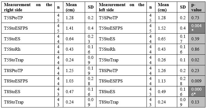
Table 2: Results of the paired t-test of the ESP measurements taken from the CT scans for age group 2.
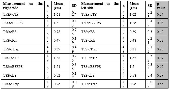
Table 3: Results of the paired t-test of the ESP measurements taken from the CT scans for age group 3.
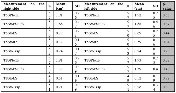
Key: n – sample size, SD – standard deviation, T5 – vertebral level T5, T8 – vertebral level T8, SPtoTP – Spinous process to the Transverse process, StoESFPS – Skin to the Erector spinae fascial plane space, StoES – Skin to the Erector spinae muscle, StoRh – Skin to the Rhomboid muscle, StoTrap – Skin to Trapezius muscle.
Addendum B
Table 4: Descriptive statistics summary after pooling the right- and left sides for the CT component scans for age group 1.
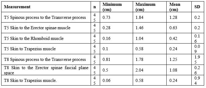
Table 5: Descriptive statistics summary after pooling the right- and left sides for the CT component scans for age group 2.
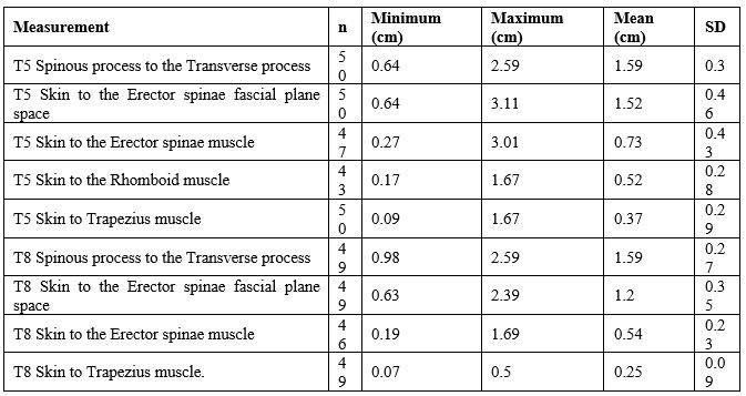
Table 6: Descriptive statistics summary after pooling the right- and left sides for the CT component scans for age group 3.
Conclusion
Although most regional blocks can be performed using ultrasound guidance, it is not always feasible when resources are limited. Landmark-based or loss of resistance techniques is the next best option. An erector spinae plane block is a novel interfascial block that can be performed in various age groups. Although the transverse process acts as an anatomical landmark for the block, it is not as evident in young children since it is less ossified and more cartilaginous. Predicted measurements for the neonatal, infant and child age groups may facilitate correct needle placement and potentially reduce the risk of complications when ultrasound guidance is not available.
Acknowledgements
The authors would like to thank Prof Lockhat and Prof Suleman from the Department of Radiology, Steve Biko Academic Hospital for their contributions to this study.
Financial assistance
The financial assistance of the National Research Foundation (NRF) towards this research is hereby acknowledged. Opinions expressed and conclusions arrived at are those of the authors and are not necessarily attributed to the NRF.
Conflicts of Interests
None.
References
- Aksu, Can, and Yavuz Gürkan. 2018. ‘Ultrasound-Guided Erector Spinae Block for Postoperative Analgesia in Pediatric Nephrectomy Surgeries’. Journal of Clinical Anesthesia 45:35–36.
- Masir, F., J. J. Driessen, K. C. Thies, M. H. Wijnen, and J. Van Egmond. 2006. ‘Depth of the Thoracic Epidural Space in Children’. 5.
- Govender, Sabashnee, Dwayne Mohr, Adrian Bosenberg, and Albert Neels Van Schoor. 2020. ‘A Cadaveric Study of the Erector Spinae Plane Block in a Neonatal Sample’. Regional Anesthesia & Pain Medicine rapm-2019-100985.
- Roy, Ritesh, Gaurav Agarwal, Chandrasekhar Pradhan, and Debasis Kuanar. 2020. ‘RACK Approach to Erector Spinae Plane Block’. Journal of Anaesthesiology, Clinical Pharmacology 36(1):120–21.
- Aksu, Can, and Yavuz Gürkan. 2020. ‘Sacral Erector Spinae Plane Block with Longitudinal Midline Approach: Could It Be the New Era for Pediatric Postoperative Analgesia?’ Journal of Clinical Anesthesia 59:38–39.
- Govender, Sabashnee, Dwayne Mohr, Albert Neels Van Schoor, and Adrian Bosenberg. 2020. ‘The Extent of Cranio-Caudal Spread within the Erector Spinae Fascial Plane Space Using Computed Tomography Scanning in a Neonatal Cadaver.’ Pediatric Anesthesia.
- De la Cuadra-Fontaine, Juan Carlos, Mario Concha, Fernando Vuletin, and Hernán Arancibia. 2018. ‘Continuous Erector Spinae Plane Block for Thoracic Surgery in a Pediatric Patient’. Pediatric Anesthesia 28(1):74–75.
- Forero, Mauricio, Sanjib D. Adhikary, Hector Lopez, Calvin Tsui, and Ki Jinn Chin. 2016. ‘The Erector Spinae Plane Block: A Novel Analgesic Technique in Thoracic Neuropathic Pain’. Regional Anesthesia and Pain Medicine 41(5):621–27.
- Kaushal, Brajesh, Sandeep Chauhan, Rohan Magoon, N. Siva Krishna, Kulbhushan Saini, Debesh Bhoi, and Akshay K. Bisoi. 2020. ‘Efficacy of Bilateral Erector Spinae Plane Block in Management of Acute Postoperative Surgical Pain After Pediatric Cardiac Surgeries Through a Midline Sternotomy’. Journal of Cardiothoracic and Vascular Anesthesia 34(4):981–86.
- Peng, Philip, Roderick Finlayson, Sang Hoon Lee, and Anuj Bhatia. 2019. Ultrasound for Interventional Pain Management: An Illustrated Procedural Guide. Springer Nature.
- Vidal, E., H. Giménez, M. Forero, and M. Fajardo. 2018. ‘Erector Spinae Plane Block: A Cadaver Study to Determine Its Mechanism of Action’. Revista Española de Anestesiología y Reanimación (English Edition) 65(9):514–19.
- Forero, Mauricio, Manikandan Rajarathinam, Sanjib Das Adhikary, and Ki Jinn Chin. 2018. ‘Erector Spinae Plane Block for the Management of Chronic Shoulder Pain: A Case Report’. Canadian Journal of Anesthesia/Journal Canadien d’anesthésie 65(3):288–93.
- karaca, ömer. 2019. ‘Efficacy of Ultrasound-Guided Bilateral Erector Spinae Plane Block for Pediatric Laparoscopic Cholecystectomy: Case Series’. Ağrı - The Journal of The Turkish Society of Algology.
- Wani, Tariq, Ralph Beltran, Giorgio Veneziano, Faris AlGhamdi, Hatem Azzam, Nahida Akhtar, Dmitry Tumin, Yasser Majid, and Joseph D. Tobias. 2018. ‘Dura to Spinal Cord Distance at Different Vertebral Levels in Children and Its Implications on Epidural Analgesia: A Retrospective Mri-Based Study’. 28(4):338–41.
- Wani, Tariq M., Mahmood Rafiq, Arif Nazir, Hatem A. Azzam, Usama Al Zuraigi, and Joseph D. Tobias. 2017. ‘Estimation of the Depth of the Thoracic Epidural Space in Children Using Magnetic Resonance Imaging’. Journal of Pain Research 10:757–62.

