Prevelance of Ovarian Cancer and its Corelation with Age in Pakistan
Muhammad Waqar Mazhar*, Abdul Manan, Muhammad Irfan, Sohail Waris, Saira Saif, Javaria Mehmood, Hira Tahir and Ahmad Raza
Department of Bioinformatics and Biotechnology, Government College University, Pakistan
University of Health Sciences, Lahore, Pakistan
Department of Biological sciences, Nuclear Institute for Agriculture & Biology, Pakistan
Received Date: 13/12/2021; Published Date: 06/01/2022
*Corresponding author: Muhammad Waqar Mazhar, Department of Bioinformatics and Biotechnology, Government College University, Faisalabad, Pakistan
Abstract
Introduction: group of diseases that is originated from the different parts of ovaries and cause production of abnormal cells that divide uncontrolled themselves in the ovaries is known as ovarian cancer. OC is mainly classified into majorly three types on the base of three components of ovaries known as epithelium, stroma and germinal cells. Approximately, 7000 women develop OC and 4200 of them die every year in UK. In Pakistan the incidence of OC is increasing at the rate of around 13.6%. Approximately 70% cases are diagnosed at later stages.
Methodology: The blood sample was collected by Layyah region. The CA125 identification through Elisa technique for their better identification on the basis of antibodies. Normal values of CA125 were considered less than 35 U/ml. the other tumor marker was also measured such as fibrinogen, prolactin, CA15.3, PT, APTT, INR, D Dimer, and CA19.9.
Results: The mean age of patients was 59.0 ± 8.1while the minimum and maximum age at which the tumor marker detection was 22 and 74 years. The no of patient was found in order of 25 >24 > 10 in 1st, 2nd and 3rd age groups, respectively. The clinical histopathological test in the ovarian cancer patients show that the tumor size 5.21 ± 3.42, fibrinogen 5.09 ± 1.29, CA-199 (U/mL) 121.17 ± 59.76, and D-dimer 0.61 ± 0.31. The CA-125 level increase in ovarian cancer patients its indication as a tumor marker.
Keywords: Ovarian Cancer; Ca-125; Transvaginal Ultrasound; Prolactin Level; CA19.9
Introduction
A group of diseases that is originated from the different parts of ovaries and cause production of abnormal cells that divide uncontrolled themselves in the ovaries is known as Ovarian Cancer (OC). OC is mainly classified into majorly three types on the base of three components of ovaries known as epithelium, stroma and germinal cells. First type of OC that arise due to malignancy in epithelial cells is known as epithelial cell carcinoma. Second type of OC that cases tumors in germinal cells is known as germ cell tumor. The cell tumors are rare that tend to occur at younger age. Third one is the sex cord- stromal tumor that occur in stromal part of the ovary that is usually diagnosed at earlier stage as compared to other type of ovarian cancer [5] Among all of the ovarian cancer, epithelial ovarian cancer is most common form of cancer in women. The epithelial ovarian cancer is the fourth main cause of women death in the developed world. Epithelial cancer has many other subtypes such as serous carcinoma and mucinous carcinoma [1].
Globally, it is estimated approximately 220,000 women develop epithelial ovarian cancer in each year. Worldwide the seven most common type of malignant neoplasm in women is the OC and eightieth major cause of mortality in women[2]. Approximately, 7000 women develop OC and 4200 of them die every year in UK. It has been estimated that, in USA round about 22,500 women develop OC and 14,000 of them die every year [3]. In Pakistan the incidence of OC is increasing at the rate of around 13.6%. Approximately 70% cases are diagnosed at later stages. The higher rate of death by developing OC is due to post symptoms that appear later. This disease remains metastatic within the abdomen at initial stages. The women of 50-70 years old have expected to be higher rate to develop OC as compared to younger ones [4].
The exact mechanism of the proliferation of the ovarian cancer is unknown to the doctors although there are some things that are identified and increase the risk of ovarian cancer. One of the main causes to develop ovarian cancer is the mutation in the DNA in the cells of ovaries or the cells near the ovaries. As the replication and mitosis of the cell is also controlled by the DNA, so mutation in DNA brings some changes and the cell divide and multiply very quickly to form mass of cancerous cells. These cancerous cells invade nearby tissues and metastasize the tumors to the other part of the body.
Small percentage of the ovarian cancers are caused by the inheritance of the gene changes by parents to offspring. But the risk of ovarian cancer with blood relatives diagnosed with ovarian cancer increase the risk in other blood relatives [6].
There are some genes that increase the risk of the ovarian cancer in women, include BRCA1 and BRCA2.These two gene also increase the risk of breast cancer in females (Mazhar 2021). There are several other gene changes that are known to increase the risk of the ovarian cancer, including gene changes associated with lynch syndrome and BRIP1, RAD51C and RAD51D genes [7]
Another cause to develop ovarian cancer is obesity. The risk of the ovarian cancer increases with the increase of weight [8].
Ovarian cancer is usually asymptomatic at early stage. Some the signs that appear as early warning signs include abdominal bloating or swelling, weight loss discomfort in pelvic area, fatigue, back pain and frequent need to urinate. Few signs that appear at later stage of the ovarian cancer include irregular menstruation, difficulty in eating and urinary issues [9].
There is a no proper treatment for the ovarian cancer although there are some preventative measures that are suggested include exercise and diet, oral contraceptives, avoiding carcinogens, avoiding pregnancy and breastfeeding and life style. By doing weekly exercise and consuming healthy diet, the risk of the ovarian cancer decreases. It is estimated that by working 30 minutes every day the ovarian cancer risk may decrease up to 20% [10]. By eating food containing vitamin D and vitamin A also decreases the chances of ovarian cancer. It has been studied the women that takes oral contraceptives have 50% lower risk of developing ovarian cancer. The substances that induce cancer are known as carcinogens e.g., Talcum powder that is found in everyday products such as baby powder, makeup and vaginal deodorants. By avoiding these carcinogens, the risk of the ovarian cancer can be reduced. Pregnancy and breastfeeding also decreases the risk of developing ovarian cancer [11]. It has been studied, women who have birthed one child before the age of 30, have low chance of ovarian cancer. The women that breastfeed their children also have low risk of ovarian cancer. Avoidance from tobacco as well as alcohol consumption also reduced the risk of ovarian cancer [12].
There are two main tests that were performed to identify and screen for ovarian cancer are transvaginal ultrasound (TVUS) and CA-125 blood test. TVUS uses sound waves to examine different parts of the ovaries such as uterus and fallopian tubes by putting an ultrasound wand into the vagina. Another test to screen out cancerous patient performed is cancer antigen (CA-125). CA-125 is a protein that’s level increases of patients effected with cancer. SO, CA-125 is tumor marker to screen out cancerous patients, hence this test is known as tumor marker test.
Materials and Method
The blood sample was collected by Layyah region. There were totally 59 patients that belong to different areas of Layyah district. The patient recommended by gynecologist and oncologist from radiology department and histopathology department. The blood sample was put into gel vials and the vials were centrifuged at 3000 rpm for 5 minutes. After that serum was extracted from all the collected blood samples individually. The CA125 identification through Elisa technique for their better identification on the basis of antibodies. Normal values of CA125 were considered less than 35 U/ml. the other tumor marker was also measured such as fibrinogen, prolactin, CA15.3, PT, APTT, INR, D Dimer, and CA19.9.
Sensitivity defined as the CA 125 identification in the patient was proportional with ovarian cancer. And the specificity of the patient proportional without ovarian cancer identified with CA125 test.
Results
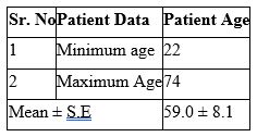
The sample of 59 patients belong to Layyah district areas were used in molecular detection of tumor markers. The mean age of patients was 59.0 ± 8.1while the minimum and maximum age at which the tumor marker detection was 22 and 74 years, respectively (Table 1).
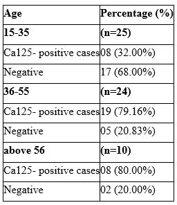
The age was categorized into 3 groups, 15-35, 36-55 and above 56 age formed 1st, 2nd and 3rd group. Number of patients in age groups 15-35, 36-55, and above 61 were, 25, 24 and 10, respectively. The no of patient was found in order of 25 >24 > 10 in 1st, 2nd and 3rd age groups, respectively. The highest no of CA-125 patient (77) were found in 1st group while the lowest patients
were found in 2nd group. The 08 (32.00%) patients showed the detection of CA-125 while 17 patients (68.00%) showed no amplification were belong to the1st age group. The 19 (79.16%) patients showed detection of CA-125 while 05 patients (20.83%) showed no detection were belong to the 2nd age group. The 08 (80.00%) patients showed detection of CA-125 while 02 patients (20.00%) showed no detection gene were belong to the 3rd age group. 35 (59.32%) out of 59 were ca125 positive cases while24 individuals were negative (Table 2).
Table 1: Minimum and maximum age of ovarian cancer patients collected from Layyah district.


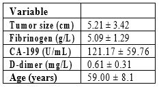
The clinical histopathological test in the ovarian cancer patients show that the tumor size 5.21 ± 3.42, fibrnigin 5.09 ± 1.29, CA-199 (U/mL) 121.17 ± 59.76, and D-dimer 0.61 ± 0.31 see in table.3.
Table 3: histopathological and clinical data of ovarian cancer patients.

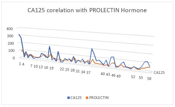
The prolactin level also increases in females that have ovarian cancer, their correlation with other discuss in next article. The CA-125 level increase in ovarian cancer patients its indication as a tumor marker. In this study results show that the CA-125 is directly proportional with prolactin level in ovarian cancer patients see in Figure1.

Discussion
In this study the patients were analyzed for the ovarian cancer. Two major tests were performed to examine the ovarian cancer that include CA-125 and TVUS (Transvaginal ultrasound). Cancer antigen test is the best marker for the screening of the ovarian cancer patient. It was seen that the patients having age above the 45 years have higher risk to develop ovarian cancer. Approximately, all the patients belong to industrial area from whose sample was collected. So, it may be due to carcinogenic chemical from industrial effluents. It may be due to DNA mutations. These mutations ultimately passe to blood relatives and can cause ovarian cancer in them.
Second test performed in this study is the TVUS that usually used to examine the size of ovarian cancer. It is usually performed with the help of ultra sound wand that uses sound waves to examine the different parts ovaries such as uterus and fallopian tubes. This test was performed to detect the epithelial ovarian cancer that is most common form of the cancer in females. The patients in which CA-125 is detected were seen to have high level of prolactin. The increase in prolactin concentration enhances the mortality rate of cancerous cells. So, we can conclude that risk of the ovarian cancer is also associated with the prolactin concentration that usually increase the life span of the cancerous cells.
Conflict of interest
The authors declared no conflict of interest.
Funding
No funds
References
- Williams TI, et al. Epithelial ovarian cancer: disease etiology, treatment, detection, and investigational gene, metabolite, and protein biomarkers. Journal of proteome research, 2007; 6(8): p. 2936-2962.
- Melikechi N, et al. Age-specific discrimination of blood plasma samples of healthy and ovarian cancer prone mice using laser-induced breakdown spectroscopy. Spectrochimica Acta Part B: Atomic Spectroscopy, 2016; 123: p. 33-41.
- Jayson GC, et al. Ovarian cancer. The lancet, 2014; 384(9951): p. 1376-1388.
- Sung H, et al. Global cancer statistics 2020: GLOBOCAN estimates of incidence and mortality worldwide for 36 cancers in 185 countries. CA: a cancer journal for clinicians, 2021; 71(3): p. 209-249.
- Shimosato Y, Mukai K, Matsuno Y. Tumors of the mediastinum. 2010: Amer Registry of Pathology.
- Xing F, Wang S, Zhou J, the expression of microRNA-598 inhibits ovarian cancer cell proliferation and metastasis by targeting URI. Molecular Therapy-Oncolytics, 2019; 12: p. 9-15.
- Andrews L, Mutch DG. Hereditary ovarian cancer and risk reduction. Best practice & research Clinical obstetrics & gynaecology, 2017; 41: p. 31-48.
- Olsen CM, et al. Obesity and risk of ovarian cancer subtypes: evidence from the Ovarian Cancer Association Consortium. Endocrine-Related Cancer, 2013; 20(2): p. 251-262.
- Allen PL. Prospective study of symptoms of ovarian cancer in postmenopausal women. 2010; UCL (University College London).
- Katzke VA, Kaaks R, Kühn T. Lifestyle and cancer risk. The Cancer Journal, 2015; 21(2): 104-110.
- Omoyajowo KO, et al. Awareness of Toxicological Impact and Risk of using Talcum Powder as a factor for Ovarian Cancer among Women in three Metropolitan Cities of Southwestern States of Nigeria.
- Modugno F, et al. Breastfeeding factors and risk of epithelial ovarian cancer. Gynecologic oncology, 2019; 153(1): p. 116-122.
- Mazhar MW. "breast cancer." Cancer Therapy & Oncology International Journal 2021; 18(4).

