Diagnostic Yield of Endoscopic Ultrasound in Intermediate Probability of Choledocholithiasis According to the ASGE Criteria
Raja Taha Yaseen*, Abbas Ali Tasneem, Manzoor ul Haq, Adeel ur rehman, Syed Mudassir Laeeq, Hina Ismail, Kiran Bajaj, Muhammad Adeel, Shoaib Ahmed Khan, Husnain Ali Metlo, Nasir Hasan Luck
Department of Gastroenterology and Hepatology, Sindh Institute of Urology and Transplantation, Karachi, Pakistan
Received Date: 22/11/2021; Published Date: 21/12/2021
*Corresponding author: Raja Taha Yaseen Khan, Department of Hepato-gastroenterology, Sindh Institute of Urology and Transplantation, Karachi, Pakistan.
Abstract
Introduction: The gold standard modality in diagnosis and therapeutic intervention for Choledocholithiasis (CL) is ERCP. Patients with intermediate probability (10-50% likelihood) of CL according to ASGE criteria were those with existence of only one strong predictor or any moderate predictor. Aim of this study is to assess the to determine the diagnostic accuracy of EUS in patients with intermediate probability for CL using ERCP as gold standard as past studies have shown the lack of accuracy of ASGE guidelines to predict CL.
Methods: It was a cross-sectional prospective study. All patients with intermediate probability criteria for CL were included in the study. Each patient underwent endoscopic ultrasound (EUS) prior to ERCP. Results were presented as means ± SD for quantitative data or as numbers with percentages for qualitative data. Continuous variables were analyzed using the Student’s t-test; while categorical variables were analyzed using the Chi-square test. A p value of ≤ 0.05 was considered statistically significant.
Results: The total number of patients included in the study were 82. Most of the patients were females49(59.8%) while males were 33(40.2%). Mean age was of 51.7 years ± 12.72. On Endoscopic ultrasound (EUS), CBD stone was present in 53 (64.6%) patients. On ERCP, 55 patients (67.1%) were found to be positive for choledocholithiasis. EUS found to be significantly associated with the presence of choledocholithiasis in the patients with intermediate probability (p-value < 0.001) with the sensitivity of 96.30%, specificity of 100 %, negative predictive value of 93.1 % and positive predictive value of 100% with diagnostic accuracy of 97.53%.
Conclusion: Performance of EUS in intermediate probability criteria for the prediction of CL was accurate in 97.6% of the patients
Keywords: Choledocholithiasis; CBD; EUS; ERCP; ASGE
Introduction
Choledocholithiasis (CL) is a common clinical problem, occurring in 10–20% of patients with symptomatic cholelithiasis, 7–14% of patients who have undergone cholecystectomy, and 18–33% of patients with acute biliary pancreatitis [1-3]. It is important to diagnose CBD stones because they are related with various severe complications, such as cholangitis and pancreatitis, especially if left untreated [4]. A combination of clinical signs and symptoms, obstructive (cholestatic) pattern of liver enzymes combined with imaging findings are necessary to establish the diagnosis of CL [5] The gold standard for the diagnosis of CL is ERCP and currently it has been the best nonsurgical therapeutic approach for CL. However, there is a 5–10% risk of complications that has been associated with it [6-8]. Due to its related risks, ERCP, as a diagnostic tool, has been replaced by less invasive diagnostic tests, such as Endoscopic Ultrasonography (EUS) and Magnetic Resonance Cholangiography (MRC) and has largely been reserved strictly as a therapeutic measure in the management of CL [9-11]. In2010, the American Society for Gastrointestinal Endoscopy (ASGE) has defined guidelines the likelihood of CL with the aim of detecting patients with the highest probability of benefiting from ERCP [12]. On the basis of presence clinical, radiological(ultrasound)and laboratorial parameters, the patients were classified into three categories. The patients with one of the following very strong predictors: CL on transabdominal ultrasound (US), clinical ascending cholangitis or bilirubin > 4 mg/dL, or those with both of the following strong predictors: dilated CBD on US (> 6 mm with gallbladder in situ) and bilirubin level between 1.8-4.0 mg/dL were classified as High probability of CL (defined as > 50% likelihood); while those with the presence of only one strong predictor or any moderate predictor (i.e. abnormal liver test, age older than 55 years or gallstone pancreatitis) were classified as intermediate probability of CL (10-50% likelihood) and those with no predictors present were classified as low probability of CL (< 10% likelihood).
The rationale of our study is that in the past, studies have shown the lack of accuracy of the ASGE guidelines for predicting CL [13].
Objective of Study
To determine the diagnostic accuracy of EUS in patients with intermediate probability (as per operational definition) for choledocholithiasis using ERCP as gold standard
Operational Definitions
Intermediate probability [11]:
Patients with intermediate probability were those have any one of the strong predictors of CBD stone i.e.
- Dilated CBD on ultrasound (>6mm with gallbladder in situ)
- Bilirubin between 1.8-4.0 mg/dl
And anyone of the moderate predictors of CBD stone i.e.,
- Abnormal Liver Function Tests (LFTs) other than bilirubin (according tour lab parameters)
- Alkaline Phosphate >130
- ALT >45mg/dl
- AST>45 mg/dl
- Gamma Glutamyl Transferase >50 mg/dl
- Age greater than 55 years
- Gall stone pancreatitis –in the presence of GB stone
- The diagnosis of acute pancreatitis requires two of the following three features:
- abdominal pain consistent with acute pancreatitis (acute onset of a persistent, severe, epigastric pain often radiating to the back);
- serum lipase activity (or amylase activity) at least three times greater than the upper limit of normal
- The diagnosis of acute pancreatitis requires two of the following three features:
Material and Methods
Study Design: Cross sectional
Setting: Department of Gastroenterology and Hepatology, Sindh Institute of Urology and Transplantation Karachi
Duration of Study: Six months from January 2019 to June 2019.
Sampling technique: Non probability simple consecutive sampling
Inclusion Criteria
Patients meeting the ASGE intermediate criteria, presence of any one of the strong predictors of high probability, or presence of CBD stone on ultrasound or ascending cholangitis
Age 25 to 55 years.
Either gender
Exclusion Criteria
- History of cholecystectomy
- Chronic Liver Disease
- Previous history of ERCP
- High and low ASGE probability criteria
Data Collection procedure
All the patients fulfilling inclusion criteria were enrolled in this study. After taking informed consent, patients demographic and clinical information were obtained including symptoms and the patients then underwent a thorough clinical examination for relevant symptoms. Then the predesigned proforma was filled by the principal investigator.
Endoscopic Ultrasound by pentax endoscopic ultrasound scope (EC-2990 Li) was performed under conscious sedation in each patient by the supervisor. Presence or absence of CBD stone was recorded in the proforma. An eight-hour fasting was advised to patient prior to the procedure. All procedures were free of cost as per institutional policy. Then ERCP was performed under general anesthesia using lateral scope (Pentax) in order to delineate biliary anatomy and to retrieve stone.
Data Analysis Procedure
All the data was entered and analyzed in SPSS Version 20. Descriptive statistics were used to summarize the continuous and categorical variables like age, duration of symptoms, and size of stone and were presented as mean (SD). Categorical variables such as gender and CBD stone on EUS and CBD stone on ERCP were reported as frequency and percentages. Stratification was done by stone on EUS and stone on ERCP. Post stratification chi square test will be applied. p -value ≤ 0.05 will be taken as significant.
Results
The total number of patients included in the study were 82. Most of the patients were females 49 (59.8%) while males were 33 (40.2%). The baseline characteristics of the patients are shown in Table 1. The mean age of 51.7 years (Range: 20 - 74 years). The Total Leucocyte Count (TLC) on admission was of 7.7±2.1(K/uL). The total bilirubin was of 1.72(mg/dl). Serum Alkaline phosphatase on admission was of 342± 136(mg/dl). The Aspartate Transaminase (AST) on admission was of 39±33(mg/dl). The Alanine transaminase (ALT) on admission was of 55±52(mg/dl). The GGT on admission was of 311±194(mg/dl) and serum amylase of 156±373(IU).
Table 1: Laboratory parameters on admission of Study population (n = 82).

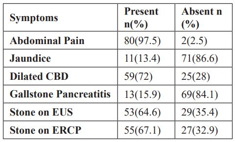
Mid Common Bile Duct (CBD) diameter was between 1.8mm-12mm with the mean of 4.35 while dilated CBD was found to be present in 59(71.9%) patients. Abdominal pain was most commonly observed in study population [80 (97.5%)] while jaundice was seen in 11(13.4%) patients. Gall stone (GB) stones were found to be present in all patients. Gall stone pancreatitis was seen in 13 (15.9%) of the patients (Table 2).
Table 2: Frequency of Categorical variables of the patients enrolled in the study.

On EUS, the stone was found in 53(64.6%) patients while on ERCP, the stone was found to be present in 55(67.1%) patients (Figure 1). 53 out of 82 were those who had stone on both EUS and ERCP while only 2 patients where those who had stone on EUS but did not found to have any stone on ERCP. There were 27 patients, who neither have stone on EUS nor on ERCP.
Post stratification, chi square test was applied which showed the sensitivity, specificity, PPV and Negative predictive value was 100%,93.1%,96.36% and 100% respectively in predicting choledocholithiasis. The diagnostic accuracy of EUS for detecting CBD stone was 97.56% (Table 3,4).
Table 3: Chi square test showing association of stone on EUS with stone on ERCP.

Table 4: Showing sensitivity, specificity, positive predictive value, negative predictive value and diagnostic accuracy.


Figure-1: Endoscopic ultrasound showing dilated CBD with Distal CBD stone causing acoustic shadow (yellow arrow).
Discussion
Previous studies although have validated American Society for Gastrointestinal Endoscopy (ASGE) guidelines for the prediction of suspected Choledocholithiasis (CL) but had lacked accuracy. An excellent tool for biliary imaging is endoscopic examination using Endoscopic ultrasonography (EUS). Radial array echoendoscopes are preferred by many endosocopists because it allows elongated views of the bile ducts. 12,13 The overall performance for the prediction of CL in our population with intermediate probability was high, 97.56% with sensitivity of 100 % and specificity of 93.1%. In order to detect CBD stones less than 5mm, EUS has been highly sensitive and decreasing stone size has no affect its accuracy [14].
In patients with biliary pancreatitis, EUS has been a safer alternative for Endoscopic Retrograde Cholangiopancreatography (ERCP) which is costly and cumbersome and requires general anesthesia. EUS has higher success rates (100 vs. 86%) and morbidity with EUS is lower (7% vs. 14%) when compared with ERCP [15]. The detection of small size (Less than 5mm) CBD stone has been effectively and excellently performed by EUS, which seems to be of highly beneficial in the context of biliary pancreatitis. Previous studies have revealed that in patients with biliary pancreatitis the risk of CL was significantly lower (36.2%) as compared to those without pancreatitis (65.9%) (OR 0.30; 95% CI, 0.17- 0.55, p < 0.001) [15]. Due to the morbidity and costs associated with ERCP, it should be limited to those who are most likely to benefit from it. In our study, thirteen patients (15.9%) had gall stone pancreatitis, while CBD stone was noted on EUS in 6(46.1%) patients while only dilated CBD was noted in 7(53.9%) patients suggestive of passage of small stone (2-3 mm) into duodenum after temporary blocking the CBD and causing pancreatitis.
In our study, the stone was found to be present in 53(64.6%) patients on EUS while on ERCP, the stone was found to be present in 55(67.1%) patients with an excellent sensitivity, specificity, PPV and Negative predictive value along with an excellent diagnostic accuracy of 97.56 % further confirming an important role of EUS in patients falling in intermediate probability criteria along with the cost effective approach of using EUS in these patients as it less time consuming and can detect CBD stones less than 5mm. There are certain limitations of our study. One of them was small sample size. This can be overcome by enrolling large number of patients falling intermediate criteria in future studies. The other limitation was that the comparison between stone dimension were not done between the two modalities i.e., EUS and ERCP. The strength of the study was that this was the pioneer study on validation of intermediate probability criteria using EUS as a diagnostic modality.
Conclusion
References
- Hunter JG. Laparoscopic trans cystic common bile duct exploration. Am J Surg 1992; 163: 53-56; discussion 57-58.
- O’Neill CJ, Gillies DM, Gani JS. Choledocholithiasis: Over diagnosed endoscopically and undertreated laparoscopically. ANZ J Surg 2008; 78: 487-491.
- Patel R, Ingle M, Choksi D, Poddar P, Pandey V, Sawant P. Endoscopic ultrasonography Can Prevent Unnecessary Diagnostic Endoscopic Retrograde Cholangiopancreatography even in patients with high Likelihood of choledocholithiasis and Inconclusive Ultrasonography: Results of a prospective Study. Clin Endosc, 2017; 50: 592-597.
- Cohen ME, Slezak L, Wells CK, Andersen DK, Topazian M. Prediction of bile duct stones and complications in gallstone pancreatitis using early laboratory trends. Am J Gastroenterol, 2001; 96: 3305-3311.
- Frossard JL, Morel PM: Detection and management of bile duct stones. Gastrointest En- dosc, 2010; 72: 808-816.
- Freitas ML, Bell RL, Duffy AJ. Choledocholithiasis: evolving standards for diagnosis and management. World J Gastroenterol, 2006; 12: 3162–3167.
- Dasari BV, Tan CJ, Gurusamy KS, Martin DJ, Kirk G, McKie L, et al: Surgical versus endoscopic treatment of bile duct stones. Cochrane Database Syst Rev, 2013; 9: CD003327.
- Aleknaite A, Simutis G, Stanaitis J, Valantinas J, Strupas K. Risk assessment of choledocholithiasis prior to laparoscopic cholecystectomy and its management options.United Europeon Gastroenterology J, 2018; 6(3): 428–438.
- Sethi S, Wang F, Korson AS, Krishnan S, Berzin TM, Chuttani R, et al. Prospective assessment of consensus criteria for evaluation of patients with suspected choledocholithiasis. Dig Endosc, 2015; 28: 75–82.
- Williams EJ, Green J, Beckingham I, Parks R, Martin D, Lombard M. Guidelines on the management of common bile duct stones (CBDS), 2008; 57: 1004–1021.
- Prachayakul V, Aswakul P, Bunthumkomal P, Deesomsak M. Diagnostic Yield of endoscopic ultrasonography in patients with intermediate or high likelihood of choledocholithiasis: a retrospective study from one university-based endoscopy center. BMC Gastroenterolo, 2014; 14: 165.
- Amouyal P, Palazzo L, Amouyal G, Ponsot P, Mompoint D, Vilgrain V, et al. Endosonography: promising method for diagnosis of extrahepatic cholestasis. Lancet, 1989; 2: 1195–1198.
- Garrow D, Miller S, Sinha D, Conway J, Hoffman BJ, Hawes RH, et al. Endoscopic ultrasound: a meta-analysis of test performance in suspected biliary obstruction. Clin Gastroenterol Hepatol, 2007; 5: 616–623.
- De Castro VL, Moura EGH, Chaves DM, Bernardo WM, Matuguma SE and Artifon ELA. Endoscopic ultrasound versus magnetic resonance cholangiopancreatography in suspected choledocholithiasis: A systematic review. Endosc Ultrasound, 2016; 5(2): 118–128.
- Rodrigo M, Narvaez-Rivera RM, Gonzialez-Gonzalez JA, Monreal-Robles R, Garcia-CompeanD, Paz-Delgadillo AA, et al. Accuracy of ASGE criteria for the prediction of choledocholithiasis. Rev Esp Enferm Dig, 2016; 108; 309-314.

