Estimation of Remaining Dentine Thickness (RDT) Under Caries Lesion with a Cone-Beam Computed Tomography and Standardized Paralleling Technique in Comparisons to Actual Measurement. In Vitro Comparative Study
Rozhyna Peshraw Kamal*, Bestoon Mohammed Faraj, Raed Fahim
Department of Dentistry, Sulaimaniyah University, Iraq
Received Date: 06/07/2021; Published Date: 04/08/2021
*Corresponding author: Rozhyna Peshraw Kamal, Department of Dermatology, Sulaimaniyah University, Iraq
Abstract
Objective: Determining a viable and reliable method for estimation of remaining dentine thickness before starting caries excavation thus reducing the incidents of post-operative sensitivities and direct pulpal exposure through finding a logistic and accurate relationship between the amounts of actual remaining dentine thickness and the radiographic remaining dentine thickness.
Methods: In three-stage experiment 60 extracted human teeth were examined by digital radiography, cone beam computed tomography, and microscopically after hand excavation of remaining caries, the results were compared and statistically analyzed for each tooth.
Result: No significant difference between actual clinical and CBCT measurements (P=0.98) but a significant difference between actual clinical and periapical radiographs (P = 0.000021).
Conclusion: Determining the remaining dentine thickness through everyday periapical radiographs is inaccurate, finding a reliable and cost-effective method remains a challenge.
Keywords: remaining dentine thickness, CBCT, challenge, caries
Introduction
With the outgrowing emphasis on the importance of minimally invasive dentistry and preservation of as much as possible of the sound tooth structure, more focus has been put on the estimation of the remaining dentin thickness below the carious lesion.
Estimating the depth of a carious lesion remains an important challenge for dentists [1].
Judging a cavity depth through the practitioner’s eyes is not always accurate and, in many cases, a shallow-looking cavity may be in fact much deeper and result in pulp exposures during cavity preparation and caries removal.
Maintaining vitality remains a major concern in restorative procedures which almost directly relates to the removing of adequate amounts of tooth structure [2].
Even with the enormous advances in technology, public dental health care, and caries preventions methods, Caries remains one of the most prevalent diseases worldwide, with an extension of caries towards the pulp chamber more and more of tooth structure is lost through bacterial byproducts losing its protective barriers against chemical and mechanical irritation [3].
The protective barrier of healthy dentine extending between the pulp chamber and pulpal floor serves the function of protection against mechanical injury and inflammation and is referred to as remaining dentine thickness3, throughout centuries there have been many arguments about the proper amount of RDT to do its function in pulp protection,
A 0.5 mm of remaining dentin in deep carious cavities were seen to induce pulpal reactions similar to pulpal exposure [4], while other authors have suggested the necessity of 2mm or 1 mm of remaining dentine thickness for pulpal protection [5,6] but a minimum thickness of 0.5mm and above should always be preserved to maintain the pulpal health [7].
As far as today the excavation of caries has been mainly through the experience of the dentists in judging the cavity depths by observing the color changes in the prepared cavity with or without the aid of radiographs,
Many parameters have been used to assist the determination of the actual RDT [8-12], the digital radiographs have been proven as the most useful [13] as the choice of appropriate treatment with proper diagnosis depends on the removal of the correct amount of tooth structure and avoiding unnecessary pulpal exposure; clinical skills in the management of deep carious lesions can be greatly enhanced by finding a correlation between the actual remaining dentine thickness and radiographic remaining dentine thickness [14].
This study is aimed towards determining a viable and reliable method for estimation of remaining dentine thickness before starting caries excavation thus reducing the incidents of post-operative sensitivities and direct pulpal exposure through finding a logistic and accurate relationship between the amounts of actual remaining dentine thickness and the radiographic remaining dentine thickness.
Material and Methods
100 extracted human teeth including teeth from incisors to molars were collected from the surgical department of shorsh teaching hospital, Sulaymaniyah City.
The teeth collected cleansed of visible blood and gross debris and maintained in a hydrated state in a glass jar of normal saline and a well-sealed lid for nearly 3 months until the required sample size was gathered, they were decontaminated prior to storage in a solution of 1:10 sodium hypochlorite for 30 minutes according to the safety guidelines of disease and infection control of CDC [15].
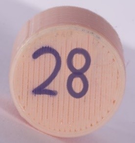
Teeth were embedded in acrylic resin to the level of the cement-enamel junction Inside custom made 3D printed cylinders using polylactic acid filaments and PRUSA MK3 3D printer (PRUSA research Czech Republic’s), After 24 hours, the acrylic resin was finished and polished with abrasive paper, each specimen is encoded with a number on the bottom of the cylinders that was used as a reference code for the Digital images and the CBCT that was taken for each tooth in the next steps.
Digital images were taken for each tooth using Sopix Ace Sensor and SoPro software at 90-degree angulation with an exposure time of 0.16ms and 70kv in buccolingual dimension.
To aid these a second image was taken for each tooth in a 20-degree mesial shift technique.
6 custom made wood box with the dimensions of (10cm x 10cm) and (2 cm height) is prepared and filled with dental plaster to standardize the position of the teeth during CBCT image acquisition.
Ten specimens are inserted inside each box consistently. Both wood boxes and acrylic resin are covered with a separating medium (Pure petroleum jelly) to facilitate their removal,
After setting the plaster, the number of each specimen is written on the upper surface of the plaster with the permanent pencil.
All the teeth were scanned by using Carestream CS9600 (care stream dental USA) CBCT machine using a field of (10 x 10) cm and 25 µsv.
Each mold is fitted horizontally to chin support in the way that the occlusal plane remains parallel to the plate [16].
For Actual clinical measurement; The tooth is sectioned in the mesiodistal plane, Diamond cutting disc (Skill-bond, High Wycombe, UK, Sintered Diamond Disc, size 400, 1/10 mm set 638R1) is used. The bur is replaced after every fifth section. Water from the 3-in-1 syringe is used as a coolant continuously,
The level of the sectioning was determined based on the location of caries in the crown and not in a standard position for all the teeth so as to gain access to the deepest area of the carious lesion so as to be hand excavated, after sectioning was completed the different zones of the carious lesions were identified with the aid of caries detection dye RED detector ( Rhodamine B, CERKAMED Poland) for 10 seconds according to the manufacturer’s instructions to facilitate distinction between differential zones of dentine [17,18].

After rinsing with water, the infected soft dentin was completely excavated by hand with small and medium excavators (Henry Schein, Kent, UK) to the point firm to moderate excavation force application and clinically accepted by the operators.
Following hand-excavation, the remaining dentine thickness is measured directly on the tooth using a translucent plastic ruler with the aid of stereomicroscope; Zumax oms1950 dental microscope ( zumax medical, China) magnification of 10x.
Measurements were taken from the thinnest point of the remaining dentine on the floor of the lesion to the pulp dentine border to the nearest 0.5 mm [19].

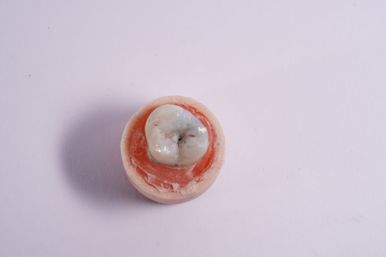
Figure 1: Tooth #28 showing occlusal caries ICADS 3 embedded in cylinder of acrylic resin up to the level of CEJ, Number of each tooth is written on the bottom of the cylinder to be used as a reference code in imaging.

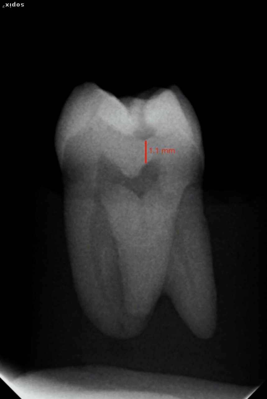
Figure 2: Peri-apical radiograph of the same tooth with measurement of the RDT through specialized software to be 1.1 mm.

Figure 3: Cone beam computed tomography showing and measuring the remaining dentine thickness in the deepest area of the lesion as 0.75 mm.

Figure 4: Tooth #28 after sectioning in Mesiodistal plane and hand excavation of remaining caries as detected by caries detection die, RED detector.
Inclusion criteria:
Visually inspected, selected teeth had carious lesions in at least one surface, without direct pulp exposure, including ICADS 20 code 3,4,5 and code 6.
Exclusion criteria:
Caries free or very initial caries not reached to dentine, that is excluding ICADS20 code 0,1 and code 2.
Statistical analysis:
All the obtained data will be collected, tabulated, and entered in MS excel and analyzed by using SPSS version 25.0.0 software 2017. Pearson’s correlation is used to assess interexaminer reliability. One-way ANOVA is used for statistical analysis. An Independent sample t-test can be used for individual evaluation of caries and dentin differences.
Result
This study consisted of 60 extracted human teeth including; 5 incisors, 10 canines, 20 premolars, and 25 molars with carious lesion distributed on different surfaces including 40 occlusal and 20 smooth surface caries.

For each tooth, three Measurements were taken including actual clinical, periapical radiographs, and CBCT.
The mean measurement for periapical radiographs was 1.23 mm with a standard deviation of 0.45mm and a range of (1.11mm -1.35 mm).
The mean measurement for CBCT was 0.82 mm with a standard deviation of 0.55 mm, and a range of (0.68mm – 0.98 mm).
The mean measurement for clinical measurements was 0.81 mm with a standard deviation of 0.31 mm and a range of (0.72mm – 0.89 mm)
The results indicated no significant difference between actual clinical and CBCT measurements (P=0.98) but a significant difference between actual clinical and periapical radiographs (P = 0.000021).
Discussion
The fact that digital radiography can be adjusted with the right software to improve the quality of the radiographs 21&22 has led to its usefulness in determining the size of the carious lesion and its proximity to the pulp aiding the clinicians in making better decisions regarding their treatment plans [21,22].
In order to obtain radiographs with great accuracy, the parallel plans approach, the utilization of the long cone in vivo, and the distance between the X-ray source and the teeth are all applied in a way that can be replicated in all circumstances [23].
Over the years various methods have been proposed in measuring the remaining dentine in the carious lesion,
For example, Everett and Fixott 24 and Krithika et al 25 recommended projecting a millimeter grid on the film, Russell and Pitts 26 developed a five-point radiographic evaluation scale depending on the extent of the radiolucency of the dental tissues to measure the depth of proximal lesions [24-26].
Valizadeh et al 27 aimed to create software that could more precisely estimate the depth of breakdown. When it comes to measuring the lesion's size, however, more challenges become apparent [27].
In this study a clear plastic ruler was used to take measurements of the teeth under 10x magnification; Other research has utilized this method to measure tooth dimensions, such as enamel, in the past [28].
This primary method of measuring was chosen since it can be easily duplicated in a practice situation with no modifications. It is a simple, quick, and easily accessible approach that is performed at the chair's side.,
It could be claimed that this magnifies the measurements, but this is true for all measures.
The decision to measure to the nearest 0.5 mm was made on the assumption that this is the minimum depth of residual dentine that can be tolerated before pulpal damage is evident [29].
For actual clinical measurement; The measures were obtained from the thinnest point of surviving dentine on the lesion's floor to the pulp dentine border [29].
Without the use of any magnification, the radiographs were read on a viewer in a calm dim room 30 to increase reliability, all measurements were taken and evaluated by two examiners to increase reliability, in addition, a measurement was taken for each carious lesion on each tooth through CBCT imaging.
In order to avoid the subjectivity in caries removal by random caries excavation depending on the operator’s judgment and tactile sensation we decided to use a caries detection dye to aid in the determination of the extent of the lesion clinically [17,18].
After sectioning of the teeth, we used microscopic magnification to measure the actual remaining dentine thickness,
After analyzing the results from these measurements, we found that although the most used method in judging the depth of the lesions, periapical radiographs is not dependable in determining the extent of caries however the CBCT measurements were more in line with the actual clinical measurements,
Although it might not be practical to take CBCT for each case in our daily routine to aid in our diagnosis and decision makings it seems to be a more valuable adjunct to our clinical judgments in assuming the depth of the carious lesions.
Conclusion
The decision to excavate remaining caries in moderate to deep carious cavities is an everyday challenge for most dental practitioners from students to specialists,
Radiographs the most commonly used mean in determining the remaining dentine thickness has proven to be misleading according to the above investigations, however, CBCT seems to be more promising in providing accurate measurements which may not be the best option due to its less accessibility and higher cost in contrast to digital radiography,
Future advancements and technologies may help to find a more reliable and accessible method in guiding dental specialists in accurately determining the remaining dentin in a both more time and cost-effective manner.
References
- Obarisiagbon A, Azodo CC, Omoaregba JO, James BO. Clinical anxiety among final year dental students: The trainers and students’ perspectives. Sahel Medical Journal 2013; 16(2): 64-70.
- Majkut P, Sadr A, Shimada Y, Sumi Y, Tagami J. Validation of Optical Coherence Tomography against Micro-computed Tomography for Evaluation of Remaining Coronal Dentin Thickness. J Endod 2015; 41: 1349-1352.
- Wegehaupt F, Betke H, Solloch N, Musch U, Wiegand A, Attin T. Influence of cavity lining and remaining dentin thickness on the occurrence of postoperative hypersensitivity of composite restorations. J Adhes Dent 2009; 11(2): 137–141.
- Smith AJ. Dentine formation and repair. In: Hargreaves KM,Goodies HE, editors. Seltzer and Bender’s dental pulp. Quintessence Publications; 2002; p. 41-62.
- Stanley HR. Pulp responses. In: Burns RC, Cohen S, editors. Pathways of the pulp. 3rd ed. St. Louis: Mosby; 1984; pp. 465-489.
- Pameijer CH, Stanley HR, Ecker G. Biocompatibility of a glass ionomer luting agent. Part II. Crown cementation. Am J Dent 1991; 4(3): 134-141.
- Murray PE, Smith AJ, Windsor LJ, Mjör IA. Remaining dentine thickness and human pulp responses. Int Endod J 2003; 36(1): 33-43.
- Lancaster PE, Craddock HL, Carmichael FA. Estimation of remaining dentine thickness below deep lesions of caries. Br Dent J 2011; 211(10): E20.
- Purton DG, Chandler NP, Monteith BD, Qualtrough AJ. A novel instrument to determine pulp proximity. Eur J Prosthodont Restor Dent 2009; 17(4): 30-34.
- Yoshida H, Tsuji M, Matsumoto H. An electrical method for examining remaining dentine thickness. J Dent 1989; 17(6): 284-286.
- Pilo R, Corcino G, Tamse A. Residual dentin thickness in mandibular premolars prepared with hand and rotatory instruments. J Endod 1998; 24(6):401-404.
- Katz A, Wasenstein-Kohn S, Tamse A, Zuckerman O. Residual dentin thickness in bifurcated maxillary premolars after root canal and dowel space preparation. J Endod 2006; 32(3):202-205.
- Sinescu C, Negrutiu ML, Bradu A, Duma VF, Podoleanu AG. Noninvasive Quantitative Evaluation of the Dentin Layer during Dental Procedures Using Optical Coherence Tomography. Comput Math Methods Med 2015; 2015: 709076.
- AL Jhany N, AL Hawaj B, AL Hassan A, AL Semrani Z, AL Bulowey M, Ansari S. Comparison of the Estimated Radiographic Remaining Dentine Thickness with the Actual Thickness Below the Deep Carious Lesions on the Posterior Teeth: An in vitro Study. Eur Endod J 2019; 3: 139-144.
- Centers for Disease Control and Prevention. Guidelines for Infection Control in Dental Health-Care Settings. MMWR 2003; 52 (No. RR17)
- R. S. Agarwal, J. Agarwal, P. Jain, and A. Chandra, “Comparative analysis of canal centering ability of different single file systems using cone-beam computed tomography: an in-vitro study,” Journal of Clinical and Diagnostic Research, 2015; 9: pp. 6–10,
- Lennon AM, Attin T, Martens S, Buchalla W. Fluorescence-aided caries excavation (FACE), caries detector, and conven- tional caries excavation in primary teeth. Pediatr Dent 2009; 31(4): 316-319.
- Boston DW, Graver HT. Histological study of an acid red caries-disclosing dye. Oper Dent 1989 Autumn;14(4):186-192.
- Murray P E, Smith A J, Windsor L J, Mjör I A. Remaining dentine thickness and human pulp responses. Int Endod J 2003; 36: 33–43.
- Jenson L, Budenz AW, Featherstone JD, Ramos-Gomez FJ, Spolsky VW, Young DA. Clinical protocols for caries management by risk assessment. J Calif Dent Assoc. 2007; 35(10): 714-723. PMID: 18044379.
- Miles DA. Imaging using solid-state detectors. Dental Clinics of North America. 1993 Oct;37(4):531-540
- De La Dure-Molla, M., Artaud, C. and Naulin-Ifi, C., 2016. Approches diagnostiques des lésions carieuses. EMC Médecine Buccale, 11(1), pp.1-9.
- Neena I E, Ananthraj A, Praveen P, Karthik V, Rani P. Comparison of digital radiography and apex locator with the conventional method in root length determination of primary teeth. J Indian Soc Pedod Prev Dent 2011; 29: 300-304.
- Everett FG, Fixott HC. The incorporated millimeter grid in oral roentgenography. Quintessence International, Dental Digest. 1975; 6(5): 53-58.
- Krithika, A.C., Kandaswamy, D., Velmurugan, N. and Krishna, V.G. (2008), Non-metallic grid for radiographic measurement. Australian Endodontic Journal, 2008; 34: 36-38.
- Russell M, Pitts N, B: Radiovisiographic Diagnosis of Dental Caries: Initial Comparison of Basic Mode Videoprints with Bitewing Radiography. Caries Res 1993; 27: 65-70. doi: 10.1159/000261518
- 27. Valizadeh, S., Goodini, M., Ehsani, S., Mohseni, H., Azimi, F., & Bakhshandeh, H. (2015). Designing of a Computer Software for Detection of Approximal Caries in Posterior Teeth. Iranian journal of radiology: a quarterly journal published by the Iranian Radiological Society, 12(4), e16242. https://doi.org/10.5812/iranjradiol. 2015; 12(2): 16242.
- Alvesalo L, Tammisalo E. Enamel thickness in 45, X females' permanent teeth. Am J Hum Genet1981; 33: 464–469.
- Murray P E, Smith A J, Windsor L J, Mjör I A. Remaining dentine thickness and human pulp responses. Int Endod J2003; 36: 33–43.
- Carmichael F. The consistent image – how to improve the quality of dental radiographs: 2. The image receptor, processor and darkroom/film-handling. Dent Update2006; 33: 39–42.

