Short-Term Analgesic Effects of Focused Shockwave Therapy in Common Orthopedic Diagnoses
Neckař P*, Kadrmasová Z, Klementová R
Centrum sportovní ortopedie a medicína, U Radnice 736/4, 41501 Teplice, Czech republic
Received Date: 15/07/2021; Published Date: 30/07/2021
*Corresponding author: Pavel Neckař, MD, Centrum sportovní ortopedie a medicína, U Radnice 736/4, 41501 Teplice, Czech republic
Abstract
Background: Common orthopedic diagnoses are considered to be the cause of pain for a great number of patients. Current options for pain management include medication, injection or surgery. Recently, Focused Shockwave Therapy (FSWT) has gained popularity due to its clinical efficacy and non-invasive application.
Objective: The aim of this study was to evaluate the short-term efficacy of FSWT in pain management of patients diagnosed with epicondylitis, rotator cuff syndrome, plantar fasciitis, patellar tendinopathy and Achilles tendinosis.
Methods: Patients underwent 3,5 ± 0,4 treatment sessions on average, depending on their state of health. All patients were treated with a FSWT device BTL-6000 FSWT (BTL Industries Ltd.) and their perception of pain was evaluated prior to the beginning of treatment (baseline) and after the last treatment using the Visual Analogue Scale (VAS).
Results: A significant (p<0.01) difference was found between VAS baseline and VAS after the last treatment in all diagnoses. The most significant pain reduction according to the VAS score was found in the group of patients with Achilles’ tendinosis (60.9%), followed by the patellar tendinopathy (57.8%) group.
Conclusion: FSWT was found to be an effective modality in immediate pain reduction in patients with epicondylitis, rotator cuff syndrome, plantar fasciitis, patellar tendinopathy and Achilles’ tendinosis.
Keywords: Focused shockwave therapy; Visual analogue scale; Pain relief; Orthopedics; Epicondylitis; Rotator cuff syndrome; Plantar fasciitis; Patellar tendinopathy; Achilles’ tendinosis; BTL-6000 FSWT
Introduction
Common ways to treat tendon and muscle disorders of musculoskeletal system include physiotherapy, standard medication (analgesics and non-steroidal anti-inflammatory drugs), injections (corticosteroid, platelet-rich plasma), physical modalities, taping and bracing, rest, intramuscular dry needling and surgery (usually the last treatment option for patients who did not respond to non-invasive methods) [1,14-18]. Recently, focused shockwave therapy (FSWT) has become a popular alternative to mentioned approaches thanks to being considered as an effective, safe, repeatable and noninvasive therapy for the treatment of many musculo-skeletal diseases [17,19].
FSWT uses the technology of extracorporeal shockwave which was introduced in 1980, when high energy extracorporeal shockwaves served as a means of treating kidney stones. Gradually, the principle of regenerative effects was discovered and shockwave therapy was introduced in the field of orthopedics [14]. Since then, ESWT has become the preferred choice in the treatment of many orthopedic disorders including plantar fasciitis of the heel, epicondylitis of the elbow or tendinitis of the shoulder. This therapy has also proven to be useful in the treatment of nonunion of long bone fractures. Prospective studies using ESWT on patellar and Achilles’ tendinopathies indicate good results as well. Rare studies done by Asian authors also show positive results in the treatment of avascular necrosis of the femoral head [20].
A shockwave is defined as an acoustic wave which produces a short (microsecond duration) three-dimensional pressure pulse. There are two modalities for shockwave therapy: focused (convergence) and radial (divergence). In FSWT, the source of energy is either electrohydraulic, electromagnetic or piezoelectric and shockwaves are concentrated into small focal areas at selected depths in the body tissues to ensure optimal therapeutic effect. On the other hand, in Radial Shock Wave Therapy (RSWT), the focal point is not centered on the target zone, as in case of FSWT. Instead, shockwave is generated through the acceleration of a projectile inside the handpiece of the treatment device and thus the focal point is on the tip of the applicator. The pressure wave then penetrates the body radially and can not be focused in the deeper layers, the area of treatment is more superficial [21-23].
Therapeutic effects are considered to be dependent on the energy delivered to a focal area (the energy flux density), the focal zone size and on the tissue penetration. Although the subject is still under study, it is known that ESWT stimulates the local biological response and is able to relieve pain, as well as positively regulate inflammation. Moreover, shockwaves improve tissue regeneration and healing by neoangiogenesis and stem cells activities. ESWT can be presented as an alternative to chirurgic therapy in some chronic tendinopathies and healing disorders. Its advantages are safety and non-invasivity. In the time of Covid pandemic, the use of ESWT is coming to the fore even more [19,23].
Clinical Indication of ESWT
The International Society for Medical Shockwave Treatment (ISMST) provides a list of recommended indications which includes epicondylitis, rotator cuff syndrome, plantar fasciitis, patellar tendinopathy and Achilles’ tendinosis.
2.1. Epicondylitis
Epicondylitis is a widespread disorder of the upper limb manifested by chronic pain and functional impairment in the region of elbow joint, precisely in the region of lateral (“tennis elbow”), as well as medial (“golfer’s elbow”) epicondyle. Epicondylitis characteristically affects the middle-aged population. Patients suffer from inflammation of the arm muscles' tendon insertions which typically causes pain during resisted extension/flexion of wrist [1,2]. Any activity involving excessive and repetitive use of forearm muscles, which could lead to microtears in tendons and graver degenerative changes in the future, may cause epicondylitis [3].
- Rotator cuff syndrome
Rotator cuff syndrome typically appears in athletes or workers performing repetitive overhead motions during which they expose their shoulder/s to high repetitive forces. This often leads to compression of the rotator cuff tendon underneath the acromion (impingement syndrome), calcified/noncalcified tendinitis of the cuff or partial rotator cuff tear appears [4,5]. Patients usually experience pain while elevating the arm or when lying on the affected side. In general, shoulder pain is considered to be the third most prevalent musculoskeletal disorder in orthopedics [5].
- Plantar fasciitis
The plantar fascia extends from the calcaneus to the distal part of each metatarsophalangeal joint sustaining the medial longitudinal arch of the foot. Plantar fasciitis is characterised by sharp pain manifested at the insertion of plantar fascia at the medial tubercle of the calcaneus [6]. The pain appears when performing activities that involve prolonged weight-bearing and has a tendency to worsen with the first walking steps in the morning or after a long period of rest. Usually either professional or recreational athletes are affected by plantar fasciitis, as the disorder is associated with physical activities which include overuse, prolonged standing, running, incorrect training and inadequate footwear. Intrinsic risk factors include obesity, pes planus, pes cavus, reduced range of ankle dorsiflexion and tight calf muscles or Achilles’ tendon [6-8].
- Patellar tendinopathy
Patellar tendinopathy, also known as jumper’s knee, is a common pathological condition caused by overload which can appear already during adolescence. Disorder affects the patellar tendon and results in progressive anterior knee pain and patellar tendon dysfunction. The patellar tendon extends distally from the patella and attaches the quadriceps muscle to the tibia. This overuse disorder typically affects athletes, especially in sports involving high-impact jumping such as volleyball and basketball or running [9-11].
- Achilles’ tendinosis
The etiology behind an Achilles tendinosis remains unclear but there are many theories indicating the cause of the disease. Causes can be divided into infective (infective pathogens proliferating within the tendon sheaths) and non-infective (autoimmune, overuse and repetitive movements, idiopathic, decreased blood supply and tensile strength with aging, muscle imbalance or weakness, insufficient flexibility, and even malalignment such as hyperpronation). Some theories also suggest that genetics, endocrine disorders, and free-radical production can also lead to tendinosis. Biopsies of diseased tendons revealed that there exists a cellular activation which has been proven by an increase in cell numbers and ground substance, collagen disarray and neovascularization. Neurovascular in-growth and glutamate, which is a modulator of pain, have been detected in the diseased tendons and they have been marked to be the source of pain in patients with Achilles’ tendinosis. [12,13].
The aim of this study was to evaluate the immediate pain relief effect of FSWT among patients with epicondylitis, rotator cuff syndrome, plantar fasciitis, patellar tendinopathy and Achilles’ tendinosis that came to our medical center.
Materials and Methods
- Inclusion criteria
Prior to starting the study, patients' orthopedic disorders (epicondylitis, rotator cuff syndrome, plantar fasciitis, patellar tendinopathy and tendinosis of insertion portion of Achilles tendon) were diagnosed and confirmed by medical doctors using the SonoScape ultrasound system and supplemented by health records.
- Exclusion criteria
Patients were excluded from the study if they had severe coagulopathy, polyneuropathy, acute inflammation and/or infection, any unstable medical or psychiatric conditions, used anticoagulants or were pregnant. Additionally, patients who experienced recent impact trauma (such as a fall) or had a local corticosteroid applied in the treated area prior to or during the study were also excluded as to avoid misinterpreted data. Common ESWT contraindications also include lung tissue in the treatment area, malignant tumor in the area, epiphyseal plate in the area, fetus in the area and the area of the brain or spine.
- Study design
The study is a retrospective evaluation of data about short-term effects of FSWT in pain management. It is based on the collection of patients' evaluations of pain which are commonly obtained before the following FSWT treatment. Despite the fact that the highest efficacy of FSWT is known to be after several months [28-41].after finishing with therapy, in this study we wanted to focus on immediate, short-term effects of the technology after the last therapy.
- Ethical standards
Each of the participants has signed a written informed consent for medical treatment. Data were collected retrospectively and all was in accord with the ethical guidelines of the 1975 Declaration of Helsinki adopted by the General Assembly of the World Medical Association (1997-2000) and by Convention on Human Rights and Biomedicine of the Council of Europe (1997) [24,25].
- Therapy device
For all participants, FSWT was applied with the device BTL-6000 FSWT (BTL Industries Ltd.). This therapy device has an intensity of up to 0.65 mJ/mm2 (the energy flux density), frequency range 1-25 Hz and adjustable penetration depth due to different coupling pads.
- Therapy procedure
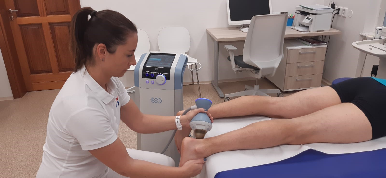
A patient’s positioning during application depends on the therapeutic area to be treated. For epicondylitis, the patient is either sitting with the affected arm placed on the couch or lying in a supine position with upper extremities along the body. For rotator cuff syndrome, the patient is lying on back or on the unaffected side. For plantar fasciitis, the patient is lying in a prone position and his treated leg is supported under the ankle. For patellar tendinopathy, the patient is lying in a supine position with a slightly flexed treated knee which is supported by an orthopaedic knee pillow. For tendinosis, the patient's positioning depends on the treated area. A suitable coupling pad was chosen based on the necessary penetration depth. Ultrasound gel was applied on the lens surface as well as in the treated area. The therapy was always initiated outside of the patient's body with low intensity. FSWT was applied in the most painful spot.

Figure 1: Therapy procedure for Achilles’ tendon (courtesy of “Centrum sportovní medicíny”)
- Therapy parameters
All patients underwent from at least 3 up to 6 treatment sessions, depending individually on each patient and on his subjective evaluation of pain. Application of FSWT continued until there was no subjective change from the last therapy. Therapies were performed once a week with the total amount of 2000 shocks per treatment and frequency between 4-8 Hz. The intensity was adjusted individually according to patients' feedback. After each session, the treated area was at rest for 48 hours.
- Pain evaluation
To evaluate and measure the subjective perception of pain intensity, Visual Analogue Scale (VAS) was used. The scale consists of a 10 cm line divided into 10 equal sections, with 0 representing “no pain” and 10 representing “unbearable pain” or in other words “the worst imaginable pain”. Each participant was asked to indicate on this scale the level of pain in the affected area before the initial intervention (baseline) and after the last treatment [26]. For some patients, data concerning final intensity (%) of FSWT were also collected.
Results
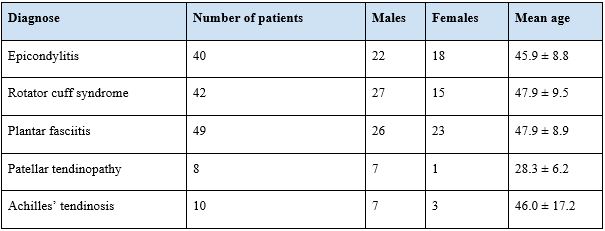
A total of 149 patients received up to 6 treatments (3,5 ± 0,4 on average), depending on their diagnosis and state of health. Baseline characteristics of the patients are shown in Table 1. Results will be presented for each diagnosis separately. A significant (p<0,01) difference was found between VAS baseline and VAS after the last treatment in all diagnoses.
Table 1: Baseline characteristics of the patients.

- Epicondylitis
For the patients diagnosed with epicondylitis, the VAS baseline results were 6.6 ± 1.3. From the baseline, VAS decreased to 4.5 ± 1.8 (Chart 1). The difference of 2.1 ± 1.7 represents 31.9% reduction in pain (Table 2). For 30 patients, data including the final intensity of 29.5 ± 7.1 were available (Table 3).
3.10. Rotator cuff syndrome
In patients with rotator cuff syndrome, the perception of pain reduced from 6.4 ± 1.3 to 3.8 ± 1.6 (Chart 1). The decrease corresponds with 41.5% (2.7 ± 1.6) (Table 2). The final intensity data of 38.0 ± 5.7 were obtained for 36 patients (Table 3).
- Plantar fasciitis
The VAS baseline for the group of patients with plantar fasciitis was 6.7 ± 1.7 and after the last therapy, the obtained VAS results were 3.7 ± 1.7 (Chart 1). The difference between both VAS values is 44.8% (3.0 ± 1.8) (Table 2). For 39 patients of this group, there were available data about the final intensity of 34.6 ± 6.4 (Table 3).
- Patellar tendinopathy
In the patellar tendinopathy group, VAS baseline results were 7.3 ± 1.0. A difference of 57.8% (4.2 ± 2.0) was found between VAS baseline and VAS after the last treatment (Table 2). From baseline, VAS was reduced to 3.1 ± 1.5 (Chart 1). The final intensity data were taken for 3 patients (38.7 ± 10.1) (Table 3).
- Tendinosis of Achilles tendon
In patients diagnosed with Achilles’ tendinosis, the pain evaluation decreased from 5.8 ± 1.8 to 2.3 ± 1.6 (Chart 1). The difference between VAS results represents 60.9% (3.5 ± 1.7) reduction in pain perception (Table 2). For 10 patients, the final intensity data of 35.9 ± 6.7 were included (Table 3).

Chart 1: VAS evaluation.
Table 2: VAS difference prior and post treatment.

Table 3: Final intensity.


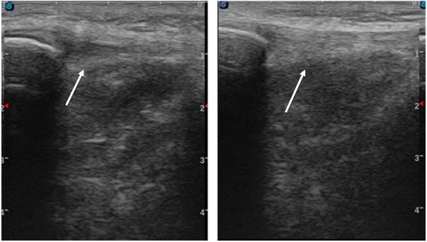
Figure 2: Patellar tendinitis prior and 6 weeks post treatment - edema reduction and widening of ligamentum patellae (arrow)

Figure 3: Rotator cuff syndrome prior, after 3rd therapy and 6 weeks post treatment - reduction in the effusion of subacromial bursa (arrow)
Discussion
The aim of this study was to evaluate the short-term effects of FSWT on pain perception, using a FSWT device BTL-6000 FSWT (BTL Industries Ltd.), in the treatment of patients diagnosed with epicondylitis, rotator cuff syndrome, plantar fasciitis, patellar tendinopathy and Achilles’ tendinosis. Our measurement results provide evidence that treatment with this FSWT device had a significant effect on decreasing the VAS score of pain in the affected area in all orthopedic diagnoses included in this study. This further supports the notion that FSWT could be an important modality for treating orthopedics patients due to a significant reduction in pain. We acknowledge that the following study has certain limitations, which include the absence of comparing FSWT to other regular physiotherapy/medical options or unequal distribution and number of participants in each diagnostic group.
In this study, the most significant change in the VAS score was found in the group of patients with Achilles’ tendinosis (60.9%), followed by the patellar tendinopathy (57.8%) group. These results could be influenced by the smallest sample of patients participating in both of these groups when compared to other diagnoses. From the data available in this study, it seems that the final intensity is not important for the effect. However, it is necessary to further research this notion.
There are several studies which report the favorable effect of ESWT in patients with epicondylitis. For example, the success rate of 59,89% in reduction of pain was found in the study by Rogoveanu et al. [2] and Tesla et al. [27] reported progressive decrease in pain perception during one, six- and twelve-months follow-up. On the other hand, in some studies, ESWT is marked as ineffective or even less effective than placebo [28]. The reasons behind the different outcomes could be understood by analyzing the methods of application in various studies. Factors such as the use of different ESWT devices, lack of standard treatment protocol (and thus variable frequency, number of sessions per week, number of impulses for each session, type of shockwave), different follow-up times and evaluation methods affect the outcomes of the studies. Furthermore, application of local anesthesia or non-steroidal anti-inflammatory drugs, stretching and eccentric exercises and the use of ice before and after every session could influence the results [2,27,28]. We might expect that best results will be obtained by ESWT and the combination of other treatment techniques performed by experienced physiotherapists or doctors.
ESWT has emerged as a strong therapeutic tool for shoulder pathology. As it was mentioned previously, rotator cuff syndrome can have multiple causes. While high quality evidence supports the efficiency of ESWT in the treatment of rotator cuff calcifications, clinical efficacy of ESWT in non-calcific tendinopathies and other shoulder pathologies remains controversial [29]. In case of calcification, Wang [30] has achieved excellent or good results in 90.9% of patients in his study and complete disappearance of calcification in 57.6% of patients. The mechanism of improved calcium reabsorption post ESWT treatment is based on the neo-lymphangiogenesis phenomenon. Regarding energy levels, there are numerous publications that have shown a high level of energy to be more effective. To achieve the same results when using low energy devices, more treatment sessions are required. For non-calcific tendinopathies and partial tendon ruptures, ESWT is able to improve vascularization of the rotator cuff and stimulate the release of growth factors [29]. However, Kolk et al. [31] or Schmitt et al. [32] found statistically not significant reduction in pain and do not recommend ESWT as a treatment option. This could be due to use of low energy parameters in both studies.
The treatment of plantar fasciitis by ESWT has been previously investigated in various clinical studies. Although there exists strong evidence proving its safety and effectiveness, there is still divergence in the characteristics of the treatment protocols and therapeutic parameters vary across different studies [14]. Findings have shown that one of the effects of ESWT is also neovascularization. This effect induces improved blood supply in the affected area, in this case at the insertion of the plantar fascia. Neovascularization is then crucial in the explanation of mechanisms of long-term improvement in chronic painful conditions, such as plantar fasciitis [33]. With regards to the results obtained in our study, in the work of Hench and Seppel [14] and multiple other studies, we can conclude that ESWT is an effective modality in patients with plantar fasciitis in the short-time period as well. There are also studies in which shock waves were directed to anatomical landmarks rather than to the point of greatest tenderness, which used lower energy levels or local analgesia. That could be the reason why these studies failed to show superiority of extracorporeal shock wave therapy over a placebo [34-36].
There is conflicting evidence regarding the effectiveness of ESWT for patellar tendinopathy. These differences in evidence may have several reasons, such as lack of objective diagnostic criteria for patellar tendinopathy, proven effectiveness of ESWT only during certain stages of tendinopathy (late degeneration phase) or, as it was already discussed, various instrumental settings [23]. Moreover, rest seems to be important in the first phase after ESWT treatment and heavy physical activity should be avoided. This is further supported by a study that showed no effect of ESWT in the early stage of patellar tendinopathy in actively competing athletes [37]. In accord with results found in our study, Wang et al. [38] achieved significantly better results for the study group treated with ESWT than for the control (placebo) group. He also followed recurrence of symptoms, which occurred in 13% of the patients of the study group and in 50% of the control group and ultrasonographic examination, which showed a significant increase in the vascularity of the patellar tendon in the ESWT group.
For the ESWT treatment of Achilles tendinosis, only a small number of studies is available. Nevertheless, authors have obtained positive results concerning the reduction in pain perception in all of these studies, which provides initial evidence that ESWT is an effective treatment option for tendinosis. Malliaropoulos et al. [39] found a functional improvement and a statistically significant decrease in VAS scores between baseline and 1-month, 3-month and 1-year follow-up in 93.1% of cases of patients with trigger finger. Similar results were achieved in the study about ESWT treatment of the primary long bicipital tendinosis [40]. In the systematic review by Ferarra et al. [41], ESWT turned out to be the most effective means of conservative treatment used for functional improvement and pain control in trigger finger.
Conclusion
FSWT is an effective modality in the treatment of patients diagnosed with epicondylitis, rotator cuff syndrome, plantar fasciitis, patellar tendinopathy and Achilles’ tendinosis based on its analgesic properties. The results of the present study proving short-term effects on pain reduction are encouraging but other studies with larger samples or comparisons with other conservative interventions should be implemented, to better understand the effects of FSWT and to unify optimal treatment parameters. For these reasons, continued research in this area is therefore of great importance.
References
- Tarpada SP, Morris MT, Lian J, Rashidi S. Current advances in the treatment of medial and lateral epicondylitis. Journal of Orthopaedics 2018; 15: 107-110.
- Rogoveanu OC, Mușetescu AE, Gofiță CE, Trăistaru MR. The Effectiveness of Shockwave Therapy in Patients with Lateral Epicondylitis. Current Health Sciences Journal 2018; 44: 368-373.
- Vaquero-Picado A, Barco R, Antuña SA. Lateral epicondylitis of the elbow. EFORT Open Reviews 2017; 13: 391-397.
- Chou WY, Wang CJ, Wu KT, Yang YJ, Cheng JH, Wang SW. Comparative outcomes of extracorporeal shockwave therapy for shoulder tendinitis or partial tears of the rotator cuff in athletes and non-athletes: Retrospective study. International Journal of Surgery 2018; 51: 184-190.
- Garving C, Jakob S, Bauer I, Nadjar R, Brunner UH. Impingement Syndrome of the Shoulder. Deutsches Ärzteblatt International 2017; 114: 765-776.
- Petraglia F, Ramazzina I, Costantino C. Plantar fasciitis in athletes: diagnostic and treatment strategies. A systematic review. Muscles, ligaments and tendons journal 2017; 7: 107–118.
- Lim AT, How CH, Tan B. Management of plantar fasciitis in the outpatient setting. Singapore Medical Journal 2016; 57: 168-171.
- Lee TL, Marx BL. Noninvasive, Multimodality Approach to Treating Plantar Fasciitis: A Case Study. Journal of Acupunctures and Meridian Studies 2018; 11: 162-164.
- Reinking MF. CURRENT CONCEPTS IN THE TREATMENT OF PATELLAR TENDINOPATHY. International journal of sports and physical therapy 2016; 11: 854–866.
- Muaidi QI. Rehabilitation of patellar tendinopathy. Journal of musculoskeletal & neuronal interactions 2020; 20: 535–540.
- López-Royo MP, Gómez-Trullén EM, Ortiz-Lucas M, Galán-Díaz RM, Bataller-Cervero AV, Al-Boloushi Z, Hamam-Alcober Y, Herrero P. Comparative study of treatment interventions for patellar tendinopathy: a protocol for a randomised controlled trial. British Medical Journal Open 2020; 10: e034304.
- Ray G, Sandean DP, Tall MA. Tenosynovitis. StatPearls Publishing [Internet]. 2020. PMID: 31335044.
- Vuillemin V, Guerini H, Bard H, Morvan G. Stenosing tenosynovitis. Journal of Ultrasound 2012; 15: 20-28.
- Hench M, Seppel G. Evaluation of the Therapeutic Effect of Extracorporeal Shockwave Therapy in Chronic Plantar Fasciitis. Clinical Research on Foot & Ankle 2019; 7: 292.
- Lenoir H, Mares O, Carlier Y. Management of lateral epicondylitis. Orthopaedics & Traumatology: Surgery & Research 2019; 105: S241-S246.
- Weiss LJ, Wang D, Hendel M, Buzzerio P, Rodeo SA (2018) Management of Rotator Cuff Injuries in the Elite Athlete. Current Reviews in Musculoskeletal Medicine 2018; 11: 102-112.
- Li X, Zhang L, Gu S, Sun J, Qin Z, Yue J, et al. Comparative effectiveness of extracorporeal shock wave, ultrasound, low-level laser therapy, noninvasive interactive neurostimulation, and pulsed radiofrequency treatment for treating plantar fasciitis: A systematic review and network meta-analysis. Medicine (Baltimore) 2018; 97: e12819.
- Sisk D, Fredericson M. Taping, Bracing, and Injection Treatment for Patellofemoral Pain and Patellar Tendinopathy. Current Reviews in Musculoskeletal Medicine 2020; 13: 537-544.
- d'Agostino MC et al. Shockwave as biological therapeutic tool: From mechanical stimulation to recovery and healing, through mechanotransduction. International Journal of Surgery 2015; 24B: 147-153.
- Wang CJ, Extracorporeal shockwave therapy in musculoskeletal disorders. Journal of Orthopaedic Surgery and Research 7, 2012. PMID: 22433113
- Speed C. A systematic review of shockwave therapies in soft tissue conditions: focusing on the evidence. British Journal of Sports Medicine 2014; 48: 1538–1542.
- Ioppolo F, Rompe JD, Furia JP, Cacchio A. Clinical application of shock wave therapy (SWT) in musculoskeletal disorders. European journal of physical and rehabilitation medicine 2014; 50: 217-230.
- van der Worp H, van den Akker-Scheek I, van Schie H, Zwerver J. ESWT for tendinopathy: technology and clinical implications. Knee surgery, sports traumatology, arthroscopy 2013; 21: 1451-1458.
- World Medical Association Declaration of Helsinki. JAMA 1997; 277: 925.
- Council of Europe. Convention for Protection of Human Rights and Dignity of the Human Being with Regard to the Application of Biology and Biomedicine: Convention on Human Rights and Biomedicine. Kennedy Institute of Ethics Journal 1997; 7: 277-290.
- Sung YT, Wu JS. The Visual Analogue Scale for Rating, Ranking and Paired-Comparison (VAS-RRP): A new technique for psychological measurement. Behavior Research Methods 2018; 50: 1694-1715.
- Testa G, Vescio A, Perez S, et al. Functional Outcome at Short and Middle Term of the Extracorporeal Shockwave Therapy Treatment in Lateral Epicondylitis: A Case-Series Study. Journal of Clinical Medicine 2020; 9: 633.
- Staples MP, Forbes A, Ptasznik R, Gordon J, Buchbinder R. A randomized controlled trial of extracorporeal shock wave therapy for lateral epicondylitis (tennis elbow). Journal of Rheumatology 2008; 35: 2038-2046.
- Moya D et al. Current knowledge on evidence-based shockwave treatments for shoulder pathology. International Journal of Surgery 2015; 24: 171-178.
- Wang CJ, Yang KD, Wang FS, Chen HH, Wang JW. Shock wave therapy for calcific tendinitis of the shoulder: a prospective clinical study with two-year follow-up. American Journal of Sports Medicine 2003; 31: 425-426.
- Kolk A, Yang KG, Tamminga R, H van der Hoeven. Radial extracorporeal shock-wave therapy in patients with chronic rotator cuff tendinitis: a prospective randomised double-blind placebo-controlled multicentre trial, Bone and Joint Journal 2013; 95-B: 1521-1526.
- Schmitt J, Haake M, Tosch A, Hildebrand R, Deike B, Griss P, Low-energy extracorporeal shock-wave treatment (ESWT) for tendinitis of the supraspinatus: a prospective, randomised study, Journal of Bone Joint and Surgery. British 2001; 83: 873-876.
- Rompe JD, Meurer A, Nafe B, Hofmann A, Gerdesmeyer L. Repetitive low-energy shock wave application without local anesthesia is more efficient than repetitive low-energy shock wave application with local anesthesia in the treatment of chronic plantar fasciitis. Journal of Orthopaedic Research 2005; 23: 931-941.
- Buchbinder R, Ptasznik R, Gordon J, Buchanan J, Prabaharan V, Forbes A. Ultrasound-guided extracorporeal shock wave therapy for plantar fasciitis: a randomized controlled trial. JAMA 2002; 288: 1364-72.
- Haake M, Buch M, Schoellner C, Goebel F, Vogel M, Mueller I, et al. Extracorporeal shock wave therapy for plantar fasciitis: randomised controlled multicentre trial. BMJ 2003; 327: 75.
- Speed CA, Nichols D, Wies J, Humphreys H, Richards C, Burnet S, et al. Extracorporeal shock wave therapy for plantar fasciitis. A double blind randomised controlled trial. Journal of Orthopaedic Research 2003; 21: 937-40.
- Zwerver J, Hartgens F, Verhagen E, van der Worp H, van den Akker-Scheek I, Diercks RL. No effect of extracorporeal shockwave therapy on patellar tendinopathy in jumping athletes during the competitive season: a randomized clinical trial. American Journal of Sports Medicine 2011; 39: 1191–1199.
- Wang CJ, Ko JY, Chan YS, Weng LH, Hsu SL. Extracorporeal shockwave for chronic patellar tendinopathy. American Journal of Sports Medicine 2007; 35: 972-8.
- Malliaropoulos N, Jury R, Pyne D et al. Radial extracorporeal shockwave therapy for the treatment of finger tenosynovitis (trigger digit). Open Access Journal of Sports Medicine 2016; 7: 143-151.
- Liu S, Zhai L, Shi Z, Jing R, Zhao B, Xing G. Radial extracorporeal pressure pulse therapy for the primary long bicipital tenosynovitis a prospective randomized controlled study. Ultrasound Med Biol. 2012; 38: 727-735.
- Ferrara PE, Codazza S, Cerulli S, Maccauro G, Ferriero G, Ronconi G. Physical modalities for the conservative treatment of wrist and hand's tenosynovitis: A systematic review. Semin Arthritis Rheum. 2020; 50: 1280-1290.

