Clinicopathological Evaluation of Benign Laryngeal Lesions
MTarhun Yosunkaya*
Lokman Hekim University Faculty of Medicine Department of ENT, Ankara/ Türkiye
Received Date: 15/10/2020; Published Date: 16/11/2020
*Corresponding author: M Tarhun Yosunkaya, Lokman Hekim Akay Hospital. Büklüm cad. No:4 Çankaya/Ankara/Türkiye. Tel: +905323564997; E-mail: tarhun.yosunkaya@lokmanhekim.edu.tr, Orcid ID: 0000-0002-8504-5317.
Abstract
Most benign vocal cord lesions occur as a result of ulceration and changes in healing processes caused by mechanical vocal trauma caused by abuse and misuse of the voice. Many factors such as social and occupational vocal use characteristics, medical conditions, psychological structures, smoking and reflux of individuals also disrupt the epithelial defense system on the vocal cord. These patients have symptoms ranging from dysphonia to life-threatening respiratory distress. In our study, the age, gender, clinical findings and pathological features of the lesions of patients who were admitted to the hospital with the complaint of dysphonia and diagnosed with polyps, nodules, Reinke edema, vocal cord cysts, granulomas and papilloma by videolaryngoscopic examination were investigated. It was found that the majority of the patients (86%) were between the ages of 30-59. According to the study, it was found that polyps (71%) were the most common lesions in males, nodules and Reinke edema were observed more frequently in females. These lesions were most common in the 40-49 age group in both sexes. While the most common symptom (89%) observed in the patients was hoarseness / change in voice, difficulty in breathing was the symptom detected at least (2%).
The resulting vocal disorder disrupts the health of individuals and has significant effects on their professional, social and emotional states. Therefore, early diagnosis and treatment of lesions is very important. Voice therapy should be applied before and after endolaryngeal surgery, which is a treatment option in treatment. Patients should also be informed about vocal hygiene, smoking and reflux.
Keywords : Benign Laryngeal Lesions; Dysphonia; Age; Gender; Pathologic Features
Introduction
Dysphonia is one of the common symptoms of ENT clinics. Approximately 30% of the adult population experiences a dysphonia during their lifetime. Although many vocal diseases occur in conditions due to acute and limited infections, benign larynx lesions are chronic. Most benign vocal cord lesions occur as a result of ulceration and changes in healing processes caused by mechanical vocal trauma caused by abuse and misuse of the voice. Many factors such as social and occupational vocal use characteristics, medical conditions, psychological structures, smoking and reflux of individuals also disrupt the epithelial defense system on the vocal cord. These patients have symptoms ranging from dysphonia to life-threatening respiratory distress [1]. The resulting vocal disorder disrupts the health of individuals and has significant effects on their professional, social and emotional states [2]. When these diseases are not diagnosed and treated early, the lesions enlarge and respiratory distress occurs and treatment becomes difficult. While most of benign laryngeal lesions can be treated with medical treatment or voice therapy, some diseases require endolaryngeal microsurgery [3]. In our study, the age, gender, clinical findings and pathological features of the lesions of patients who were admitted to the hospital with the complaint of dysphonia and diagnosed with polyps, nodules, Reinke edema, vocal cord cysts, granulomas and papilloma by videolaryngoscopic examination were investigated.
Material and Method
Our study included patients who presented to the Otorhinolaryngology Clinic between 2014 and 2017 with complaints of hoarseness, voice change, feeling-pain in the throat, difficulty in breathing, difficulty in speaking, fatigue in voice, cough, and diagnosed with benign laryngeal lesion by videolaryngoscopic examination. Age, gender and clinical symptoms of a total of 136 patients, 75 males and 61 females, between the ages of 21-68 were analyzed retrospectively. Patients with inflammatory lesions, premalignant and malignant laryngeal lesions, speech disorders due to central nervous system, and patients with nasal, nasopharyngeal, oral and pharyngeal pathologies were excluded from the study. Patients with inflammatory lesions, premalignant and malignant laryngeal lesions, speech disorders due to central nervous system, and patients with nasal, nasopharyngeal, oral and pharyngeal pathologies were excluded from the study. Age, gender, clinical symptoms and pathological features of the lesions were evaluated.
Results
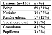
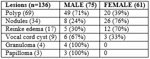
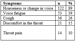
In our study, a polyp was the most detected lesion in 69 patients (51%) in 136 patients who were followed and treated with a diagnosis of benign laryngeal lesion as a result of videolaryngoscopic examination. Nodule 34 (25%), Reinke edema 17 (12.5%), vocal cord cyst 9 (7%), granuloma 4 (3%), and papilloma 3 (2%) were observed (Table 1). Of the 136 patients, 75 (55%) were male and 61 (45%) were female (Table 2). The youngest age was 21, while the oldest age was 68. It was found that the lesions were most common (41%) between the ages of 40-49. In the 20-29 age range, it was observed to be the least (2.5%). It was found that the majority of the patients (86%) were between the ages of 30-59. According to the study, it was found that polyps (71%) were the most common lesions in males, nodules and Reinke edema were observed more frequently in females (Table 3,4). these lesions were most common in the 40-49 age group in both sexes. While the most common symptom (89%) observed in the patients was hoarseness/change in voice, difficulty in breathing was the symptom detected at least (2%) (Table 5).
Table 1: Distribution of patients with benign laryngeal lesions.

Table 2 : Distribution of patients with benign laryngeal lesions by gender.

Table 3 : Distribution of benign laryngeal lesions in males by age.

Table 4: Distribution of benign laryngeal lesions in females by age.

Table 5: Distribution of symptoms of benign laryngeal lesions.

Discussion
The rate of referrals to ENT clinics due to dysphonia is 1% and 11% of them constitute benign vocal cord lesions [4]. The underlying etiology of benign laryngeal lesions is vocal trauma due to misuse of the voice. Secondarily, smoking, allergy, and acid reflux also play a major role in the formation of diseases by causing mucosal damage [5,6]. Voice disorders due to benign laryngeal lesions negatively affect both the social and work-life of individuals, thus causing functional, physical, psychological and economic impairments in the quality of life [7]. In our study, in which we evaluated the age, gender, clinical symptoms and pathological features of the lesions in patients with benign laryngeal lesions, it was found that the majority of the patients (86%) were between the ages of 30-59. The most common symptom (89%) seen in the patients was found to be hoarseness/change in voice. According to the study, a polyp was the most common lesion in males (71%), while the nodule was found mostly in females (76%). The nodule and polyp which are among the benign laryngeal lesions and detected most frequently are similar to each other [8]. According to Marcatullio et al., Changes in the lamina propria with mechanical trauma begin earlier in vocal cord nodules compared to polyps. Polyps occur with the later stages of wound healing [9]. Generally, bilateral and small nodules in women are common in professional voice users. Too much talking, shouting, nervous, aggressive women are nodule candidates [10,11]. Studies show that women are more vulnerable to vocal disorders. Female vocal cords anatomically have a weaker tissue mass to absorb vocal trauma. Hyaluronic acid, which is found in excess in shock-absorbing places in the body and plays a major role in wound repair, is less in the vocal cord lamina propria of females [12]. In the nodules, there is edema and fibromyxoid stroma caused by epithelial hyperplasia, edema and vascular cell infiltration in the early period due to vocal trauma. With the continuing use of false voice, fibrosis in the lamina propria and thickening in the basement membrane are observed in the late period. Unlike nodules, polyps are seen more unilaterally and larger in males. In polyp, changes occur after vascular bleeding in the submucosal area due to vocal trauma as a result of irritating factors such as smoking, infections, allergy, gastroesophageal reflux, throat clearing habit, as well as misuse and abuse of the voice [13]. In their research on the etiology of polyps, Wang et al. Reported that the amount of pepsin in the cell fluid of polyps increased and laryngopharyngeal reflux also played a potential role in the development of the disease [14]. Hoarseness-changes and voice fatigue are common symptoms in polyps and nodules. In the treatment of nodules and polyps, vocal hygiene ve voice therapy which includes the elimination of wrong vocal habits and teaching correct techniques is the first choice. Endolaryngeal microsurgery is applied in nodules and polyps that are not treated for a long time and do not respond to voice therapy. To prevent recurrence, vocal hygiene should be ensured after the operation and voice therapy should also be applied to eliminate the habit of misuse voice [15-17]. Reinke's edema and vocal cord cyst are also seen in people who misuse their voices, have a history of reflux, smoke, talk too much. In Reinke edema, mucinous and gelatinous material accumulation occurs in the place known as the Reinke space between the superficial lamina propria under the mucosa. In a vocal cord cyst, an accumulation of serous or mucous fluid is formed in the Reinke space surrounded by a thin wall. Vocal cord cysts are thought to occur as a result of obstruction of the mucous gland ducts as a result of inflammation caused by abuse of voice, gastroesophageal reflux and upper respiratory tract infections, or the covering of the newly healing mucosa over the damaged epithelium after vocal trauma. Flexibility in the vocal cords decreases and vibration is forced [18-21]. Granuloma occurs with the effect of secondary trauma due to abuse of voice, chronic cough, throat clearing habit, intubation and gastroesophageal reflux. In a study showing that reflux plays an active role in granuloma formation, reflux was found in 30-76% of granuloma cases. Ulceration with desquamation in the epithelium after vocal trauma and inflammatory cell infiltration in the lamina propria, ulceration and hyperplasia in the surface epithelium, and excessive granulation tissue beneath it are observed. The voices of the patients may not be affected due to the granuloma originating from the posterior and afonary part of the vocal cord. Large granulomas can cause breathing difficulties by closing the glottic distance [22,23]. Papilloma is a benign, cauliflower-looking lesion of the larynx caused by the HPV virus. In infectious lesions, papillary or acanthotic growth pattern covered with squamous epithelium is observed. Cells contain well-differentiated squamous epithelium. Maturation is regular, Koilocytic atypia and frequent mitotic activity are seen. While the adult type occupies a limited place in the larynx and does not recur if completely surgically removed, the juvenile form manifests itself as extensive, large lesions, causing respiratory distress, does not respond to treatment, and recurs very frequently [24,25].
Conclusion
Benign laryngeal lesions have clinical features ranging from dysphonia to life-threatening respiratory distress. The resulting vocal disorder can create emotional and mental tension in people's lives and also disrupts the very important social communication. In addition to its negative effects on the quality of life and health, especially for professional voice users, it also causes a loss of workforce and productivity. Therefore, early diagnosis and treatment of lesions is very important. Voice therapy should be applied before and after endolaryngeal surgery, which is a treatment option in treatment. Patients should also be informed about vocal hygiene, smoking and reflux.
References:
- Roy N, Merril MR, Gray SD, Smith EM. Voice Disorders in the General Population: Prevalence, Risk Factors, and Occupational Impact. Laryngoscope 2005;115:1988-95.
- Lierde KV, Heule SV, Ley SD,Mertens E, Claeys S. Effect of Psychological Stress on Female Vocal Quality. Folia Phoniatr Logop. 2009;61:105-111.
- Yosunkaya MT. Basic principles of microlaryngeal surgery in benign larynx lesions. Arch Otolaryngol Rhinol 2020;6(2):12-15.
- Cohen SM, Kim J, Roy N, Asche C, Courney M. Prevalence and causes of dysphonia in a large treatment-seeking population. Laryngoscope. 2012;122(2):343-348.
- Wani A, Rehman A, Hamid S, et al. Benign Mucosal Fold Lesion as a Cause of Hoarseness of Voice. A Clinical Study. Otolaryngology. 2012;2(3):120
- Schwartz SR, Cohen SM, Dailey SH, et al. Clinical practice guideline: Hoarseness (Dysphonia). Otolaryngol Head Neck Surg. 2009;141:1-31.
- Nelson R, Bless MD, Heisey D. Personality and voice disorders: a super factor trait analysis. Journal of Speech-Language Hearing Research. 2000;43:749-768.
- Rosen CA, Gartner-Schmidt J, Hathaway B, et al. A nomenclature paradigm for benign midmembranous vocal fold lesions. Laryngoscope. 2012;122:1335-1341.
- Marcotullio D, Magliulo G, Pietrunti S, et al. Exudative laryngeal diseases of Reinke's space: a clinicohistopathological framing J Otolaryngol. 2002;(1):376-380.
- Karkos PD, McCornick M. The etiology of vocal fold nodules in adults. Curr Opin Otolaryngol Head Neck Surg. 2009;17(6):420-423.
- Sharma DK, Sohal BS, Bal MS, Aggarwal S. Clinico-pathological Study of 50 Cases of Tumours of Larynx. Indian Journal of Otolaryngology and Head&Neck Surgery. 2013;65(1):29-35.
- Ward PD, Thibeault SL, Gray SD. Hyaluronic acid: its role in voice. J Voice. 2002; 16:303-309.
- Kristen L, Kraimer B, Husain I. Updated Medical and Surgical Treatment for Common Benign Laryngeal Lesions. Otolaryngol Clin N Am 52. 2019:745-757.
- Wang L, Tan J, Wu T, et al. Association between laryngeal pepsin levels and the presence of vocal fold polyps. Otolaryngol Head Neck Surg. 2017;156:144-151.
- Uloza V, Saferis V, Uloziene I. Perceptual and acoustic assessment of voice pathology and efficacy of endolaryngeal phonomicrosurgery. J Voice. 2005;19:138-145.
- Bequignon E, Bach C, Fugain C, et al. Long-term results of surgical treatment of vocal fold nodules. Laryngoscope. 2013;123:1926-1930.
- Barillari MR, Volpe U, Mirra G, et al. Surgery or rehabilitation: a randomized clinical trial comparing the treatment of vocal fold polyps via phonosurgery and traditional voice therapy with “voice therapy expulsion training” J Voice. 2017;31(3):13-20.
- Kambic V, Gale N, Radsel Z. Anatomical markers of Reinke's space and the etiopathogenesis of Reinke edema. Laryngorhinootologie. 1989;68(4):231-235.
- Hantsagos A, Remacle M, Dikkers FG et al. Exudative lesions of Reinke's space: a terminology proposal. Eur Arch Otorhinolaryngol. 2009;266(6): 869-878.
- Martins RH, Santana MF, Tavares EL. Vocal cysts: clinical, endoscopic and surgical aspects. J Voice 2011;25:107-110.
- Tibbets KM, Dominguez LM, Simpson CB. Impact of perioperative voice therapy on outcomes in the surgical management of vocal cysts. J Voice 2018; 32:347-351.
- Devaney KO, Rinaldo A, Ferlito A. Vocal process granuloma of the larynx-recognition, differential diagnosis and treatment. Oral Oncol. 2005;41:666-669.
- Karkos PD, George M, Van Der Veen J, et al. Vocal process granulomas: a systematic review of treatment. Ann Otol Rhinol Laryngol. 2014;123:314-320.
- Martins RH, Dias N, Gregorio E, Alencar M, et al. Laryngeal papillomatosis: morphological study by light and electron microscopy of the HPV-6. Rev Bras Otorhinolaryngol 2008;74(4): 539-543.
- Escopedo AR, Santillana KM, Gonzales JL. Recurrent Respiratory Papillomatosis: Review and Treatment Update. J Otolaryngol Rhinol. 2018;4: 40-44.

