Double J stent Migration: When, Where, and Why?
Daghdagh Yassine, Mahmoud Alafifi*, Alae eddine Seffar, Abdi El Mostapha, Amine Moataz, Mohammed Dakir, Adil Debbagh, Rachid Aboutaieb
Department of Urology, CHU Ibn Rochd, Morocco
Received Date: 01/01/2022; Published Date: 21/01/2022
*Corresponding author: Mahmoud Alafifi, Department of Urology, CHU Ibn Rochd, Casablanca, Morocco
Abstract
A double J stent is a standard therapy for drainage during ureteral obstruction. Complications such as antegrade bladder migration or retrograde ureter migration are well-known. Despite the fact that this is a safe and minimally invasive treatment, serious cases of double J stent migration have been reported, albeit seldom. Following ureteral stent placement, radiologic imaging is critical for avoiding the complication. We report a rare case of a double-J stent migrating into the inter duodenal caval zone, which was retracted laparoscopically.
Keywords: Double J stent, inter duodenal caval, migration, complication
Introduction
For more than 40 years, the double J stent has been an essential internal bypass device utilised in urology. It drains the urinary upper excretory tract, relieves obstructions, promotes ureter healing, and manages urine leaks.
Despite the fact that this is a safe and minimally invasive therapy, it can have serious short and long-term complications.
Although the double J placement is simple, it can cause hematuria, bacteriuria, urosepsis, migration, fragmentation, and encrustation.
We report a rare case of a double-J stent migrating into the inter duodenal caval zone as an unusual complication of ureteral stent placement, which was retracted laparoscopically.
Case Report
Y.B, a 40-year-old patient, had been treated for lithiasis disease two weeks prior with left laser ureteroscopy and complete lithiasis fragmentation, followed by placement of a double J stent. Before being referred to our department for pain in the epigastric and right hypochondrium.
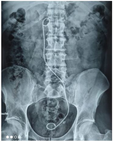
Unexpectedly, the kidney, ureters, and bladder (KUB) x-ray revealed that the DJS was not in the proper site, with the proximal third of the double J stent in the right latero-vertebral region and the distal two thirds in the iliac and pelvic lateralized on the left (Figure1).

Figure 1: (KUB) X-ray revealed the proximal third of the double J stent in the right latero-vertebral region and the distal two thirds in the iliac and pelvic lateralized on the left.
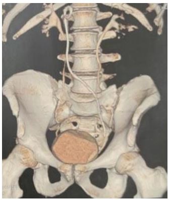
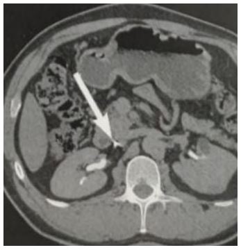
An abdominal CT scan revealed an unusual false path of the double J stent, with the proximal end situated between the duodenum and the inferior vena cava (Figure 2,3).


Figure 3: The proximal end (white Arrow) situated between the duodenum and the inferior vena cava.
The double J stent was successfully removed laparoscopically. A new DJS was placed, and KUB x-ray confirmed its position. His post-operative care was simple, and he was discharged on the third day.
Discussion
DJS placement is a standard urological technique. However, this approach may cause complications such as dysuria, hematuria, bacteriuria, fragmentation, stone formation, and migration. The stent migrating is one of the most serious complications.
The double J stent may migrate during or after placement.
Double J stent migration is a reported complication that can occur proximally into the pelvicaliceal system or ureter and distally into the urinary bladder. Proximal migration of the double J stent is uncommon, occurring in 0.6-3.5 percent of patients [1].
It has been observed that the incidence of ureteral stent migration is less than 5% [2].
FALAHATKAR and AL. reported an unexpected complication after double J stent placement ; intravascular migration and malposition of a double J stent in the inferior vena cava [3].
Another case was reported by Ian Wall and Al of a ureteral stent that migrated and perforated the duodenum [4].
In our situation, this is the first report of ureteral stent migration into the interduodenal caval zone.
Endoscopic surgery, laparoscopic surgery, endovascular surgery, and open surgery have all been used to remove a migrating double J stent. The position of the stent, the patient's condition, the hemorrhagic condition, the formation of thrombus, and the surgeon's expertise all influence treatment options.
In this case, we performed a laparoscopic method to remove the migrating double J stent.
There is no one, commonly approved, or recommended approach for double J stent placement. We conclude that while placement a double J stent, peroperative real-time radioscopy should always be performed to check correct placement.
If there is any uncertainty about the position of the stent after placement, or if a large urethral obstruction was identified on preoperative imaging, an ascending ureterography should be performed.
The ideal double J stent should be biocompatible, inexpensive, resistant to migration, have great flow properties, be resistant to encrustation and infection, be readily insertable, non-refluxing, and radiopaque. Despite advancements in implanted device design and material, no currently existing device meets all of the criteria for a perfect stent [5].
Conclusion
References
- Smith MJV. Ureteral Stent: Their use and miss use. Monogr. Urol, 1993: 14: 1.
- Breau RH. Optimal prevention and management of pro ximal ureteral stent migration and remigration ; Sep ; J Urol, 2001; 166(3): pp. 890-893.
- SiavashFalahatkar, Hossein Hemmati, et al. Intracaval Migration: An Uncommon Complication of Ureteral Double-J Stent Placement, Journal of Endourology, 2012; 26(2).
- Ian Wall, Robin Baradarian, et al. Spontaneous perforation of the duodenum by a migrated ureteral stent. Gastrointestinal Endoscopy, 2008; 68(6).
- Bansal N, Bhangu G, Bansal D. Postoperative complications of double-J ureteral stenting: a prospective study. Int Surg J, 2020; 7(5): 1397-1403.

