
Email id: info@ijclinmedcasereports.com
First Case of Parathyroidectomy with Intraoperative Parathyroid Hormone Monitoring at King Abdullah University Hospital: A Case Report
Osama K Shattarah1,*, Fuad T Alshogerrat1, Madani T Alshogerrat1, Rasha Al-Ebbini2, Johnathan Hubbard3, Ivan Tomasi3, Anoud Ramadan4, Bayan Bani Issa4, Ahmad Eyadeh4, Ayat Aloqaily5, Reham Jaradat5, Lina Alshadfan6 and Saleh Abu-alhaj6
1Department of General Surgery and Urology, Faculty of Medicine, Jordan University of Science and Technology, Irbid, 22110, Jordan
2Department of Diagnostic Radiology, Faculty of Medicine, Jordan University of Science and Technology, Irbid, 22110, Jordan
3Department of Upper GI Surgery, Guy's and St Thomas' NHS Foundation Trust, London, United Kingdom
4Department of Internal Medicine, Faculty of Medicine, Jordan University of Science and Technology, Irbid, 22110, Jordan
5Department of Pathology and Microbiology, Faculty of Medicine, Jordan University of Science and Technology, Irbid, 22110, Jordan
6Faculty of Medicine, Al-Balqa Applied University, Al-Salt, Jordan
Received Date: 14/10/2026; Published Date: 11/12/2026
*Corresponding author: Osama K Shattarah, Department of General Surgery and Urology, Faculty of Medicine, Jordan University of Science and Technology, Irbid, Jordan; Department of Endocrine Surgery, King Abdullah University Hospital (KAUH), Irbid, Jordan
Abstract
Background: Intraoperative parathyroid hormone (IOPTH) monitoring is now a standard adjunct to parathyroid surgery. It provides real-time biochemical confirmation that hyperfunctioning parathyroid tissue has been completely removed and facilitates minimally invasive, image-guided procedures. To our knowledge, Miami-criterion-guided IOPTH monitoring had not previously been used for parathyroidectomy at King Abdullah University Hospital (KAUH).
Case presentation: A 35-year-old man with recurrent nephrolithiasis, osteoporosis, and morbid obesity had hypercalcemia, markedly elevated parathyroid hormone (PTH), and hypophosphatemia, with normal renal function and vitamin D. His weight exceeded the load capacity of the gamma-camera imaging table, preventing technetium-99m sestamibi scintigraphy. Neck ultrasonography and contrast-enhanced neck CT concordantly localized a left inferior parathyroid adenoma. Focused, minimally invasive left inferior parathyroidectomy was performed with serial IOPTH monitoring. PTH decreased from 1385 pg/mL pre-excision to 320 pg/mL at 5 minutes (77% drop) and 138 pg/mL at 10 minutes (90% drop) after excision, meeting the Miami criterion for biochemical cure. Histopathological examination confirmed a solitary parathyroid adenoma.
Conclusion: This case illustrates the feasibility, safety, and clinical utility of introducing Miami-criterion-guided IOPTH monitoring within a newly established endocrine surgery program. Structured implementation may improve operative precision and limit unnecessary bilateral neck exploration in comparable centers.
Keywords: Primary hyperparathyroidism; Parathyroid adenoma; Intraoperative parathyroid hormone; Miami criterion; Minimally invasive parathyroidectomy; Endocrine surgery; Case Report
Abbreviations: IOPTH - Intraoperative Parathyroid Hormone; PHPT - Primary Hyperparathyroidism; PTH - Parathyroid Hormone; MIP - Minimally Invasive Parathyroidectomy; KAUH - King Abdullah University Hospital
Introduction
Primary Hyperparathyroidism (PHPT) is among the most common endocrine disorders and is the leading cause of hypercalcemia in outpatients. Its population prevalence has increased substantially as serum calcium screening has become widely available [1]. A single parathyroid adenoma is responsible for 80–85% of cases, whereas multigland hyperplasia accounts for 10–15% and parathyroid carcinoma for less than 1% [2]. Surgical removal remains the only definitive cure.
Nussbaum et al. first described intraoperative parathyroid hormone (IOPTH) measurement in 1988 [3]. Irvin and colleagues subsequently developed it into a practical adjunct that provides real-time guidance during surgery [4,5]. Circulating PTH has a short biological half-life of approximately 3–5 minutes. Consequently, a rapid decrease in serum PTH after removal of hyperfunctioning tissue provides immediate biochemical confirmation of surgical success, supporting focused, image-guided parathyroidectomy without routine bilateral neck exploration [6].
The Miami criterion is the most widely used definition of operative success: PTH must decrease by more than 50% from the highest pre-incision or pre-excision value at 10 minutes after removal of all grossly hyperfunctioning tissue [7,8]. In prospective series, this criterion has predicted cure with approximately 98% sensitivity and 97% accuracy [7]. Compared with localization imaging alone, IOPTH-guided surgery has also improved cure rates in patients undergoing minimally invasive parathyroidectomy (MIP) [9]. Of the proposed intraoperative criteria, including Miami, Halle, Rome, and Vienna, the Miami criterion continues to provide the most favorable balance between sensitivity and specificity [10].
Although IOPTH monitoring is well established in high-volume endocrine surgery centers worldwide, many Middle Eastern centers have introduced it only recently. Historically, patients with primary hyperparathyroidism in this region have presented later and with more severe skeletal and renal manifestations than Western cohorts [19,20]. Body habitus may further complicate preoperative localization. Technetium-99m sestamibi scintigraphy requires the patient to lie on a gamma-camera table with a fixed weight limit; morbid obesity may therefore make the examination technically impossible, leaving ultrasonography and cross-sectional imaging as the available localization methods. To our knowledge, this is the first report of parathyroidectomy with Miami-criterion-guided IOPTH monitoring at King Abdullah University Hospital (KAUH). The lesion was localized by ultrasound and CT without sestamibi scintigraphy. We describe the case and the introduction of this technique within an emerging endocrine surgery program. The report follows the CARE (CAse REport) guidelines for case report development.
Case Presentation
Patient Information and History:
A 35-year-old man with morbid obesity was referred for assessment of recurrent nephrolithiasis and osteoporosis. His medical and surgical history was otherwise unremarkable. He reported no family history of parathyroid or other endocrine disease and was not receiving any medication known to influence calcium metabolism.
Clinical Findings:
Physical examination revealed no abnormalities, palpable neck mass, or clinical signs of hypercalcemic crisis. The patient was clinically euthyroid, with no cervical lymphadenopathy.
Diagnostic Assessment:
Biochemistry: Elevated serum calcium and markedly increased parathyroid hormone were accompanied by hypophosphatemia. Renal function and serum vitamin D were normal. Urine Calcium-to-Creatinine ratio (UCCR) was elevated; thus, Familial hypocalciuric hypercalcemia (FHH) was excluded. This biochemical pattern supported primary hyperparathyroidism rather than a secondary or vitamin D-driven process.
Neck ultrasonography: Grayscale and color Doppler examination demonstrated a discrete, hypoechoic, well-vascularized lesion inferior and posterior to the left thyroid lobe. Its peripheral vascular pattern was typical of a parathyroid adenoma (Figure 1).
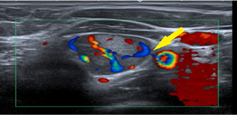
Figure 1: Color Doppler neck ultrasound showing a hypoechoic left inferior parathyroid lesion with a peripheral feeding vessel (yellow arrow), a vascular pattern typical of a parathyroid adenoma.
Thyroid/parathyroid scintigraphy: Technetium-99m sestamibi scintigraphy was planned but could not be undertaken because the patient's weight exceeded the maximum load capacity of the gamma-camera imaging table. The examination was therefore omitted for technical reasons; it was neither declined nor non-diagnostic.
Cross-sectional imaging: As scintigraphy could not be obtained, contrast-enhanced CT of the neck was used for complementary localization. CT showed an avidly enhancing oval soft-tissue nodule posterior to the left thyroid lobe, at the expected site of the left inferior parathyroid gland. This finding agreed with the ultrasound localization (Figure 2).
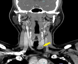
Figure 2: Contrast-enhanced axial CT of the neck showing an enhancing left inferior parathyroid adenoma posterior to the left thyroid lobe(empty yellow arrow), concordant with the ultrasound localization.
After multidisciplinary discussion, the patient was scheduled for focused, minimally invasive parathyroidectomy with intraoperative PTH monitoring. This decision was supported by concordant ultrasound and CT localization of a left inferior parathyroid adenoma despite the unavailability of Sestamibi scintigraphy.
Diagnosis:
Primary hyperparathyroidism attributable to a solitary left inferior parathyroid adenoma, identified through biochemical assessment and localized by ultrasound and CT without sestamibi scintigraphy, was diagnosed. The findings supported a focused surgical approach.
Therapeutic Intervention:
Focused left inferior parathyroidectomy was performed under general anesthesia. The suspected adenoma was identified and excised en bloc. Serial PTH sampling included baseline and pre-excision measurements, followed by measurements at 5 and 10 minutes after excision and a confirmatory level on postoperative day 1 (Table 1).
Table 1: Intraoperative and postoperative parathyroid hormone (PTH) values, showing a 90% decline from the pre-excision baseline within 10 minutes, fulfilling the Miami criterion for biochemical cure.
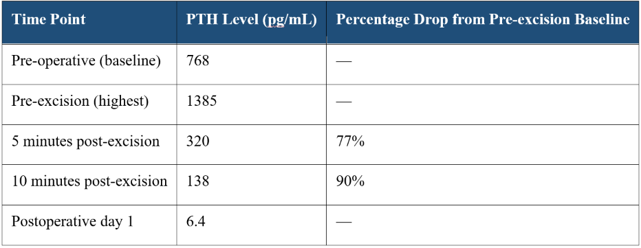
The Miami criterion defines successful excision as a PTH decrease of more than 50% from the highest baseline value within 10 minutes of gland removal [7,8]. In this case, the 90% decrease at 10 minutes strongly indicated complete excision of the hyperfunctioning gland and excluded residual multigland disease, permitting completion of the procedure without bilateral exploration. The specimen was a well-circumscribed, encapsulated, reddish-brown gland (Figure 3) that corresponded to the lesion identified on preoperative ultrasound and CT.
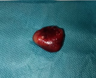
Figure 3: Gross specimen photograph of the excised left inferior parathyroid adenoma, showing a well-circumscribed, encapsulated, reddish-brown gland.
Follow-up and Outcomes:
There were no intraoperative complications, and histopathological examination confirmed a parathyroid adenoma. Serum calcium normalized after surgery. Mild, transient hypocalcemic symptoms were successfully treated with oral calcium carbonate and vitamin D supplementation. Recovery was uneventful, and the patient was discharged in stable condition. At outpatient follow-up six months after the index surgery, he remained normocalcemic, and his presenting symptoms had resolved completely.
Discussion
IOPTH monitoring has substantially changed the surgical management of primary hyperparathyroidism by providing objective, real-time evidence of adequate resection [3–6]. The rapid clearance of circulating PTH means that a sustained decrease after gland excision distinguishes cure of true single-gland disease from persistent multigland disease, without depending solely on visual assessment of the glands or frozen section [7]. In this patient, technetium-99m sestamibi scintigraphy could not be performed because his weight exceeded the certified load limit of the gamma-camera table. This examination is one of the two standard first-line localization studies and has a reported sensitivity of approximately 79% when used alone [17]. The table weight limit is a practical constraint of nuclear medicine imaging that disproportionately affects patients with morbid obesity. Localization instead depended on ultrasonography, which has a reported sensitivity of approximately 76% alone [17], and corroborating contrast-enhanced CT. Both studies identified a left inferior parathyroid adenoma. Concordant localization and the 90% intraoperative PTH decrease at 10 minutes provided strong support for the adequacy and safety of a focused procedure. The case illustrates that minimally invasive parathyroidectomy does not invariably require a diagnostic sestamibi scan when concordant anatomical imaging and IOPTH monitoring are available.
Morbid obesity creates a specific and under-recognized obstacle to conventional nuclear medicine localization. Most gamma-camera and SPECT/CT tables have manufacturer-specified weight limits, commonly 150–200 kg. Above these limits, the examination cannot be performed safely, irrespective of its clinical indication. High-resolution ultrasound and cross-sectional imaging, either CT or four-dimensional CT where available, then become the principal and sometimes the only feasible methods of preoperative localization. Surgeons and radiologists serving populations with a high prevalence of obesity should identify this constraint early in the diagnostic pathway, rather than discovering it after an unsuccessful attempt at scintigraphy.
Focused minimally invasive parathyroidectomy (MIP) guided by IOPTH achieves cure rates statistically comparable to those of conventional bilateral neck exploration, with shorter operating times, less extensive dissection, and reduced risk to nerves and parathyroid glands outside the surgical target [14–16]. A 2020 Cochrane systematic review reported operative success within six months in approximately 97% of patients undergoing MIP and 99% undergoing bilateral exploration. When adequate localization and IOPTH monitoring were used, the difference between approaches was not statistically significant [15]. Current international guidelines, including those of the American Association of Endocrine Surgeons and the Fifth International Workshop on Primary Hyperparathyroidism, support either approach for appropriately selected patients with well-localized disease and recommend adjunctive IOPTH monitoring when a focused procedure is planned [11,13].
The Miami criterion remains the most extensively validated of the proposed intraoperative biochemical criteria and was used in this case [7,8]. It requires a PTH decrease of more than 50% from the highest pre-incision or pre-excision value within 10 minutes of excision. Comparisons of the Miami, Halle, Rome, and Vienna criteria have demonstrated a favorable balance of sensitivity and specificity for the Miami criterion. Nevertheless, double adenomas or asymmetric multigland disease may occasionally produce falsely reassuring results. Biochemical findings must therefore be considered alongside preoperative imaging and intraoperative gland inspection, rather than interpreted as an isolated measurement [10].
Reports from our region have historically described later presentation of primary hyperparathyroidism and a greater burden of skeletal and renal complications than in cohorts routinely screened for asymptomatic hypercalcemia [19,20]. The nephrolithiasis and osteoporosis in this patient were consistent with that pattern. Our institution already has an experienced parathyroid surgery practice [18]. Incorporating Miami-criterion-guided IOPTH monitoring into this practice provides a structured, evidence-based means of offering focused, minimally invasive surgery to patients who might otherwise undergo routine bilateral exploration.
The observations are subject to the inherent limitations of a single case, and longer follow-up is needed to establish durable biochemical cure. We nevertheless consider this first successful institutional experience useful as a local benchmark and as an example for emerging endocrine surgery programs in comparable settings of how IOPTH monitoring can be safely incorporated into standard preoperative localization pathways.
Conclusion
This case illustrates the feasibility, safety, and clinical effectiveness of incorporating Miami-criterion-guided intraoperative PTH monitoring into focused parathyroidectomy within a newly established endocrine surgery program. Concordant preoperative localization and a rapid, substantial intraoperative PTH decrease supported confident, minimally invasive treatment of a solitary parathyroid adenoma, with an excellent outcome. Wider use of IOPTH monitoring in comparable centers may improve operative precision and lessen the need for more extensive bilateral neck exploration.
Author Contributions:
Osama K. Shattarah: Conceptualization, Project administration, article writing, final approval, and accountability for all aspects of the work.
Fuad T Alshogerrat: article writing, Investigation, Visualization.
Madani T Alshogerrat: article writing, Investigation.
Rasha Al-Ebbini: Conceptualization, article writing, investigation, Visualization, final approval.
Johnathan Hubbard: Review & editing.
Ivan Tomasi: Review & editing.
Anoud Ramadan: Review & editing.
Bayan Bani Issa: Review & editing.
Ahmad Eyadeh: Data curation, writing-review & editing. Ayat Aloqaily: Data curation, writing-review & editing.
Reham Jaradat: Data curation.
Lina Alshadfan: Writing-review & editing.
Ahmad Eyadeh: Review & editing.
Conflicts of Interest: The author reports no conflicts of interest.
References
- Yeh MW, Ituarte PHG, Zhou HC, Nishimoto S, Liu IA, Harari A, et al. Incidence and prevalence of primary hyperparathyroidism in a racially mixed population. J Clin Endocrinol Metab, 2013; 98(3): 1122–1129. https://doi.org/10.1210/jc.2012-4022.
- DeLellis RA. Parathyroid tumors and related disorders. Mod Pathol, 2011; 24(Suppl 2): S78–S93. https://doi.org/10.1038/modpathol.2010.132.
- Nussbaum SR, Thompson AR, Hutcheson KA, Gaz RD, Wang CA. Intraoperative measurement of parathyroid hormone in the surgical management of hyperparathyroidism. Surgery, 1988; 104(6): 1121–1127.
- Irvin GL 3rd, Dembrow VD, Prudhomme DL. Operative monitoring of parathyroid gland hyperfunction. Am J Surg, 1991; 162(4): 299–302. https://doi.org/10.1016/0002-9610(91)90135-z.
- Irvin GL 3rd, Deriso GT 3rd. A new, practical intraoperative parathyroid hormone assay. Am J Surg, 1994; 168(5): 466–468. https://doi.org/10.1016/s0002-9610(05)80101-x.
- Irvin GL 3rd, Prudhomme DL, Deriso GT, Sfakianakis G, Chandarlapaty SK. A new approach to parathyroidectomy. Ann Surg, 1994; 219(5): 574–581. https://doi.org/10.1097/00000658-199405000-00015.
- Irvin GL 3rd, Solorzano CC, Carneiro DM. Quick intraoperative parathyroid hormone assay: surgical adjunct to allow limited parathyroidectomy, improve success rate, and predict outcome. World J Surg, 2004; 28(12): 1287–1292. https://doi.org/10.1007/s00268-004-7708-6.
- Carty SE, Worsey J, Virji MA, Brown ML, Watson CG. Concise parathyroidectomy: the impact of preoperative SPECT 99mTc sestamibi scanning and intraoperative quick parathormone assay. Surgery, 1997; 122(6): 1107–1114.
- Chen H, Pruhs Z, Starling JR, Mack E. Intraoperative parathyroid hormone testing improves cure rates in patients undergoing minimally invasive parathyroidectomy. Surgery, 2005; 138(4): 583–587. https://doi.org/10.1016/j.surg.2005.06.046.
- Barczynski M, Konturek A, Hubalewska-Dydejczyk A, Cichon S, Nowak W. Evaluation of Halle, Miami, Rome, and Vienna intraoperative iPTH assay criteria in guiding minimally invasive parathyroidectomy. Langenbecks Arch Surg, 2009; 394(5): 843–849. https://doi.org/10.1007/s00423-009-0510-z.
- Wilhelm SM, Wang TS, Ruan DT, Lee JA, Asa SL, Duh QY, et al. The American Association of Endocrine Surgeons guidelines for definitive management of primary hyperparathyroidism. JAMA Surg, 2016; 151(10): 959–968. https://doi.org/10.1001/jamasurg.2016.2310.
- Bilezikian JP, Brandi ML, Eastell R, Silverberg SJ, Udelsman R, Marcocci C, et al. Guidelines for the management of asymptomatic primary hyperparathyroidism: summary statement from the Fourth International Workshop. J Clin Endocrinol Metab, 2014; 99(10): 3561–3569. https://doi.org/10.1210/jc.2014-1413.
- Bilezikian JP, Khan AA, Silverberg SJ, El-Hajj Fuleihan G, Marcocci C, Minisola S, et al. Evaluation and management of primary hyperparathyroidism: summary statement and guidelines from the Fifth International Workshop. J Bone Miner Res, 2022; 37(11): 2293–2314. https://doi.org/10.1002/jbmr.4677.
- Udelsman R. Six hundred fifty-six consecutive explorations for primary hyperparathyroidism. Ann Surg, 2002; 235(5): 665–670. https://doi.org/10.1097/00000658-200205000-00008.
- Ahmadieh H, Kreidieh O, Akl EA, El-Hajj Fuleihan G. Minimally invasive parathyroidectomy guided by intraoperative parathyroid hormone monitoring (IOPTH) and preoperative imaging versus bilateral neck exploration for primary hyperparathyroidism in adults. Cochrane Database Syst Rev, 2020; 10(10): CD010787. https://doi.org/10.1002/14651858.CD010787.pub2.
- Westerdahl J, Bergenfelz A. Unilateral versus bilateral neck exploration for primary hyperparathyroidism: five-year follow-up of a randomized controlled trial. Ann Surg, 2007; 246(6): 976–980. https://doi.org/10.1097/SLA.0b013e31815c3ffd.
- Cheung K, Wang TS, Farrokhyar F, Roman SA, Sosa JA. A meta-analysis of preoperative localization techniques for patients with primary hyperparathyroidism. Ann Surg Oncol, 2012; 19(2): 577–583. https://doi.org/10.1245/s10434-011-1870-5.
- Algargaz W, Abushukair HM, Odat H, Hamouri S, Abuashour R. Giant versus regular parathyroid adenoma: a retrospective comparative study. Ann Med Surg (Lond), 2021; 66: 102454. https://doi.org/10.1016/j.amsu.2021.102454.
- Younes NA, Al-Trawneh IS, Albesoul NM, Hamdan BR, Sroujieh AS. Clinical spectrum of primary hyperparathyroidism. Saudi Med J, 2003; 24(2): 179–183.
- Al-Saleh Y, AlSohaim A, AlAmoudi R, AlQarni A, Alenezi R, Mahdi L, et al. Primary hyperparathyroidism in Saudi Arabia revisited: a multi-centre observational study. BMC Endocr Disord, 2022; 22(1): 155. https://doi.org/10.1186/s12902-022-01059-7.