
Email id: info@ijclinmedcasereports.com
Laryngeal Low-Grade Chondrosarcoma: When Anatomy Rather Than Grade Dictates Total Laryngectomy — A Case Report and Updated Review of The Literature
Hajar El Agouri1,*, Mohamed Tbouda1, Marouane Balouki2 and Sanaa Abbaoui3
1Pathology department, Military Hospital Oued Eddahab, Agadir Morocco, Faculty of Medicine and Pharmacy, University Ibn Zohr, Agadir, Morocco
2Otorhinolaryngology department, Military Hospital Oued Eddahab, Agadir Morocco, Faculty of Medicine and Pharmacy, University Ibn Zohr, Agadir, Morocco
3Pathology and Laboratory department, Mohammed VI University Hospital, Faculty of Medicine and Pharmacy, University Ibn Zohr, Agadir, Morocco
Received Date: 24/09/2026; Published Date: 20/11/2026
*Corresponding author: Hajar El Agouri, Pathology department, Military Hospital Oued Eddahab, Agadir Morocco, Faculty of Medicine and Pharmacy, University Ibn Zohr, Agadir, Morocco
Abstract
Background: Chondrosarcoma is the commonest non-epithelial malignancy of the larynx but remains a rare tumor. It arises predominantly from the cricoid hyaline cartilage, is low grade in the great majority of cases, and behaves indolently, with local persistence rather than metastatic spread. Its submucosal growth and its morphological overlap with benign cartilaginous lesions make the diagnosis one of clinical, radiological and pathological correlation. As management has shifted towards function-preserving surgery, defining the minority of patients in whom total laryngectomy remains unavoidable has become correspondingly important.
Case presentation: A 72-year-old man presented with hoarseness and dysphonia of six months' duration, subsequently complicated by dyspnea. Laryngoscopy showed a firm, smooth submucosal swelling of the right laryngeal wall under intact mucosa, with an immobile right vocal cord. Computed tomography showed an expansile, heterogeneously enhancing mass centered on the cricoid cartilage, containing punctate calcification and disrupting the cricoid ring, with supraglottic, subglottic and paraglottic extension. Biopsy showed a mildly hypercellular hyaline cartilaginous proliferation without mitoses or necrosis, reported without obvious evidence of malignancy, definitive classification being deferred to complete excision. Because the cricoid framework had been destroyed and no functional airway could be reconstructed, total laryngectomy with neck dissection was performed. The specimen showed a multinodular hyaline cartilaginous proliferation with moderately increased cellularity, occasional binucleated chondrocytes and mildly enlarged hyperchromatic nuclei; mitoses, necrosis, myxoid change, a dedifferentiated component and permeation of intertrabecular spaces were absent; margins were free and fifteen dissected lymph nodes were negative. The final diagnosis was conventional well-differentiated chondrosarcoma of the cricoid cartilage (grade 1). Adjuvant radiotherapy was given on the tumor board's recommendation; the patient was free of recurrence and metastasis two years after surgery.
Conclusion: Laryngeal chondrosarcoma is recognized by integrating endoscopic, radiological and histological data, and its separation from chondroma is reliable only on a generously sampled resection specimen. Function-preserving surgery is the contemporary standard whenever complete excision leaves a reconstructable airway; total laryngectomy is reserved, as here, for tumors that have consumed the cricoid ring, and as salvage after failure of conservative surgery. The decision is therefore governed by the anatomy of the laryngeal framework rather than by histological grade.
Keywords: Chondrosarcoma; Cricoid cartilage; Laryngeal neoplasms; Laryngectomy; Airway obstruction; Organ-preserving surgery; Case report
Background
Cancer of the larynx is overwhelmingly epithelial. Mesenchymal malignancies represent a small residue in which chondrosarcoma is the commonest entity, accounting for roughly 0.2% of head and neck malignancies and about 1% of laryngeal malignant tumors, and for less than 1% of laryngeal cancers in population-based data [1-5]. Chondrosarcoma is by contrast one of the commonest primary sarcomas of the skeleton; only 2–5% of all chondrosarcomas arise in the head and neck, where the larynx is among the more frequently affected sites [1,6,7].
Three features give laryngeal chondrosarcoma a clinical identity of its own. First, it has a strong topographical preference for the cricoid cartilage — 77 of 111 tumors (69%) in the series of Thompson and Gannon and 271 of 377 (72%) in the pooled literature — most often the posterior lamina, the region where ossification of the laryngeal hyaline cartilages is most pronounced in the aging larynx [1,8]. Second, it is low grade in the majority of cases, with a correspondingly indolent natural history and a metastatic rate of a few per cent in surgical series and long survival [1,4,8]. And, most importantly for the pathologist, its histological separation from benign cartilaginous lesions of the larynx is genuinely difficult and its diagnosis on a small endoscopic biopsy is unreliable [6].
The therapeutic corollary of this indolence has been a progressive shift, over the past three decades, from total laryngectomy towards function-preserving surgery, confirmed in national registry data showing that partial resections now outnumber total laryngectomy by about two to one [4,9-14]. That shift is not unconditional: it presupposes a lesion whose complete excision leaves a reconstructable airway.
We report a case of low-grade chondrosarcoma of the cricoid cartilage, in a 72-year-old man, in whom the extent of the tumor and the degree of airway obstruction dictated a total laryngectomy, and we use it as a starting point for an updated review of the clinical, radiological, pathological and therapeutic aspects of this unusual tumor.
Case Presentation
Clinical presentation
A 72-year-old man was referred to the outpatient department for progressive hoarseness and dysphonia of six months' duration, subsequently complicated by dyspnea. There was no dysphagia and no weight loss. He was a chronic alcohol user and ex-smoker with a 20 pack-year history. There was no history of cervical irradiation or prolonged intubation.
Ear, nose and oral cavity examination was normal and there was no palpable cervical lymphadenopathy or laryngeal deformity. Flexible and direct laryngoscopy showed a right-sided, firm, smooth submucosal swelling of the laryngeal wall, covered by intact and non-ulcerated mucosa, extending to the ipsilateral anterior commissure and false vocal fold and displacing the adjacent endolaryngeal structures, with immobility of the right vocal cord, associated with decreased mobility of the left vocal cord. The airway lumen was markedly narrowed, but the obstruction was partially tolerated and no tracheostomy was required before definitive surgery.
Imaging
Contrast-enhanced computed tomography of the neck showed a heterogeneously enhancing expansile mass, measuring 4 × 2.5 × 2 cm, centered on the cricoid cartilage and containing punctate intralesional calcifications. The cricoid ring was expanded and disrupted. The adjacent laryngeal structures were displaced and the airway was narrowed, with superior extension to the supraglottic region and inferior extension to the subglottic region, without extension beyond the cricoid cartilage. Laterally, the lesion extended into the paraglottic space, with no infiltration of the adjacent surgical planes and no enlarged cervical nodes. Thoraco-abdominal computed tomography showed no distant metastasis. The overall findings were staged as cT3N0M0.
Biopsy
Biopsy under direct laryngoscopy yielded numerous firm fragments with semi-translucent cut surfaces. Histological examination showed hyaline cartilaginous proliferation with increased cellularity, without mitotic activity or necrosis. The final report was deliberately restricted to a cartilaginous neoplasm without obvious evidence of malignancy, with definitive classification pending complete excision of the entire lesion.
Treatment
Following multidisciplinary team discussion, and based on the clinical and radiological TNM classification and the histological nature of the tumor, a total laryngectomy was performed, associated with bilateral functional jugulocarotid neck dissection despite the absence of enlarged cervical nodes, definitive tracheostomy and reconstruction of a neopharynx. The postoperative course was uneventful.
Pathological findings
Macroscopic examination:
The laryngectomy specimen contained a firm submucosal mass with a lobulated, translucent cut surface, measuring 4 cm in greatest dimension and containing areas of calcification (Figure 1). The mass arose from the cricoid cartilage and pushed against the adjacent submucosal structures and soft tissues. The resection margins were macroscopically free.
Microscopic examination:
Multiple sections showed a well-delimited cartilaginous proliferation arranged in confluent nodules of hyaline cartilage, separated by thin fibrous septa and bounded peripherally by a dense fibrous perichondrial band (Figure 2). Cellularity was slightly to moderately increased and unevenly distributed, being highest at the periphery of the lobules. The neoplastic chondrocytes were embedded in lacunae and occasional binucleate nuclei were seen throughout the lesion (Figure 3). Nuclei were mildly to moderately enlarged, round to ovoid and hyperchromatic, with coarsely clumped chromatin and inconspicuous nucleoli; occasional nuclei had slightly irregular contours (Figure 4). Multinucleated giant chondrocytes, mitotic figures, tumor necrosis, myxoid change, high-grade areas and dedifferentiated component were all absent. No permeation of intertrabecular spaces was identified. Fifteen cervical lymph nodes were excised, all free of metastasis. Immunohistochemistry was not performed, since none of the differential diagnoses raised by the morphology required it. On these grounds, the final pathological diagnosis was well differentiated conventional chondrosarcoma, low grade (grade 1), completely excised.
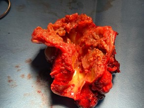
Figure 1: Macroscopic appearance of the total laryngectomy showing a translucent tumor of the larynx, arising from the cricoid cartilage and containing areas of calcification.
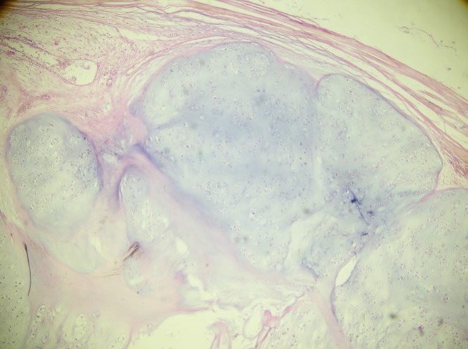
Figure 2: Low-power view of the multinodular growth of the proliferation, showing confluent nodules of hyaline cartilage separated by thin fibrous septa (Hematoxylin and eosin stain, original magnification ×40).
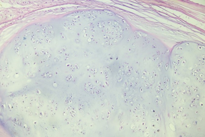
Figure 3: Neoplastic nodular cartilaginous proliferation showing moderately increased cellularity, with numerous lacunae containing two or more chondrocytes (Hematoxylin and eosin stain, original magnification ×100).
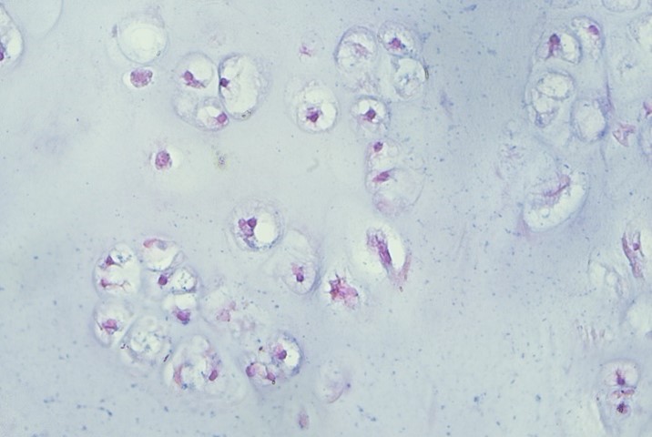
Figure 4: High-power view of the neoplastic chondrocytes showing mild atypia with some binucleated chondrocytes within one lacuna (Hematoxylin and eosin stain, original magnification ×400).
Follow-up
After a 10-day hospital stay, the patient underwent a swallowing test to assess the integrity of the reconstruction and healing; the results were normal, with no evidence of a fistula. Subsequently, and according to the tumor board meeting, the patient received 30 sessions of preventive adjuvant radiotherapy (50 Gy) targeting the surgical site and lymph node regions. Follow-up examinations, performed at one month, three months, and then every six months for two years, showed no local recurrence or distant metastasis.
Table 1: Selected series and case reports of laryngeal chondrosarcoma, with the present case.
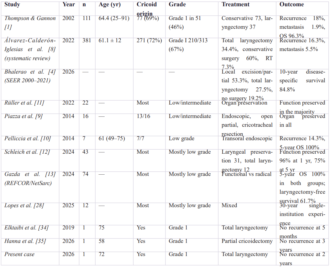
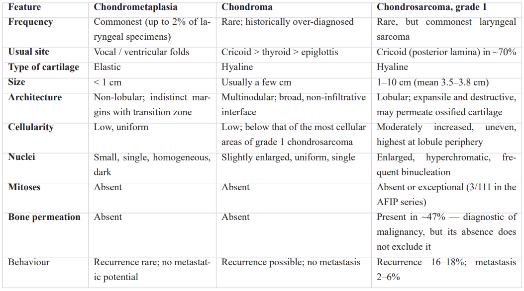
Table 2: Differential diagnosis of cartilaginous lesions of the larynx [1,6].
Discussion
Epidemiology
Laryngeal chondrosarcoma is a tumor of the older male larynx. In the 111-case series of Thompson and Gannon the male-to-female ratio was 3.6:1 and the mean age at presentation 64.4 years (range 25–91) [1]; the systematic review of Álvarez-Calderón-Iglesias and colleagues, which pooled 381 published cases, found 74% men and a mean age of 61.1 ± 12 years [8]. The recent analysis of the Surveillance, Epidemiology and End Results database, covering patients diagnosed between 2000 and 2021, confirms both the rarity of the tumor — less than 1% of laryngeal cancers — and its favorable long-term behavior, with a ten-year disease-specific survival of 84.8% [4]. Our patient sits within this typical demographic.
Unlike squamous cell carcinoma of the larynx, no convincing etiological link with tobacco or alcohol has been established: about one third of Thompson and Gannon's patients were smokers, a proportion that does not differ meaningfully from the background population of that age [1]. Our patient's smoking and alcohol history is best regarded as incidental, and it is worth emphasizing that it should not be allowed to bias the clinician towards a mucosal carcinoma when the endoscopic picture reveals a submucosal mass under intact mucosa.
Etiopathogenesis
To date, no definite cause has been known. The most widely accepted hypothesis attributes the tumor to disordered endochondral ossification of the laryngeal hyaline cartilages [1,6]. Two observations support it: the peak age of presentation coincides with the period at which ossification of the laryngeal cartilages is most active, and the posterior lamina of the cricoid — where ossification is most pronounced and where the posterior cricoarytenoid muscle inserts — is by far the commonest point of origin [1]. Competing or complementary hypotheses invoke persistent embryonic cartilaginous rests, transformation of pluripotent mesenchymal stem cells, and ischemic change within a pre-existing chondroma [1,6]. The last deserves emphasis: Thompson and Gannon identified a benign chondroma intimately juxtaposed to the chondrosarcoma in 60.4% of their cases, and 61.2% of those chondromas showed ischemic degeneration, which they interpreted as evidence of a developmental continuum rather than of coincidence [1]. Ferlito and colleagues take the alternative view that such lesions represent a single neoplasm of varying differentiation rather than malignant transformation of a benign tumor, and acknowledge that the question remains open [6].
Isolated chondrosarcomas of the larynx have been described after Teflon injection and after therapeutic irradiation, but these remain anecdotal [1]. From a molecular standpoint, the IDH1/IDH2 mutations that characterize a substantial proportion of conventional central chondrosarcomas of the skeleton are distinctly uncommon at this site: in the series of 88 head and neck chondrosarcomas of Tallegas and colleagues, skull base tumors showed an IDH1 mutation rate of 85.7%, whereas laryngotracheal tumors showed a rate of only 11.8% and craniofacial tumors none at all [15]. IDH testing is therefore of limited diagnostic value in the larynx and was not performed in our case.
Clinical Presentation
Clinical symptoms are those of a slowly enlarging submucosal mass, and their nature depends on the direction of growth. Endolaryngeal expansion narrows the airway and produces dyspnea and stridor; fixation of the cricoarytenoid joint or involvement of the vocal fold produces hoarseness and dysphonia; extralaryngeal growth produces dysphagia or a neck mass [1,16,17,18]. Hoarseness was the presenting complaint in 64.9% of Thompson and Gannon's patients [1], whereas in the pooled published cases dyspnea was the most frequently reported symptom (42.8%), followed by hoarseness (35.7%), dysphonia (27.6%), dysphagia (14.4%) and a sensation of a neck mass (10.8%) [8].
The most clinically important feature of the presentation is its duration: the mean interval between the first symptom and diagnosis was 28.2 months in the AFIP series, with a range extending to 20 years [1]. Our patient followed this pattern, dysphonia preceding the respiratory symptoms by several months; the airway obstruction that eventually brought him to consult was the endpoint of a long and silent submucosal growth rather than an acute event. Persistent dysphonia in an older patient, particularly when laryngoscopy shows a smooth submucosal mass with intact overlying mucosa rather than a mucosal lesion, should therefore raise the possibility of a cartilaginous neoplasm early rather than late. The endoscopic appearance in our patient is worth underlining in this respect: although the swelling reached the anterior commissure and the false vocal fold, it did so as a submucosal bulge under intact mucosa, which is characteristic of a cartilaginous tumor expanding from the laryngeal framework and quite unlike the ulcerated, exophytic mucosal lesion of a squamous carcinoma.
Imaging
Computed tomography is the gold standard for evaluating the cartilaginous nature of laryngeal tumor and providing reliable tumor-node-metastasis classification. The characteristic appearance is an expansile, well-circumscribed, lobulated mass arising within a laryngeal cartilage, containing coarse, stippled, punctate or "ring-and-arc" chondroid matrix calcification, with expansion and destruction of the involved cartilage and intact overlying mucosa [1,6,19]. Coarse or stippled intratumoral calcification is, in Thompson and Gannon's judgement, the single most helpful radiological feature for identifying a laryngeal mass as cartilaginous, and it is present in nearly all cases irrespective of grade [1]; the systematic imaging review of Baba and colleagues confirms the combination of an expansile cricoid mass, chondroid calcification and marked T2 hyperintensity with peripheral and septal enhancement as the typical profile, magnetic resonance imaging delineating soft-tissue extent while depicting the calcified matrix far less well than computed tomography [19].
However, imaging is still unable to grade the tumor or separate a grade 1 chondrosarcoma from a chondroma, both being expansile cartilaginous masses with matrix calcification [1,6]. Its role is to establish the chondroid nature of the lesion and to map its extent — in particular the proportion of the cricoid ring involved, the number of cartilages affected and any extension beyond the external perichondrium — and thereby to determine the indication of organ preservation. These are precisely the variables that have emerged as predictors of eventual total laryngectomy in multicenter data [13], and in our patient the disruption of the cricoid ring and the degree of luminal compromise were the findings that drove the surgical decision. It should be noted that the radiological "destruction" of the cricoid in such cases denotes expansion, thinning and disruption of the ring by a tumor growing within the cartilage; it is not synonymous with the histological permeation of intertrabecular spaces.
Pathology and grading
Macroscopically, laryngeal chondrosarcoma is a lobulated, submucosal, gray-white to blue-white translucent mass of firm, rubbery or gritty consistency, usually confined within the laryngeal cartilage and only rarely extending beyond the external perichondrium except in recurrent lesions [6]. Reported sizes range from under 1 cm to 10 cm, with a mean of 3.5 cm in the AFIP series and 3.8 ± 1.7 cm in the pooled literature [1,8].
Microscopically, the diagnosis rests on the criteria established for cartilaginous tumors of the skeleton: increased cellularity, nuclear enlargement and hyperchromasia, binucleation and multinucleation, irregularity in the size of cells and nuclei, and invasion with destruction of surrounding structures [1,6]. Grading follows the conventional three-tier O'Neal and Ackerman system, based on cellularity, nuclear size and atypia, and mitotic activity. Grade 1 tumors, such as that in the present case, show nodular growth and a slight to moderate increase in cellularity, chondrocytes with small to mildly enlarged hyperchromatic nuclei, occasional binucleation, absent or inconspicuous mitoses and no tumor necrosis. Grade 2 tumors show diffuse growth with higher cellularity, greater nuclear enlargement and often a myxoid component; grade 3 tumors show marked atypia with pleomorphism, readily identified mitoses and necrosis [1,6,20].
Two ancillary observations deserve comment. The first is the diagnostic weight of bone invasion — neoplastic chondrocytes permeating and entrapping the pre-existing lamellar bone of the ossified laryngeal cartilage — which was present in 47% of the AFIP cases and is regarded by Thompson and Gannon as diagnostic of malignancy irrespective of the nuclear features, since it was never seen in the associated chondromas [1]. In our specimen, there was no permeation of intertrabecular spaces. Its absence carries no negative weight, since it was equally absent in more than half of the AFIP tumors, but it does mean that the diagnosis here rested entirely on the combination of architecture, cellularity, binucleation and nuclear enlargement, supported by tumor size and by the radiological expansion and disruption of the cricoid ring — a reminder that in this tumor morphology is weighed together with the clinical and radiological context rather than in isolation. The second is that mitotic figures are of little help at this site: they were identified in only 3 of 111 tumors [1].
Immunohistochemistry has no role in establishing the final diagnosis. Chondrosarcomas express S100 protein, SOX9 and vimentin, which confirm chondroid differentiation but do not distinguish benign from malignant cartilage [6]. MDM2 immunohistochemistry or MDM2 fluorescence in situ hybridization may be used to exclude a low-grade osteosarcoma or a dedifferentiated liposarcoma with cartilaginous differentiation when that differential arises, and Ki-67 has been proposed as an adjunct in the chondroma versus low-grade chondrosarcoma distinction without having become a reliable discriminator in practice [6]. None of these situations arose in our case, which is why no ancillary study was performed.
Differential diagnosis
The practical differential diagnosis, as delimited by both Thompson and Gannon and Ferlito and colleagues, comprises three entities [1,6].
Chondrometaplasia is the commonest cartilaginous lesion of the larynx, found in up to 2% of laryngeal specimens. It forms small nodules, usually under 1 cm, typically of the vocal or ventricular folds, composed of elastic rather than hyaline cartilage, with small uniform nuclei, no atypia and no mitoses. It lacks the lobular architecture of hyaline cartilage, tends to be eosinophilic, and has indistinct margins with a peripheral zone of transition to the surrounding soft tissue. Its multifocality can be mistaken for recurrence [1,6].
Chondroma is the genuinely difficult differential, and one that has historically been over-diagnosed: many lesions reported as laryngeal chondromas in the older literature were in retrospect low-grade chondrosarcomas, a fact revealed only when they recurred [6]. Chondroma resembles normal cartilage with slightly larger nuclei, has a cellularity lower than that of the most cellular areas of a low-grade chondrosarcoma, shows no nuclear enlargement or atypia and no mitoses, and displays a broad rather than infiltrative interface with the adjacent tissues. Crucially, binucleation alone is not a criterion of malignancy: it occurs in benign cartilaginous lesions and must be weighed together with cellularity, nuclear size and the pattern of growth [6]. In our case, the argument for malignancy rested on the conjunction of a 4 cm tumor expanding and disrupting the cricoid ring, with slight to moderate hypercellularity and occasional binucleated chondrocytes with enlarged hyperchromatic nuclei — no single one of which would have been sufficient alone.
Tracheopathia osteoplastica, the third entity, produces multiple submucosal osteocartilaginous nodules projecting from the inner surface of the tracheal and occasionally laryngeal cartilages; the bony spicules have thin walls enclosing fatty marrow, and the diagnosis is usually evident radiologically and endoscopically [1].
Beyond these three, chondroblastic osteosarcoma must be considered whenever osteoid is produced directly by the neoplastic cells — its presence reclassifies the tumor as osteosarcoma — and, depending on the morphology, spindle cell (sarcomatoid) squamous carcinoma with cartilaginous metaplasia, chordoma, pleomorphic adenoma with chondroid stroma and myxoid sarcomas may enter the differential [6,20]. None of these diagnoses was a consideration in the present case, in which the proliferation was purely hyaline-cartilaginous, bland and low grade.
The corollary for practice is a cautionary one. Cellularity and atypia vary from field to field within a chondrosarcoma, so a small endoscopic biopsy from a large cartilaginous lesion that shows no malignant features does not exclude malignancy. Ferlito and colleagues recommend that the pathologist report it as "cartilaginous lesion of the larynx, not further subclassified", with an explicit comment that complete excision is required for definitive classification [6]; Thompson and Gannon reach the same conclusion from the other direction, arguing that because chondroma and chondrosarcoma coexist in some 60% of cases a laryngeal chondroma should be managed as though it were a chondrosarcoma [1]. Our biopsy report strictly followed that recommendation, and the definitive grade was established only on the laryngectomy specimen, which is the usual sequence.
Treatment
The only effective treatment of laryngeal chondrosarcoma is surgery, with the main concern for function conservation. The place of radiotherapy is not yet very clear. For many authors, conventional chondrosarcomas are radio- and chemoresistant [1,6]. The beneficial use of radiation therapy is restricted to palliative situations: residual, recurrent or inoperable tumors. According to most authors, radiotherapy should be performed only for dedifferentiated chondrosarcoma as adjuvant therapy after surgery [21,22]. In our case, despite the clinical and radiological absence of enlarged cervical lymph nodes and the free surgical margins, the tumor board recommended preventive adjuvant radiotherapy, at a low dose of 50 Gy, targeting the surgical site and lymph node regions.
Chemotherapy has no place in curative treatment and is reserved for palliative care in aggressive tumors with local invasion. In the pooled literature adjuvant radiotherapy was delivered in 7.3% and chemotherapy in 0.5% of cases [8]. Particle therapy (carbon ion or proton) has been explored for unresectable laryngeal malignancies but remains investigational at this site [23].
The real debate concerns the extent of resection, and it has now been settled in favor of conservation wherever it is anatomically possible. Historically about one third of patients underwent total laryngectomy [6,8,24,25,26]; in the SEER cohort of 2000–2021, 53.3% underwent local excision or partial laryngectomy against 27.5% total laryngectomy, a clear national shift towards organ preservation [4]. The contemporary consensus, supported by every recent series and by two systematic reviews of surgical practice, is that initial management should be as conservative as the anatomy allows: transoral laser or robotic resection, laryngofissure excision, partial or supratracheal laryngectomy, hemicricoidectomy or total cricoidectomy with reconstruction, all aimed at complete removal of the tumor together with the external perichondrium while preserving a functional airway and voice [9,10,11,14,27,28,29]. Piazza and Pelliccia and their colleagues documented good oncological and functional results with organ-preserving surgery in low- and intermediate-grade tumors [9,10], Rüller and colleagues reached the same conclusion in a multicenter cohort [11], and Gandhi and colleagues removed a large low-grade cricoid chondrosarcoma entirely transorally with a CO₂ laser [30]. Most recurrences reported in the older literature followed piecemeal excision that spared the external perichondrium [6], and salvage total laryngectomy for recurrence does not compromise survival, which is the central argument for attempting conservation first [1,8].
Two recent multicenter studies give this argument quantitative form. In the French REFCOR and NetSarc-ResOs study of 74 patients, five-year overall survival was 100% in both the conservative and the radical groups after propensity matching, five-year total-laryngectomy-free survival was 61.7%, and the factors that predicted eventual total laryngectomy were involvement of more than half of the cricoid ring, involvement of more than one cartilage and an ASA score of 3 or 4, tumor-related dyspnea predicting reoperation [13]. In the multicenter series of Schleich and colleagues, laryngeal function was preserved in 96% of conservatively treated patients at one year and 75% at five years [12].
Total laryngectomy therefore retains two indications: extensive or high-grade disease in which complete excision would leave no reconstructable airway, and salvage after failure of conservative surgery. Our patient fell squarely into the first category, and did so on every criterion that the recent literature identifies: the tumor had expanded and disrupted the cricoid ring — the keystone of the laryngeal airway — over more than half of its circumference, and it had produced severe obstruction. It should be stated plainly that the functional cost of that decision is permanent, and that it was accepted for anatomical rather than histological reasons: a grade 1 tumor that has consumed the cricoid ring is as much an indication for laryngectomy as a higher-grade one, and conversely a high-grade tumor confined to a limited segment may still be amenable to conservation.
Prognosis and follow-up
The prognosis of low-grade laryngeal chondrosarcoma is excellent. Thompson and Gannon reported an overall survival of 96.3% at a mean follow-up of 10.9 years, with a five-year disease-free survival of 78.9% and a ten-year disease-free survival of 47.8%, only 3.7% of patients dying of disease [1]; the SEER analysis gives a ten-year disease-specific survival of 84.8% [4]. Recurrence occurred in 18% of the AFIP patients and in 16.3% of the pooled published cases, sometimes as late as 13 years after the original diagnosis [1,8,31]. Metastasis is distinctly uncommon — 1.9% in the AFIP series and 5.5% in the pooled literature — and involves the lung most often [1,8].
The behavior of the rare aggressive variants is different and should not be extrapolated from the low-grade majority. In the systematic review of 38 high-grade, dedifferentiated and myxoid laryngeal chondrosarcomas by Piazza and colleagues, recurrence reached 33.3% at a mean of 21.6 months, 42.8% of patients treated by a primary organ-sparing approach developed locoregional recurrence, and 19.5% died of disease [32]. The companion review of metastatic cases by Montenegro and colleagues found that these aggressive subtypes accounted for 54.3% of all reported metastatic laryngeal chondrosarcomas, that distant metastases — involving the lung in more than half — were present in 71.7% and regional nodal involvement in 37.7%, that metastasis was detected on average 23.8 months after diagnosis, and that 49% of those patients had died of disease at last follow-up [33]. Grade therefore governs biological risk even though, as discussed above, it does not govern the extent of surgery.
Two nuances complete the picture. In the AFIP series tumor grade did not significantly affect overall survival (p = 0.210) although higher grade did predict recurrence (p = 0.009); neither location, size, bone invasion, necrosis nor type of initial treatment affected survival, whereas a myxoid histology worsened outcome (p = 0.044) and age over 60 years at presentation was associated with a poorer outcome (p = 0.002) [1]. The pooled systematic review found a survival difference by grade, with an estimated mean survival of 253.8 months for grade I against 118.7 months for dedifferentiated tumors, and a worse outcome when more than one cartilage was involved [8].
The practical consequence is that follow-up must be long. Recurrences appearing more than a decade after treatment mean that a five-year surveillance horizon is inadequate for this disease; clinical and cross-sectional imaging surveillance should be maintained indefinitely, at intervals that can be lengthened but not abandoned [1,8]. Our patient is followed every six months on that basis. The two-year disease-free interval in our patient is reassuring but, by the standards of this tumor's natural history, still short.
Conclusion
Laryngeal chondrosarcoma is a rare, predominantly low-grade malignancy of the aging male larynx, arising in the great majority of cases from the cricoid cartilage. It grows slowly beneath an intact mucosa, and its symptoms — hoarseness first, then airway obstruction — are present for a mean of more than two years before diagnosis. The diagnosis is made by correlation: an expansile cartilaginous mass with matrix calcification on computed tomography, a smooth submucosal mass on endoscopy, and a hypercellular hyaline cartilaginous proliferation with binucleated atypical chondrocytes on histology. Separation from chondroma cannot be made reliably on a small biopsy and definitive diagnosis requires complete excision of the entire lesion. Treatment is surgical and should be as conservative as the anatomy permits; total laryngectomy remains necessary when, as here, the tumor has consumed the cricoid framework and no functional airway can be reconstructed. The extent of surgery is thus dictated by the anatomy of the laryngeal skeleton rather than by histological grade, while grade governs the risk of recurrence and metastasis and hence the intensity of surveillance. Prognosis after complete excision is excellent, but late recurrence is well documented and follow-up must be lifelong.
Declarations
Ethics approval: The manuscript respects ethical publication standards, it does not contain any personal information of patient.
Consent for publication: Written informed consent was obtained from the patient for the publication of this case report and the accompanying images.
Availability of data and materials: Not applicable.
Competing interests: The authors declare no competing interests.
Funding: This research received no specific grant from any funding agency in the public, commercial or not-for-profit sectors.
Authors' contributions and acknowledgements: All authors have participated in the preparation of the draft, have read and agreed to the published version of the manuscript.
References
- Thompson LD, Gannon FH. Chondrosarcoma of the larynx: a clinicopathologic study of 111 cases with a review of the literature. Am J Surg Pathol, 2002; 26(7): 836–851.
- Mireștean CC, Simionescu CE, Iancu RI, Stan MC, Iancu DPT, Bădulescu F. Head and neck low grade chondrosarcoma — a rare entity. Diagnostics (Basel), 2023; 13(19): 3026.
- Chin OY, Dubal PM, Sheikh AB, Unsal AA, Park RC, Baredes S, et al. Laryngeal chondrosarcoma: a systematic review of 592 cases. Laryngoscope, 2017; 127(2): 430–439.
- Bhalerao RV, Huppert Steed A, Kelbert J, Hintze JM, Lott DG, Jategaonkar AA. Laryngeal chondrosarcoma: a SEER database analysis. Laryngoscope, 2026; 136(7): 3034–3038.
- Dubal PM, Svider PF, Kanumuri VV, Patel AA, Baredes S, Eloy JA. Laryngeal chondrosarcoma: a population-based analysis. Laryngoscope, 2014; 124(8): 1877–1881.
- Ferlito A, Devaney KO, Mäkitie AA. Differing characteristics of cartilaginous lesions of the larynx. Eur Arch Otorhinolaryngol, 2019; 276(10): 2635–2647.
- Moerman M, Kreps B, Forsyth R. Laryngeal chondrosarcoma: an exceptional localisation of a not unfrequent bone tumor. Sarcoma, 2009; 2009: 394908.
- Álvarez-Calderón-Iglesias O, Pérez-Sayáns M, Hurtado-Ruzza R, Lorenzo-Pouso AI, Chamorro-Petronacci CM. Survival outcomes in laryngeal chondrosarcoma: a systematic review. Acta Otorhinolaryngol Ital, 2022; 42(6): 502–515.
- Piazza C, Del Bon F, Grazioli P, Mangili S, Barbieri D, Nicolai P, et al. Organ preservation surgery for low- and intermediate-grade laryngeal chondrosarcomas: analysis of 16 cases. Laryngoscope, 2014; 124(4): 907–912.
- Pelliccia P, Del Pero MM, Mercier G, Klersy C, Makeieff M, Garrel R, et al. Transoral endoscopic resection of low-grade, cricoid chondrosarcoma: endoscopic management of a series of seven patients. Ann Surg Oncol, 2014; 21(8): 2767–2772.
- Rüller K, Sittel C, Kölmel JC, Burghartz M, Steimer J, Fiz F, et al. Organ preservation strategies in laryngeal chondrosarcoma. Laryngoscope, 2022; 132(4): 838–843.
- Schleich M, Laccourreye L, Marianowski R, Dufour X, Babin E, Bastit V, et al. Treatment strategy in laryngeal chondrosarcoma: a multicenter study of 43 cases. Eur Arch Otorhinolaryngol, 2024; 281(2): 883–890.
- Gazda P, Baujat B, Sarini J, Gomez-Brouchet A, Philouze P, Moya-Plana A, et al. Functional or radical surgical treatment of laryngeal chondrosarcoma, analysis of survival and prognostic factors: a REFCOR and NetSarc-ResOs multicenter study of 74 cases. Eur J Surg Oncol, 2024; 50(2): 107315.
- Cronkite DA, Moffatt C, Tam K, Yu AC, Chhetri DK, St John M. Contemporary surgical approaches in managing laryngeal chondrosarcoma: a scoping review. Laryngoscope, 2025; 135(7): 2241–2248.
- Tallegas M, Miquelestorena-Standley É, Labit-Bouvier C, Badoual C, Francois A, Gomez-Brouchet A, et al. IDH mutation status in a series of 88 head and neck chondrosarcomas: different profile between tumors of the skull base and tumors involving the facial skeleton and the laryngotracheal tract. Hum Pathol, 2019; 84: 183–191.
- Hernández-Brito C, Salazar-Álvarez MA, Álvarez-Bojórquez ME, Cisneros-Juvera FC, López-Gómez J, Elizalde-Méndez Á, et al. Laryngeal chondrosarcoma, case report and literature review. Int J Surg Case Rep, 2018; 51: 62–66.
- Vučković L, Klisic A, Filipović A, Popović M, Ćulafić T. Low-grade chondrosarcoma of the larynx: a case report. World J Clin Cases, 2021; 9(26): 7805–7810.
- Policarpo M, Taranto F, Aina E, Aluffi PV, Pia F. Chondrosarcoma of the larynx: a case report. Acta Otorhinolaryngol Ital, 2008; 28(1): 38–41.
- Baba A, Kurokawa R, Kurokawa M, Ota Y, Lobo R, Srinivasan A. Imaging features of laryngeal chondrosarcomas: a case series and systematic review. J Neuroimaging, 2022; 32(2): 213–222.
- WHO Classification of Tumours Editorial Board. Head and Neck Tumours. 5th ed. Lyon: International Agency for Research on Cancer, 2022.
- Galletti B, Costanzo D, Gazia F, Galletti F. High-grade chondrosarcoma of the larynx: treatment and management. BMJ Case Rep, 2019; 12(9): e230918.
- Waters DK, Elliott JA, O'Duffy F, O'Dwyer T. High-grade chondrosarcoma of the larynx: a case report and literature review. J Surg Case Rep, 2018; 2018(8): rjy222.
- Akbaba S, Lang K, Held T, Bulut OC, Mattke M, Uhl M, et al. Accelerated hypofractionated active raster-scanned carbon ion radiotherapy (CIRT) for laryngeal malignancies: feasibility and safety. Cancers (Basel), 2018; 10(10): 388.
- Oliveira JF, Branquinho FA, Monteiro AR, Portugal ME, Guimarães AM. Laryngeal chondrosarcoma — ten years of experience. Braz J Otorhinolaryngol, 2014; 80(4): 354–358.
- Buda I, Hod R, Feinmesser R, Shvero J. Chondrosarcoma of the larynx. Isr Med Assoc J, 2012; 14(11): 681–684.
- Rinaldo A, Howard DJ, Ferlito A. Laryngeal chondrosarcoma: a 24-year experience at the Royal National Throat, Nose and Ear Hospital. Acta Otolaryngol, 2000; 120(6): 680–688.
- de Vincentiis M, Greco A, Fusconi M, Pagliuca G, Martellucci S, Gallo A. Total cricoidectomy in the treatment of laryngeal chondrosarcomas. Laryngoscope, 2011; 121(11): 2375–2380.
- Lopes PF, Nascimento J, Rocha G, Tinoco C, Hebe A, Montalvão P, et al. Laryngeal chondrosarcoma, a 30-year series. Acta Otorrinolaringol Esp (Engl Ed), 2025; 76(1): 20–24.
- Ahmadzada S, Sritharan N, Smith M, Palme CE, Riffat F. Laryngeal chondrosarcoma of the cricoid cartilage — a case series towards conservative management. Aust J Otolaryngol, 2020; 3: 22.
- Gandhi S, Ganesuni D, Desai SM, Bhatta S, Ghanpur GBKAD. Low grade laryngeal chondrosarcoma: clinical presentation, management and short term outcome. Indian J Otolaryngol Head Neck Surg, 2022; 74(Suppl 2): 1893–1895.
- Zhou HW, Wang J, Liu Y, Zhang HM. Recurrent chondrosarcoma of the larynx: a case report and literature review. Medicine (Baltimore), 2016; 95(27): e4118.
- Piazza C, Montenegro C, Tomasoni M, Leivo I, Stenman G, Agaimy A, et al. Aggressive subtypes of laryngeal chondrosarcoma and their clinical behavior: a systematic review. Oncol Ther, 2025; 13(1): 49–67.
- Montenegro C, Piazza C, Ferlito A. Regional and distant metastases from laryngeal chondrosarcoma: a systematic review. Acta Otorhinolaryngol Ital, 2026; 46(1): 1–11.
- Elktaibi A, Rharrassi I, Hammoune N, Darouassi Y, Hanine MA, Ammar H. A rare case of malignant tumor of the larynx with good prognosis: laryngeal chondrosarcoma. Case Rep Oncol Med, 2019; 2019: 9468194.
- Hanna J, Ayad M, Preveau B, Izadian Bidgoli A, Shams AK, Jahoda J, et al. Chondrosarcoma of the larynx: case report and brief literature review. GSC Adv Res Rev, 2026; 27(1): 137–143.