
Email id: info@ijclinmedcasereports.com
Leiomyosarcoma of the Inferior Vena Cava: A Rare Case Report and Review of the Literature
Mohamed Fadil*, El Mehdi Ait Belhaj, Rachida Saouab, Jamal El Fenni and ZakariaToufga
Radiology department, Mohamed V military hospital, Rabat, Morocco
Received Date: 09/08/2026; Published Date: 14/10/2026
*Corresponding author: Mohamed Fadil, Radiology department, Mohamed V military hospital, Rabat, Morocco
Abstract
Leiomyosarcoma of the Inferior Vena Cava (IVC) is a rare malignant tumor originating from the smooth muscle cells of the vessel wall. It presents diagnostic and therapeutic challenges due to its rarity, insidious progression, and anatomical complexity. We report a case of a 67-year-old woman diagnosed with IVC leiomyosarcoma. Through imaging and histopathological confirmation, this report highlights the key diagnostic features and discusses therapeutic strategies. A review of recent literature is integrated to contextualize this case.
Keywords: Leiomyosarcoma; Inferior Vena Cava; Retroperitoneal Tumor; Vascular Sarcoma; Case Report
Introduction
Leiomyosarcoma of the inferior vena cava is an extremely rare neoplasm, accounting for less than 2% of all soft tissue sarcomas. It is the most common primary malignancy of the IVC and predominantly affects women in their fifth to sixth decades [1]. Its insidious onset and non-specific symptoms often delay diagnosis until advanced stages. We present a case of a 67-year-old woman with IVC leiomyosarcoma, aiming to contribute to the limited pool of case reports and enrich the discussion with a review of recent open-access literature.
Case Report
A 67-year-old woman was referred to our department after experiencing six months of vague abdominal discomfort, accompanied by swelling in both legs and increasing fatigue. Despite these symptoms, her medical history was otherwise unremarkable. On examination, mild tenderness was noted in the abdomen along with bilateral lower limb edema. Laboratory investigations revealed that biological markers were within normal limits.
A contrast-enhanced CT scan of the abdomen and pelvis revealed a heterogeneous enhancing mass encasing and expanding the inferior vena cava, extending from the infrarenal segment up to the suprahepatic region (Figure 1 & 2).
To establish a definitive diagnosis, a percutaneous biopsy of the mass was carried out. Histopathological analysis confirmed the presence of leiomyosarcoma originating from the inferior vena cava, and the patient was referred for systemic chemotherapy.
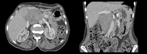
Figure 1: Abdominal enhanced CT in axial and coronal plan, demonstrating a large and heterogenous mass encasing the inferior vena cava, with masse effect on the adjacent aorta.
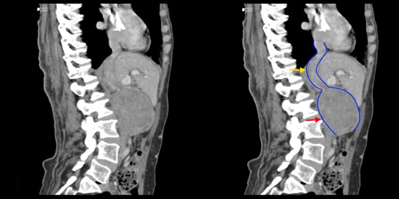
Figure 2: Abdominal enhanced CT in sagittal plan, demonstrating clearly the continuity of the mass (red arrow) with inferior vena cava (yellow arrow).
Discussion
Leiomyosarcoma of the inferior vena cava poses a significant clinical challenge due to its rarity, deep anatomical location, and often nonspecific presentation. Imaging modalities such as contrast-enhanced CT and MRI are essential for determining the extent of vascular involvement and for preoperative planning [1,2]. Typical radiological findings include a lobulated, enhancing mass, often with signs of vascular compression or invasion.
The tumor is classified by its location along the IVC into infrarenal, inter-renal and suprahepatic segments, which influences surgical planning and prognosis [2]. In this case, the lesion spanned a long segment from the infrarenal to the suprahepatic region, necessitating complex resection and vascular reconstruction. Radical surgical excision with negative margins remains the cornerstone of treatment and offers the best chance of long-term survival [3].
Histologically, leiomyosarcomas exhibit spindle-shaped cells with eosinophilic cytoplasm and cigar-shaped nuclei, often demonstrating immunoreactivity for SMA, desmin, and h-caldesmon. The mitotic index and degree of necrosis can provide prognostic information.
Although adjuvant chemotherapy or radiotherapy has been used in some cases, there is no clear consensus on their efficacy, and decisions are often individualized based on margin status, tumor grade, and patient performance status [3,4].
The prognosis for IVC leiomyosarcoma remains guarded, with five-year survival rates varying between 30% and 60% depending on completeness of resection and tumor biology [4]. Recurrence is common, particularly in cases with incomplete resection or high-grade histological features.
Conclusion
Leiomyosarcoma of the IVC is a rare and aggressive tumor requiring a high index of suspicion and a multidisciplinary approach. Imaging plays a crucial role in diagnosis and preoperative planning. Radical resection with vascular reconstruction offers the best chance for survival. This case underscores the importance of early recognition and contributes to the growing body of literature on this rare pathology.
References
- Kuker RA, et al. Leiomyosarcoma of the inferior vena cava: clinical and imaging features. Radiographics, 2015; 35(6): 1549-1557. doi: 10.1148/rg.2015140225.
- Mingoli A, et al. Leiomyosarcoma of the inferior vena cava: analysis of a world literature of 281 cases. Journal of Vascular Surgery, 1996; 23(3): 556-563. doi: 10.1016/S0741-5214(96)70206-6.
- Wachtel H, et al. Outcomes after surgical resection of IVC leiomyosarcomas: a 30-year experience. Annals of Surgical Oncology, 2015; 22(1): 297-303. doi: 10.1245/s10434-014-3997-1.
- Hines OJ, et al. Leiomyosarcoma of the inferior vena cava: prognosis and comparison with leiomyosarcoma of other locations. Cancer, 1999; 85(5): 1077-1083. doi: 10.1002/(SICI)1097-0142(19990301)85:5<1077::AID-CNCR22>3.0.CO;2-0.