
Email id: info@ijclinmedcasereports.com
Right Non-Recurrent Laryngeal Nerve Associated with an Aberrant Right Subclavian Artery Encountered During Total Thyroidectomy for Graves’ Disease: A Case Report
Osama K Shattarah1,*, Rasha Al-Ebbini2, Anh N Vu3, Hasan Aljawder3, Johnathan Hubbard3, Saleh Abu-alhaj4, Ahmad Eyadeh5 and Ahmad suleihat6
1Department of general Surgery and Urology, Faculty of Medicine, Jordan University of Science and Technology, Irbid, Jordan
2Department of Diagnostic radiology, Faculty of Medicine, Jordan University of Science and Technology, Irbid, Jordan
3Department of Endocrine Surgery, Guy's and St Thomas' NHS Foundation Trust, London, United Kingdom
4Faculty of Medicine, Al-Balqa Applied University, Al-Salt, Jordan
5Department of Internal medicine, Faculty of Medicine, Jordan University of Science and Technology, Irbid, Jordan
6Department of endocrine surgery, Oxford University Hospitals NHS Trust, Oxford, United Kingdom
Received Date: 25/07/2026; Published Date: 30/09/2026
*Corresponding author: Osama K Shattarah, Department of general Surgery and Urology, Faculty of Medicine, Jordan University of Science and Technology, Irbid, Jordan
Abstract
Background: The Non-Recurrent Laryngeal Nerve (NRLN) is a rare congenital anomaly of the inferior laryngeal nerve, occurring almost exclusively on the right side and strongly associated with an aberrant right subclavian artery (ARSA, arteria lusoria). Failure to anticipate this anomaly during thyroid surgery substantially raises the risk of iatrogenic nerve injury. To our knowledge, an NRLN encountered specifically during thyroidectomy performed for Graves’ disease has not been previously reported.
Case presentation: A 24-year-old woman with a 5-year history of Graves’ disease on carbimazole presented with dysphagia for solids and dyspnea on lying flat, due to a diffuse, tense, moderately enlarged goiter. Biochemistry confirmed thyrotoxicosis with a markedly elevated TSH-receptor antibody. Neck ultrasound showed a bulky, heterogeneous, hypervascular gland, and thyroid scintigraphy showed diffusely increased homogeneous uptake, both consistent with Graves’ disease. A previously obtained contrast-enhanced neck and chest computed tomography (CT), reviewed retrospectively, revealed an aberrant right subclavian artery. She underwent total thyroidectomy for definitive control of Graves’ disease and relief of compressive symptoms. Intraoperatively, a right NRLN arising directly from the cervical vagus nerve and coursing above and parallel to the right inferior thyroid artery toward the larynx was identified, traced, and preserved intact. Her voice remained normal postoperatively and at follow-up, and she was discharged on postoperative day 1.
Conclusion: This case illustrates the well-described but easily overlooked association between ARSA and right NRLN, and demonstrates that careful preoperative imaging review and meticulous intraoperative visual identification allow safe nerve preservation even in the technically demanding setting of a hypervascular Graves’ gland. Surgeons should routinely screen any available preoperative cross-sectional imaging for ARSA before right-sided neck dissection and maintain a low threshold for anticipating an NRLN, regardless of the underlying thyroid pathology.
Keywords: Non-recurrent Laryngeal Nerve; Aberrant Right Subclavian Artery; Arteria Lusoria; Graves’ Disease; Total Thyroidectomy; Case Report
Abbreviations: NRLN - Non-Recurrent Laryngeal Nerve; ARSA - Aberrant Right Subclavian Artery; IONM - IntraOperative Neuromonitoring; CT - Computed Tomography
Introduction
Injury to the recurrent laryngeal nerve remains the most feared neurological complication of thyroid and parathyroid surgery. A rare but critical anatomic variant that predisposes to such injury is the Non-Recurrent Laryngeal Nerve (NRLN), in which the nerve arises directly from the cervical vagus nerve and passes transversely to the larynx without first descending into the thorax and recurring around a great vessel. The anomaly was first described by Stedman in 1823 [1] and was subsequently characterized in larger surgical and cadaveric series by Henry et al. [2] and Toniato et al. [3], who proposed operative classifications still in clinical use.
The right NRLN is overwhelmingly more common than the left and is strongly linked to an aberrant right subclavian artery (ARSA, also termed arteria lusoria), a vascular anomaly resulting from abnormal involution of the right fourth aortic arch. When this association is not anticipated, the nerve is vulnerable to injury during routine paratracheal dissection, where it is mistaken for a vessel, lymphatic, or strap-muscle fascia rather than the laryngeal nerve itself.
We report a case of a right NRLN with an associated ARSA encountered intraoperatively during total thyroidectomy performed for Graves’ disease, a clinical context that is, to our knowledge, essentially unreported in the indexed literature. This report is written in accordance with the CARE (CAse REport) guidelines for case report development [4].
Case Presentation
Patient Information and History
A 24-year-old woman with a 5-year history of Graves’ disease was referred for surgical management of a goiter. She had been maintained on carbimazole 10 mg three times daily, with good medication compliance. She reported progressive dysphagia for solid food and shortness of breath when lying flat, both attributable to mechanical compression by the enlarging goiter. She specifically denied any voice change. There was no family history of thyroid disease, no known drug or food allergy, and no smoking history. She had no other relevant medical history.
Clinical Findings
Examination revealed a visible and palpable diffuse goiter that was tense, mobile, and moderate in size, without cervical lymphadenopathy or clinical evidence of thyroid eye disease. The patient was clinically euthyroid at the time of examination.
Diagnostic Assessment
Biochemistry: Free T3 and free T4 were within normal limits with a suppressed TSH. TSH-receptor antibody (TRAb) was markedly elevated at 29 IU/L, consistent with Graves’ disease. Serum vitamin D was low.
Neck ultrasound: Grayscale imaging showed a bulky, enlarged thyroid gland with coarse echotexture and patchy, diffusely heterogeneous (pseudonodular) echogenicity Figure 1. A single small right-sided nodule, 11 × 10 × 6 mm and TI-RADS 3, was noted and deemed appropriate for follow-up rather than biopsy; no suspicious cervical lymph nodes were identified. Color Doppler imaging showed markedly and diffusely increased parenchymal vascularity (the so-called “thyroid inferno” pattern), in keeping with Graves’ disease Figure 2.
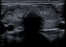
Figure 1: Preoperative neck ultrasound. Grayscale transverse image showing a bulky thyroid gland with coarse, heterogeneous, pseudonodular echotexture.
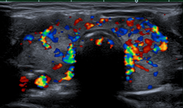
Figure 2: Color Doppler image showing markedly increased diffuse parenchymal vascularity, the “thyroid inferno” pattern characteristic of Graves’ disease.
Thyroid scintigraphy: Technetium-99m pertechnetate thyroid uptake scan showed diffusely increased, homogeneous tracer uptake throughout an enlarged, bilobed gland, confirming the diagnosis of diffuse toxic goiter (Graves’ disease) (Figure 3,4).
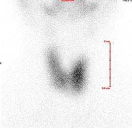
Figure 3: Technetium-99m pertechnetate thyroid scintigraphy. Printed grayscale display with linear measurement scale.
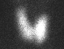
Figure 4: Intensity-windowed display (uptake shown as bright signal on a dark background). Both images show diffusely increased, homogeneous tracer uptake throughout an enlarged gland, consistent with diffuse toxic goiter.
Cross-sectional imaging: A contrast-enhanced CT of the neck and chest, originally obtained as part of the work-up for dysphagia and compressive symptoms, confirmed the enlarged thyroid gland and showed no pathological lymphadenopathy. On retrospective review following surgery, the same CT was also found to demonstrate an aberrant right subclavian artery, identifiable on axial images at the level of the thoracic inlet as a vessel arising as the most distal branch of the aortic arch and coursing posterior to the esophagus toward the right upper limb (Figure 5, 6), and confirmed on sagittal reconstruction by its oblique retroesophageal course (Figure 7). This finding had not been prospectively flagged on the original radiology report or recognized by the surgical team preoperatively.
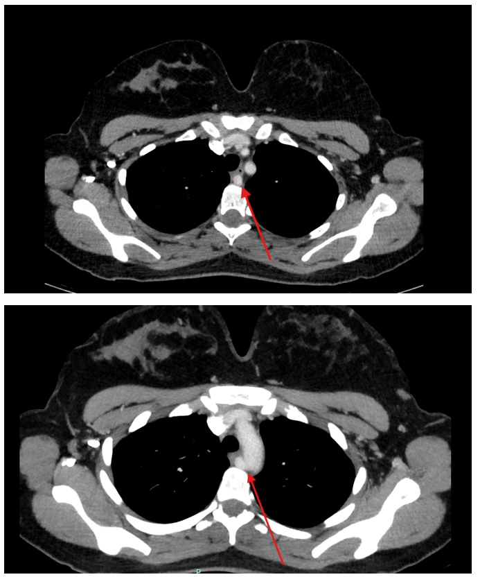
Figure 5, 6: Contrast-enhanced axial CT of the thoracic inlet. Sequential axial images (arrow) showing an aberrant right subclavian artery arising as the last branch of the aortic arch and coursing posterior to the esophagus toward the right upper limb — the radiologic hallmark of arteria lusoria.
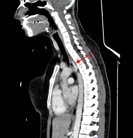
Figure 7: Sagittal reformatted CT image (arrow) showing the oblique, retroesophageal course of the aberrant right subclavian artery as it ascends toward the right upper limb.
Diagnosis
Graves’ disease (diffuse toxic goiter) with compressive symptoms refractory to medical therapy alone, with an incidentally identified aberrant right subclavian artery on cross-sectional imaging — a recognized radiologic predictor of a right non-recurrent laryngeal nerve.
Therapeutic Intervention
The patient underwent total thyroidectomy under general anesthesia to achieve definitive control of Graves’ disease and to relieve her compressive symptoms. Intraoperatively, during dissection of the right paratracheal region, a right inferior laryngeal nerve was identified that did not follow its expected recurrent course around the subclavian artery. Instead, the nerve arose directly from the cervical vagus nerve and coursed transversely, above and parallel to the right inferior thyroid artery, toward the larynx — a Toniato type 2A configuration [3]. The nerve was carefully dissected using meticulous capsular (Berry-ligament-level) technique and was traced proximally to confirm its origin from the vagus nerve within the right carotid sheath, and distally toward the larynx. Visual identification alone (without intraoperative neuromonitoring) was used to confirm and preserve the nerve, which remained macroscopically intact throughout the dissection (Figure 8, 9). The gland was removed en bloc and was diffusely enlarged and hypervascular on gross inspection, consistent with Graves’ disease (Figure 10, 11).
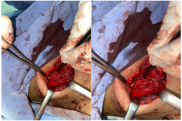
Figure 8, 9: Intraoperative photographs of the right thyroid bed showing dissection and preservation of the right non-recurrent laryngeal nerve, identified coursing directly from the cervical vagus nerve toward the larynx, above and parallel to the right inferior thyroid artery, without a recurrent loop into the mediastinum.
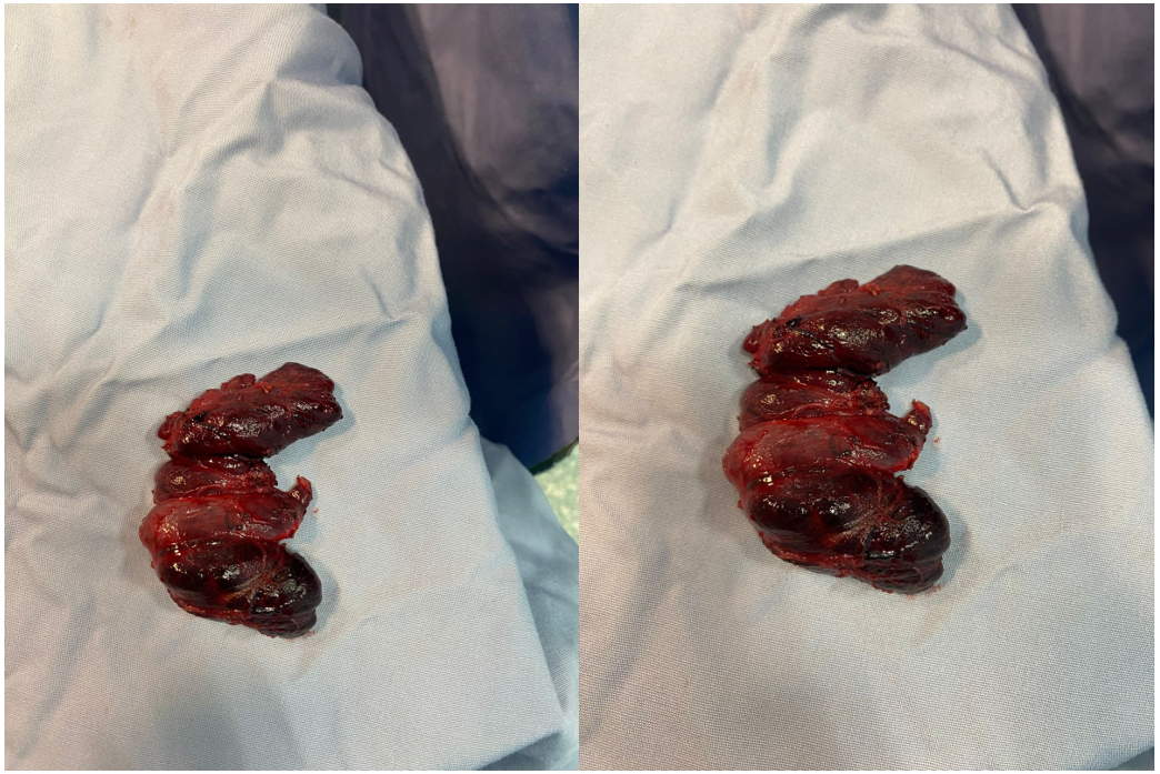
Figure 10, 11: Gross specimen photographs of the resected thyroid gland, showing a diffusely enlarged, hypervascular gland consistent with Graves’ disease.
Follow-up and Outcomes
Her postoperative course was uneventful. Voice was normal without hoarseness in the immediate postoperative period, and she was discharged on postoperative day 2. At outpatient follow-up, her voice remained normal.
Discussion
The NRLN is an uncommon but clinically important anatomic variant in which the inferior laryngeal nerve fails to follow its usual recurrent course. Pooled data from a large meta-analysis estimate the prevalence of a right NRLN at approximately 0.7% of right-sided nerves at risk (95% CI 0.6–0.9%) [5], closely matching the 0.51% operative incidence (31 of 6,000 thyroidectomies) reported in one of the largest single-institution surgical series. [3] A left-sided NRLN is exceedingly rare and is essentially confined to patients with a right-sided aortic arch or situs inversus. Toniato et al. proposed an operative classification still widely used today: type 1, in which the nerve runs with the superior thyroid pedicle; type 2A, in which it crosses above or parallel to the inferior thyroid artery; and type 2B, in which it passes below the artery or between its branches [3]. The nerve in the present case corresponded to a type 2A configuration.
Embryologically, the right NRLN results from abnormal regression of the right fourth aortic arch and the proximal right dorsal aorta. The right subclavian artery consequently arises as the last (most distal) branch of the aortic arch — the arteria lusoria — and follows a retroesophageal course to reach the right upper limb. [6] Because the vessel that would normally tether the developing inferior laryngeal nerve caudally into the thorax is absent, the nerve is not drawn down into a recurrent loop and instead passes directly from the vagus to the larynx.
The association between ARSA and right NRLN is one of the most consistent anatomic–vascular correlations in endocrine surgery. In the meta-analysis by Henry et al., 86.7% of right NRLNs were associated with an aberrant subclavian artery. [5] In a focused review of 104 NRLN cases with documented vascular variants, 97.7% were right-sided, and ARSA was present in 97% of these. [7] The converse relationship is equally strong: in a controlled ultrasound study, an NRLN was identified only among patients in whom an ARSA had first been demonstrated, and never in matched controls without ARSA (p = 0.0006). [8] In practical terms, identification of an ARSA on any preoperative imaging should be treated as a near-certain predictor of a right NRLN.
This association underlines the value of preoperative cross-sectional imaging review. CT can demonstrate the retroesophageal course of the aberrant artery directly [9], and a dedicated neck ultrasound can suggest the diagnosis by the absence of the normal brachiocephalic (innominate) bifurcation in the supraclavicular fossa. [10-12] In the present case, a contrast-enhanced CT had already been obtained for an unrelated indication (dysphagia and compressive symptoms), and the ARSA was visible on both axial and sagittal sequences, yet it was only recognized on retrospective review after surgery. This represents a missed opportunity for preoperative anticipation and is, in our view, the most actionable teaching point of this report: surgeons should routinely and specifically screen any available preoperative neck or chest cross-sectional imaging for ARSA before right-sided thyroid or parathyroid dissection, regardless of the indication for which the scan was originally obtained.
Intraoperatively, careful visual dissection remains the gold standard for identifying the NRLN, but intraoperative neuromonitoring (IONM) using a vagus-first stimulation algorithm — stimulating the vagus nerve both proximally and distally in the neck before paratracheal dissection — can predict a non-recurrent course before the nerve is put at risk, and has been shown to increase intraoperative detection rates. [13-16] IONM was not used in the present case; identification relied on meticulous capsular dissection and proximal tracing of the nerve to its vagal origin within the carotid sheath. Although the outcome was favorable, we believe that in any patient with a known or suspected ARSA, a vagus-first IONM protocol, performed in line with International Neural Monitoring Study Group standards, represents the safer default strategy and should be adopted where available [13].
The clinical stakes of failing to anticipate an NRLN are substantial. A recent systematic review and meta-analysis reported a pooled NRLN injury risk of 7%, a risk ratio of 3.8 compared with a normal recurrent laryngeal nerve [17]. In the original Toniato series, 12.9% of patients with an NRLN had a postoperative vocal cord deficit, with most injured nerves arising above the laryngotracheal junction and running with the superior thyroid vessels — analogous to type 1 nerves [3]. These figures compare unfavorably with reported nerve palsy rates of roughly 1–4% in benign thyroid surgery overall [18], and IONM use has been associated with a reduction in permanent (though not necessarily early) postoperative palsy [19]. The favorable voice outcome in our patient reflects careful anatomic dissection, but the elevated baseline risk associated with an unanticipated NRLN reinforces the case for a structured, anticipatory approach rather than reliance on intraoperative vigilance alone.
Finally, we note that the NRLN literature is dominated by case reports and series involving multinodular goiter or thyroid malignancy; reports in the specific context of Graves’ disease are essentially absent. The closest disease-matched reports we identified describe a toxic multinodular goiter [20] and a mixed surgical series in which toxic goiter/Graves’ disease accounted for a minority of NRLN-screened cases [21]. Hyperthyroid, Graves’-affected glands are typically larger, more friable, and considerably more vascular than euthyroid glands, which may compound the technical difficulty of nerve identification and increase the consequences of an unanticipated NRLN. To our knowledge, this report represents one of the first cases specifically describing a right NRLN with associated ARSA encountered during total thyroidectomy performed for Graves’ disease, and we believe it adds a clinically relevant data point to a sparsely documented intersection of thyroid pathology and surgical anatomy.
Conclusion
The non-recurrent laryngeal nerve is a rare but clinically significant anomaly that should be anticipated whenever a right thyroid or parathyroid dissection is planned, particularly when an aberrant right subclavian artery is visible on any available preoperative imaging, irrespective of the indication for which that imaging was originally obtained. Hyperthyroid, Graves’-affected glands add technical complexity to an already high-stakes scenario. Meticulous capsular dissection, deliberate proximal tracing of the nerve to its vagal origin, and — where available — a vagus-first intraoperative neuromonitoring protocol together support safe identification and preservation of the nerve, as illustrated by the favorable outcome in this case.
Author Contributions:
Osama K. Shattarah: Conceptualization, Project administration, article writing, final approval, and accountability for all aspects of the work.
Rasha Al-Ebbini: Conceptualization, article writing, Investigation, Visualization, final approval.
Anh N. Vu: Review & editing
Hasan Aljawder: Review & editing
Johnathan Hubbard: Supervision, writing- review & editing
Saleh Abualhaj: Data curation, writing-review & editing
Ahmad Eyadeh: Data curation, writing-review & editing
Ahmad Suleihat: Writing-review & editing
Ahmad Eyadeh: Review & editing
Conflicts of Interest: The authors declare that they have no conflicts of interest.
References
- Stedman GW. A singular distribution of some of the nerves and arteries in the neck, and the top of the thorax. Edinb Med Surg J, 1823; n19: n564–565.
- Henry JF, Audiffret J, Denizot A, Plan M. The nonrecurrent inferior laryngeal nerve: review of 33 cases, including two on the left side. Surgery, 1988; 104(6): 977–984.
- Toniato A, Mazzarotto R, Piotto A, Bernante P, Pagetta C, Pelizzo MR. Identification of the nonrecurrent laryngeal nerve during thyroid surgery: 20-year experience. World J Surg, 2004; 28(7): 659–661. https://doi.org/10.1007/s00268-004-7197-7.
- Gagnier JJ, Kienle G, Chrousos GP, Sox H, Riley DS. CARE Group. The CARE guidelines: consensus-based clinical case report guideline development. J Clin Epidemiol, 2014; 67(1): 46–51. https://doi.org/10.1016/j.jclinepi.2013.08.003.
- Henry BM, Sanna S, Graves MJ, Vikse J, Sanna B, Tomaszewska IM, et al. The Non-Recurrent Laryngeal Nerve: a meta-analysis and clinical considerations. Peer J, 2017; 5: e3012. https://doi.org/10.7717/peerj.3012.
- Avisse C, Marcus C, Delattre JF, Marcus C, Cailliez-Tomasi JP, Palot JP, et al. Right nonrecurrent inferior laryngeal nerve and arteria lusoria: the diagnostic and therapeutic implications of an anatomic anomaly. Review of 17 cases. Surg Radiol Anat, 1998; 20(3): 227–232. https://doi.org/10.1007/BF01628900.
- Bakalinis E, Makris I, Demesticha T, Tsakotos G, Skandalakis P, Filippou D. Non-Recurrent Laryngeal Nerve and Concurrent Vascular Variants: A Review. Acta Med Acad, 2018; 47(2): 186–192. https://doi.org/10.5644/ama2006-124.230.
- Liang RX, Wang B, Zhao WX, Xue ES, Ye Q, Chen ZY, et al. Predictive value of ultrasound diagnosis of aberrant right subclavian artery with non-recurrent laryngeal nerve. Arch Med Sci, 2024; 20(3): 719–725. https://doi.org/10.5114/aoms.2020.98942.
- Watanabe A, Kawabori S, Osanai H, Taniguchi M, Hosokawa M. Preoperative computed tomography diagnosis of non-recurrent inferior laryngeal nerve. Laryngoscope, 2001; 111(10): 1756–1759. https://doi.org/10.1097/00005537-200110000-00017.
- Iacobone M, Viel G, Zanella S, Bottussi M, Frego M, Favia G. The usefulness of preoperative ultrasonographic identification of nonrecurrent inferior laryngeal nerve in neck surgery. Langenbecks Arch Surg, 2008; 393(5): 633–638. https://doi.org/10.1007/s00423-008-0372-9.
- Citton M, Viel G, Iacobone M. Neck ultrasonography for detection of non-recurrent laryngeal nerve. Gland Surg, 2016; 5(6): 583–590. https://doi.org/10.21037/gs.2016.11.07.
- Devèze A, Sebag F, Hubbard J, Jaunay M, Maweja S, Henry JF. Identification of patients with a non-recurrent inferior laryngeal nerve by duplex ultrasound of the brachiocephalic artery. Surg Radiol Anat, 2003; 25(3-4): 263–269. https://doi.org/10.1007/s00276-003-0135-9.
- Randolph GW, Dralle H. International Intraoperative Monitoring Study Group. Electrophysiologic recurrent laryngeal nerve monitoring during thyroid and parathyroid surgery: international standards guideline statement. Laryngoscope, 2011; 121(Suppl 1): S1–16. https://doi.org/10.1002/lary.21119.
- Donatini G, Carnaille B, Dionigi G. Increased detection of non-recurrent inferior laryngeal nerve (NRLN) during thyroid surgery using systematic intraoperative neuromonitoring (IONM). World J Surg, 2013; 37(1): 91–93. https://doi.org/10.1007/s00268-012-1782-y.
- Kamani D, Potenza AS, Cernea CR, Kamani YV, Randolph GW. The nonrecurrent laryngeal nerve: anatomic and electrophysiologic algorithm for reliable identification. Laryngoscope, 2015; 125(2): 503–508. https://doi.org/10.1002/lary.24823.
- Watanabe A, Taniguchi M, Kimura Y, Ito S, Hosokawa M, Sasaki S. Efficient, effective, safe procedure to identify nonrecurrent inferior laryngeal nerve during thyroid surgery. Head Neck, 2016; 38(4): 573–576. https://doi.org/10.1002/hed.23932.
- So JT, Thompson CSG. Non-Recurrent Laryngeal Nerve: Surgical Risk of Injury in Comparison with the Normal Recurrent Laryngeal Nerve—A Systematic Review and Meta-Analysis. Clin Otolaryngol, 2025; 50(4): 654–663. https://doi.org/10.1111/coa.14312.
- Hermann M, Alk G, Roka R, Glaser K, Freissmuth M. Laryngeal recurrent nerve injury in surgery for benign thyroid diseases: effect of nerve dissection and impact of individual surgeon in more than 27,000 nerves at risk. Ann Surg, 2002; 235(2): 261–268. https://doi.org/10.1097/00000658-200202000-00015.
- Bergenfelz A, Salem AF, Jacobsson H, Nordenström E, Almquist M, Wallin GW, et al. Risk of recurrent laryngeal nerve palsy in patients undergoing thyroidectomy with and without intraoperative nerve monitoring. Br J Surg, 2016; 103(13): 1828–1838. https://doi.org/10.1002/bjs.10276.
- Gurleyik E, Dogan S, Gunal O, Pehlivan M. The Rare Coincidence: Nonrecurrent Laryngeal Nerve Pointed by a Zuckerkandl’s Tubercle. Case Rep Med, 2012; 2012: 143049. https://doi.org/10.1155/2012/143049.
- Omer AR. Abu Dhabi Neural Mapping (ADNM) during Minimally Invasive Thyroidectomy Enables the Early Identification of Non-Recurrent Laryngeal Nerve and Prevents Voice Dysfunction. J Clin Med, 2022; 11(19): 5677. https://doi.org/10.3390/jcm11195677.