
Email id: info@ijclinmedcasereports.com
Burkitt Lymphoma Masquerading as Serosal Disease in an Elderly Female: A Diagnostic Challenge Unveiled Through Integrated Cytomorphology, Flow Cytometry, PET-CT Metabolic Imaging and Cytogenetics
Kiran Ghodke1,*, Gojiri Mawalankar1, Aaishwarya Dhabe2, Hemant Khandare3, Akshita Rane1, Kiran Shetty4 and Nishant Jindal5
1Department of Hematology, Kokilaben Dhirubhai Ambani Hospital and Medical Research Institute, Mumbai, Maharashtra, India
2Department of Cytogenetics, Kokilaben Dhirubhai Ambani Hospital and Medical Research Institute, Mumbai, Maharashtra, India
3Department of Nuclear Medicine, Kokilaben Dhirubhai Ambani Hospital and Medical Research Institute, Mumbai, Maharashtra, India
4Department of Critical Care Medicine, Kokilaben Dhirubhai Ambani Hospital and Medical Research Institute, Mumbai, Maharashtra, India
5Department of Clinical Haematology, Kokilaben Dhirubhai Ambani Hospital and Medical Research Institute, Mumbai, Maharashtra, India
Received Date: 18/07/2026; Published Date: 23/09/2026
*Corresponding author: Dr. Kiran Ghodke, Department of Hematology, Kokilaben Dhirubhai Ambani Hospital and Medical Research Institute, Mumbai 400053, Maharashtra, India
ORCID: 0000-0002-0810-2349
Abstract
Background: Burkitt Lymphoma (BL) is a highly aggressive germinal-centre-derived B-cell malignancy that rarely presents as serosal effusion without peripheral lymphadenopathy or organomegaly. Its occurrence in elderly females is exceptional, with initial mimicry of cirrhotic ascites or carcinomatosis representing a formidable diagnostic pitfall.
Case Presentation: An 82-year-old immunocompetent female presented with massive ascites, bilateral pleural effusion, anaemia, bilateral lower limb oedema, and constitutional symptoms, with no lymphadenopathy or hepatosplenomegaly. Ascitic fluid cytomorphology revealed large high-grade hematolymphoid cells. Flow cytometry demonstrated a clonal B-cell neoplasm: CD19+, CD20+, CD10+, CD45+, CD38 bright, CD3−, CD5−, CD34−, BCL2−, consistent with Burkitt lymphoma. FISH using a cMYC break-apart probe demonstrated a positive signal pattern of one fused, one red, and one green signal (1F1R1G), confirming MYC gene rearrangement; the translocation partner chromosome was not determined. BCL2 and BCL6 rearrangements were not detected. Whole-body 18F-FDG PET-CT disclosed disseminated extranodal disease — FDG-avid soft tissue deposits bilaterally (SUVmax 11.9), metabolically active pleural effusion (SUVmax 3.5), cutaneous/subcutaneous deposits of the lower abdominal wall and vulva, and intramuscular lesion of the right thigh — without discrete lymph node involvement. In view of poor performance status, pre-phase chemotherapy with cyclophosphamide and dexamethasone was administered with clinical improvement; the patient was subsequently discharged at family request for home supportive care.
Conclusion: This case illustrates a rare presentation of a MYC-rearranged high-grade B-cell lymphoma, favoured as sporadic Burkitt lymphoma on integrated criteria, in an elderly female with predominant serosal and extranodal soft-tissue dissemination devoid of nodal disease. It underscores the indispensability of effusion cytomorphology, multiparameter flow cytometry, and FISH in unexplained serosal effusion in older adults, and the diagnostic primacy of integrated morphological and immunophenotypic features when the MYC translocation partner is not characterised.
Keywords: Burkitt lymphoma; flow cytometry; MYC rearrangement; serosal effusion; HGBCL; extranodal lymphoma; pre-phase chemotherapy
Introduction
Burkitt Lymphoma (BL) is one of the most rapidly proliferating human malignancies, characterised by translocation and deregulation of the MYC proto-oncogene at chromosome 8q24. The WHO 5th Edition Classification of Haematolymphoid Tumours (WHO-HAEM5, 2022) recognises BL as an aggressive mature B-cell lymphoma characterised by MYC rearrangement, germinal-centre immunophenotype, and a near-100% Ki-67 proliferative index, reclassified by EBV status as EBV-positive or EBV-negative [1,2]. In the sporadic variant, the abdomen is the most common site of involvement; however, serosal effusions as the primary presenting manifestation are uncommon, constituting fewer than 2% of all lymphoma presentations [5]. Occurrence as the dominant disease manifestation in an immunocompetent elderly female without nodal or visceral disease is exceptional and diagnostically treacherous, given its clinical mimicry of hepatic cirrhosis, carcinomatosis peritonei, or primary ovarian malignancy [3,4,6].
Case Presentation
An 82-year-old non-immunocompromised female presented with a six-week history of progressive abdominal distension, bilateral lower limb oedema, severe generalised weakness, and decreased appetite, with no fever or night sweats. There was no prior malignancy, immunosuppressive therapy, or organ transplantation. HIV serology was negative.
On examination, she appeared cachectic and pale. Blood pressure was 110/70 mmHg, afebrile, SpO2 96% on room air. Gross ascites was confirmed by fluid thrill and shifting dullness. Bilateral basal dullness and diminished breath sounds indicated pleural effusion. No peripheral lymphadenopathy was palpable in any nodal region, and there was no hepatosplenomegaly.
Laboratory investigations revealed normocytic normochromic anaemia (Hb 9.2 g/dL), total leucocyte count 6.9 × 10⁹/L with neutrophilic predominance, adequate platelets (384 × 10⁹/L), and markedly elevated LDH (829 U/L; reference 125–214 U/L). Serum albumin was 2.8 g/dL. Ascitic fluid analysis revealed total protein 2.26 g/dL and ascitic albumin 0.87 g/dL, yielding a serum-ascites albumin gradient (SAAG) of 1.93 g/dL (high SAAG, ≥1.1 g/dL). While high SAAG classically suggests portal hypertension, in this context it likely reflects hypoalbuminaemia-driven oncotic pressure reduction rather than true portal hypertension, corroborated by absent hepatosplenomegaly, normal liver function, and high ascitic protein. GeneXpert MTB/RIF assay on ascitic fluid was negative, effectively excluding tuberculous peritonitis. Tumour markers showed mildly elevated CA-125 (69 U/mL; reference 0–35) and CA 19-9 (38.3 U/mL; reference 0–37), with normal CEA (3.0 ng/mL; reference 0–4.7) and AFP (2.69 ng/mL; reference 0.89–8.78). The mild elevation in CA-125 and CA 19-9 was interpreted as a non-specific reactive phenomenon secondary to serosal inflammation rather than indicative of primary epithelial malignancy.
Investigations and Diagnostic Workup
Effusion Cytomorphology:
Ascitic tapping was performed as the initial diagnostic intervention. Smears demonstrated a hypercellular specimen comprising a monomorphic population of large to medium-sized hematolymphoid tumour cells with deeply basophilic cytoplasm, prominent lipid vacuoles, round-to-oval nuclei with finely stippled chromatin, and conspicuous nucleoli. Numerous mitotic figures and apoptotic bodies were identified, with scattered tingible body macrophages imparting a focal ‘starry-sky’ appearance. The morphology was that of a high-grade hematolymphoid neoplasm, favouring Burkitt lymphoma.
Flow Cytometric Immunophenotyping:
Multiparameter Flow Cytometry (MFC) on ascitic fluid identified a dominant clonal B-cell population comprising approximately 78% of total events (Table 1). The cells expressed CD19, CD20, CD10, and CD38 (bright), consistent with germinal-centre B-cell origin. Bright CD38 expression is a key distinguishing feature of classical BL from Burkitt-like lymphoma with 11q aberration (BLL-11q) or large B-cell non-Hodgkin lymphoma, in which CD38 intensity is typically low [7]. CD5, CD34, and BCL2 were negative. Surface immunoglobulin kappa light chain restriction confirmed clonal B-cell neoplasia. This immunophenotype effectively excluded B-lymphoblastic leukaemia/lymphoma, Diffuse Large B-Cell Lymphoma (DLBCL), and Mantle Cell Lymphoma (MCL) [8,9].
Table 1: Multiparameter Flow Cytometric Immunophenotyping of Ascitic Fluid.
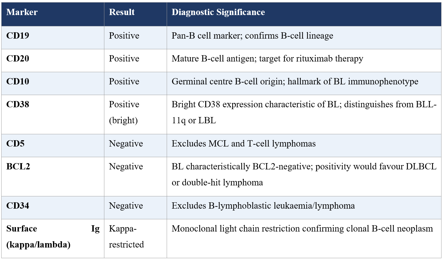
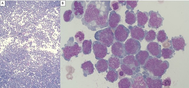
Figure 1: Cytomorphologic findings in ascitic fluid. (A) Low-power photomicrograph showing a hypercellular ascitic fluid smear extensively infiltrated by a monomorphic population of atypical lymphoid cells. (B) High-power photomicrograph demonstrating medium-sized neoplastic lymphoid cells with a high nuclear-to-cytoplasmic ratio, round nuclei with finely clumped chromatin, multiple small but conspicuous nucleoli, and intensely basophilic cytoplasm. Numerous mitotic figures and apoptotic cells are present, reflecting the high proliferative activity.
Whole-Body 18F-FDG PET-CT:
PET-CT performed for staging demonstrated highly FDG-avid ill-defined soft-tissue deposits in bilateral cervical, supraclavicular, and subpectoral-axillary regions maximum standardised uptake value (SUVmax) 11.9 without discrete lymph node uptake. Moderate bilateral pleural effusions exhibited metabolic activity (SUVmax 3.5). A well-defined FDG-avid lesion involved the subcutaneous and cutaneous layers of the lower abdominal wall extending bilaterally to the vulva, and a metabolically active intramuscular lesion was identified in the right thigh. Moderate ascites with peritoneal involvement was also evident. No FDG uptake was detected in the liver, spleen, bone marrow, or CNS (Table 2). This pattern of diffuse extranodal soft-tissue deposits with high FDG-avidity without lymph node enlargement confirmed stage IV disseminated disease with atypical extranodal predominance [10,11].
Table 2: Summary of Whole-Body 18F-FDG PET-CT Findings.
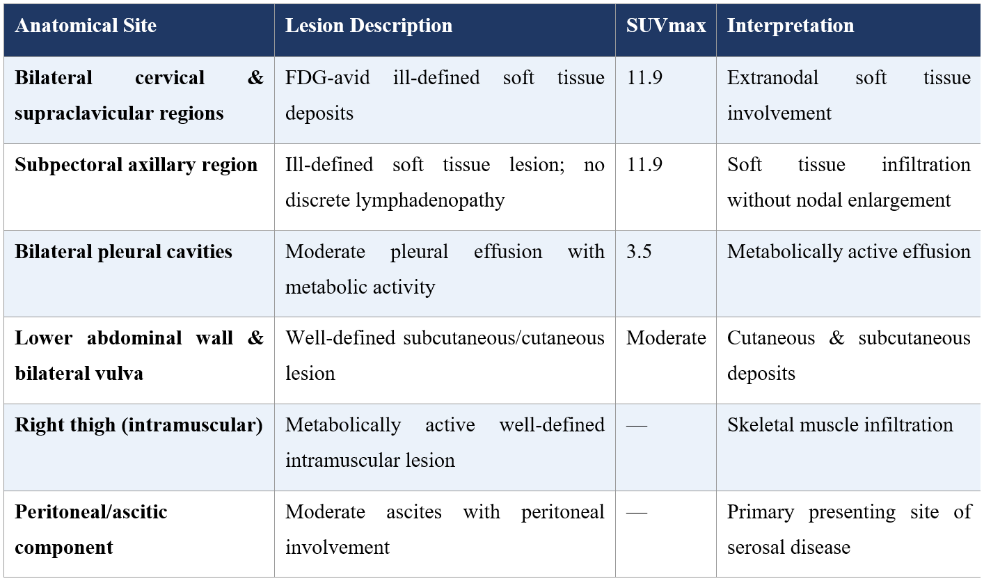
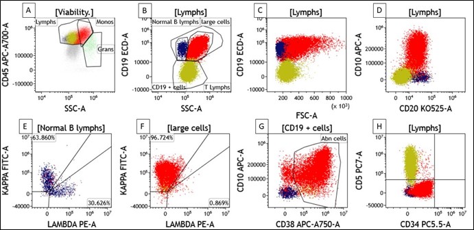
Figure 2: Flow cytometric immunophenotypic analysis of ascitic fluid demonstrating a clonal mature B-cell population consistent with Burkitt lymphoma. Sequential gating strategy was performed on viable cells. (A) Initial gating on CD45 versus side scatter (SSC) identified lymphocytes, monocytes, and granulocytes, with the neoplastic cells demonstrating relatively bright CD45 expression and increased side scatter. (B) Within the lymphocyte gate, an abnormal population of large B cells (red) was identified based on increased forward scatter (FSC) and SSC characteristics, distinct from residual normal B lymphocytes (blue) and T lymphocytes (olive). (C) The abnormal population showed increased cell size (high FSC) with uniform expression of CD19. (D) The neoplastic cells exhibited bright CD10 expression together with CD20 positivity, consistent with a germinal center B-cell phenotype. (E, F) Surface immunoglobulin light-chain analysis demonstrated restricted kappa light-chain expression in the abnormal B-cell population (red), confirming clonality, whereas the residual normal B cells (blue) displayed a polytypic kappa/lambda distribution. (G) The abnormal B-cell population demonstrated bright CD38 expression in conjunction with CD10 positivity, a characteristic immunophenotypic feature supportive of Burkitt lymphoma. (H) The neoplastic cells were negative for CD5 and CD34, excluding mantle cell lymphoma, and precursor B-lymphoblastic neoplasms.
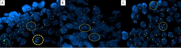
Figure 3: Fluorescence in situ hybridization (FISH) analysis performed on the ascitic fluid cell block confirming MYC rearrangement and excluding BCL2 and BCL6 rearrangements.
(A) MYC (8q24) break-apart FISH probe: Representative interphase nuclei demonstrating a positive break-apart signal pattern, with one fused (yellow), one separate red, and one separate green signal (1F1R1G), consistent with MYC gene rearrangement.
(B) BCL2 (18q21) break-apart FISH probe: Representative interphase nuclei demonstrating two intact fused signals (2F), indicating no evidence of BCL2 gene rearrangement.
(C) BCL6 (3q27) break-apart FISH probe: Representative interphase nuclei demonstrating two intact fused signals (2F), indicating no evidence of BCL6 gene rearrangement.
Fluorescence In-Situ Hybridisation (FISH):
FISH was performed on the ascitic fluid cell block using a cMYC break-apart probe. The analysis demonstrated a positive signal pattern of one fused (F), one red (R), and one green (G) signal — the 1F1R1G pattern — consistent with rearrangement of the MYC gene at 8q24. This break-apart probe methodology confirms that the MYC locus has undergone structural rearrangement but does not identify the translocation partner chromosome; accordingly, the specific partner locus (IGH, IGK, IGL, or non-immunoglobulin) was not characterised in this case. Concurrent FISH for BCL2 and BCL6 rearrangements was negative, excluding a double-hit or triple-hit high-grade B-cell lymphoma with MYC co-rearrangement. HIV serology was non-reactive. EBER-ISH was recommended to assess EBV association per WHO-HAEM5 criteria [1, 2, 12].
Discussion
Epidemiology and Atypical Age at Presentation:
BL constitutes approximately 1–2% of adult lymphomas in non-endemic regions with a median age at diagnosis of approximately 30 years [3,4]. Its occurrence beyond the eighth decade is vanishingly rare. The ERN-EuroBloodNet guidelines (2025) acknowledge sparse epidemiological data for elderly-onset BL and note that intensity-modified treatment regimens are under increasing exploration in this cohort [12]. At 82 years, the index patient occupies the extreme tail of the age distribution for sporadic BL.
Serosal Effusion, SAAG Interpretation, and Diagnostic Mimicry:
Serosal effusions as the primary manifestation of lymphoma are uncommon (~2% of cases); ascites is especially rare [5]. In this case, the high SAAG (1.93 g/dL) may have initially suggested portal hypertension; however, this is a recognised pitfall in severely hypoalbuminaemic patients, in whom the SAAG is artifactually elevated irrespective of the underlying aetiology [14]. The co-existing high ascitic protein (2.26 g/dL) and the absence of hepatosplenomegaly or cirrhotic stigmata directed reassessment toward malignant serosal infiltration. Elevated LDH (829 U/L) was the pivotal laboratory alert to a high-grade haematological malignancy. The negative GeneXpert MTB/RIF result excluded tuberculous peritonitis — a critical differential in this demographic — and the mildly elevated CA-125 (69 U/mL) and CA 19-9 (38.3 U/mL) were interpreted as non-specific serosal reactive changes, a well-documented phenomenon in haematological effusive disease [14]. Shrivastav et al. (2021) and Ismail et al. (2022) have previously described analogous diagnostic reconceptualisation in BL presenting with serosal effusion [5,13].
Immunophenotypic Diagnosis and Differential Diagnosis:
The CD19+/CD20+/CD10+/CD38-bright/BCL2−/CD34− profile is highly specific for BL. Cunningham et al. (2024) confirmed that this mature B-cell immunophenotype distinguishes BL from B-lymphoblastic leukaemia and DLBCL [8]. Gabka et al. (2023) demonstrated that bright CD38 expression specifically discriminates BL from BLL-11q [7]. The absence of BCL2 excluded the therapeutically critical double-hit lymphoma category [9].
Burkitt Lymphoma versus MYC-Rearranged High-Grade B-Cell Lymphoma: Diagnostic Distinction When the Partner Chromosome is Unknown:
The most challenging and clinically consequential diagnostic issue in this case is the distinction between Burkitt lymphoma and MYC-rearranged high-grade B-cell lymphoma not otherwise specified (HGBCL-NOS, MYC-R). FISH using the cMYC break-apart probe confirmed MYC gene rearrangement via the 1F1R1G signal pattern. However, because a break-apart probe detects separation of the MYC locus without identifying the partner gene, the translocation partner chromosome remained undetermined. This is a common and practically important diagnostic limitation when FISH is performed on cytological cell blocks or effusion specimens rather than paraffin-embedded tissue with full interphase cytogenetics. This distinction is diagnostically and therapeutically significant. Classical BL harbours MYC translocations involving immunoglobulin loci — most commonly IGH [t(8;14)], and less frequently IGK [t(2;8)] or IGL [t(8;22)] — and this IG::MYC configuration results in constitutive plasmacytic MYC deregulation. In contrast, HGBCL-NOS may harbour MYC rearrangements with non-immunoglobulin partners (e.g., BCL6, PAX5, FCGR2B), and such tumours may differ in their morphological features, immunophenotype, and clinical behaviour compared to classical BL [1,2]. From a treatment perspective: BL with IG-partnered MYC is highly curable in younger adults with dose-intensive regimens (DA-EPOCH-R, modified BFM); HGBCL-NOS with non-IG MYC partners is generally treated along DLBCL lines (R-CHOP or R-EPOCH), with evidence suggesting inferior outcomes even with intensive regimens [1,9].
In the present case, despite the partner chromosome being undetermined, the integrated features strongly and collectively favour Burkitt lymphoma as the correct diagnosis over HGBCL-NOS. Specifically: (i) the cytomorphology demonstrated classic BL features — medium-to-large cells with deeply basophilic vacuolated cytoplasm, finely stippled chromatin, multiple nucleoli, and a prominent starry-sky pattern; (ii) the flow cytometric immunophenotype showed bright CD38 expression — a marker that Gabka et al. (2023) demonstrated is expressed at substantially higher intensity in BL than in BLL-11q or HGBCL-NOS, reflecting the plasmacytic differentiation state driven by IG-partnered MYC [7]; (iii) BCL2 negativity is a hallmark of BL and is characteristically retained even with MYC rearrangement in the IG-driven context, whereas HGBCL-NOS may co-express BCL2; and (iv) CD10, CD38 positivity and clonal kappa light chain restriction complete a germinal-centre B-cell phenotypic constellation that is highly consistent with BL. [1,2,8] In the elderly geriatric context, while definitive partner characterisation using next-generation sequencing (NGS) or dual-colour fusion FISH would provide molecular certainty, the integrated morphological and immunophenotypic evidence is sufficient to support a working diagnosis of Burkitt lymphoma and guide therapeutic decision-making [12].
PET-CT Dissemination Pattern:
BL is uniformly FDG-avid and PET-CT is the gold standard for staging [10]. The high SUVmax (11.9) and dissemination to cutaneous, subcutaneous, vulvar, and intramuscular compartments without nodal involvement is highly unusual in published BL series. Albano et al. (2024) demonstrated that wider metabolic disease dissemination (Dmaxbsa) predicts inferior outcomes [11]. Formal bone marrow trephine biopsy and CSF cytology remain mandatory for complete staging, as PET-CT sensitivity for bone marrow microinvolvement is suboptimal [3].
Treatment and Clinical Outcome
In view of the patient’s poor performance status and advanced age, standard dose-intensive BL regimens (DA-EPOCH-R, modified BFM) were deemed unsuitable. Pre-phase chemotherapy — a widely adopted cytoreductive strategy in elderly or unfit lymphoma patients prior to definitive therapy — was initiated with cyclophosphamide and dexamethasone from 11 April 2026 to 15 April 2026. Prior to commencing chemotherapy, rasburicase was administered as prophylaxis against tumour lysis syndrome (TLS), given the high tumour burden, markedly elevated LDH, and clinical risk profile. The patient demonstrated clinical improvement following pre-phase treatment. However, in view of the patient’s overall condition and the family’s informed decision, she was discharged at the family’s request for home-based supportive care.
This approach aligns with published experience in elderly BL: Ader et al. (2024) reported meaningful clinical responses with reduced-intensity regimens in an elderly with serosal BL, and the ERN-EuroBloodNet guidelines (2025) explicitly recommend pre-phase chemotherapy as a bridging strategy in frail patients awaiting definitive therapy [6,12]. The decision to prioritise supportive care reflects the profound unmet need for validated geriatric BL treatment protocols and the essential role of shared decision-making in this setting.
Conclusion
We describe an exceptionally rare presentation of a MYC-rearranged high-grade B-cell lymphoma — diagnosed as sporadic Burkitt lymphoma on integrated criteria — in an 82-year-old immunocompetent female with dominant serosal disease, massive ascites, and bilateral pleural effusion without peripheral lymphadenopathy or hepatosplenomegaly. FISH using a cMYC break-apart probe confirmed MYC gene rearrangement (1F1R1G signal pattern); the translocation partner was not determined. The diagnosis of Burkitt lymphoma over HGBCL-NOS was supported by the convergence of cytomorphology, bright CD38 expression, BCL2 negativity, and CD10 positivity — features that collectively reflect an IG-driven MYC deregulation consistent with BL. A high SAAG, mildly elevated CA-125 and CA 19-9, and negative GeneXpert were among the key findings requiring careful contextual interpretation. Pre-phase chemotherapy with rasburicase prophylaxis achieved clinical stabilisation. This case reinforces that high-grade B-cell lymphoma, including BL, can present in a clinically atypical guise, and that haematopathological evaluation — integrating morphology, flow cytometry, and FISH — is mandatory in all unexplained serosal effusions in elderly patients.
Ethics Statement and Consent: This case report was prepared in accordance with the Declaration of Helsinki (revised 2013). Written informed consent was obtained from the patient’s legally authorised family representative for publication of this case and accompanying data. All data have been fully anonymised. The case did not involve experimental intervention and was managed per standard clinical practice. Ethics Committee review was waived per institutional policy for single case reports; the case was discussed at the institutional Tumour Board.
Acknowledgements: None
Funding: No external funding was received.
Conflicts of interest: None declared.
References
- Alaggio R, Amador C, Anagnostopoulos I, Attygalle AD, Araujo IBO, Berti E, et al. The 5th edition of the World Health Organization Classification of Haematolymphoid Tumours: Lymphoid Neoplasms. Leukemia, 2022; 36(7): 1720-1748. doi: 10.1038/s41375-022-01620-2.
- Roschewski M, Dunleavy K, Abramson JS, Wilson WH. Burkitt Lymphoma. N Engl J Med, 2022; 387(12): 1111-1122. doi: 10.1056/NEJMra2107338.
- Jacobson C, LaCasce A. How I treat Burkitt lymphoma in adults. Blood, 2014; 124(19): 2913-2920. doi: 10.1182/blood-2014-06-538504.
- Ribrag V, Koscielny S, Bosq J, Leguay T, Casasnovas O, Fornecker LM, et al. Rituximab and dose-dense chemotherapy for adults with Burkitt's lymphoma: a randomised, controlled, open-label, phase 3 trial. Lancet, 2016; 387(10036): 2402-2411. doi: 10.1016/S0140-6736(15)01317-3.
- Ismail S, Abdulrahman SA, Muhammad I, Ghanem A, Daoud A, Alshehabi Z. Primary intestinal Burkitt’s lymphoma in a Syrian child with a challenging initial presentation: A case report and literature review. Ann Med Surg (Lond), 2022; 76: 103453. doi: 10.1016/j.amsu.2022.103453.
- Adan Rios, Kok Hoe Chan, Binoy Yohannan, Asad Haider, Brian Dinh, Pushan P, et al. Burkitt’s lymphoma responding to rituximab-cyclophosphamide-vincristine-dexamethasone-lenalidomide: time for a paradigm shift? Clin Lymphoma Myeloma Leuk, 2024; 24(S1): 692-693. doi: 10.1016/j.clml.2024.07.285
- Gabka A, Grzybowska-Izydorczyk O, Zielinska-Krawczyk M, Blonski JZ, Smolewski P, Kowalczyk J, et al. A comprehensive flow-cytometry-based immunophenotypic characterization of Burkitt-like lymphoma with 11q aberration. Mod Pathol, 2023; 36(1): 100005. doi: 10.1016/j.modpat.2022.100005.
- Cunningham MT, Sedivy R, Siddiqui MT. Burkitt lymphoma with aberrant expression of cytoplasmic terminal deoxynucleotidyl transferase: a case report. Cureus, 2024; 16(2): e54231. doi: 10.7759/cureus.54231.
- Swerdlow SH, Campo E, Pileri SA, Harris NL, Stein H, Siebert R, et al. The 2016 revision of the World Health Organization classification of lymphoid neoplasms. Blood, 2016; 127(20): 2375-2390. doi: 10.1182/blood-2016-01-643569.
- Mo YW, Xiao ZZ, Wei Y, Li XL, Zhang X, Fan W. The clinical accuracy and risk stratification in end of therapy 18F-FDG PET/CT in Burkitt lymphoma. Front Oncol, 2021; 11: 625436. doi: 10.3389/fonc.2021.625436.
- Albano D, Calabro A, Talin A, Giubbini R, Dondi F, Pagani C, et al. 2-[18F] FDG PET/CT dissemination features in adult Burkitt lymphoma are predictive of outcome. Ann Hematol, 2024; 103(6): 1983-1992. doi: 10.1007/s00277-024-05672-5.
- Ribrag V, Bron D, Rymkiewicz G, Hoelzer D, Jørgensen J, de Armas-Castellano A, et al. Diagnosis and treatment of Burkitt lymphoma in adults: clinical practice guidelines from ERN-EuroBloodNet. Lancet Haematol, 2025; 12(2): e138-e150. doi: 10.1016/S2352-3026(24)00351-X.
- Shrivastav M, Bhargava P, Guleria P, Kumar P. Burkitt lymphoma with disseminated pleuroperitoneal and serosal involvement: a rare presentation. Indian J Pathol Microbiol, 2021; 64(1): 189-192. doi: 10.4103/IJPM.IJPM_200_20.
- Hines OB, Scardino PT, Atkins MB, Smith MR, Woda BA. A case series of diffuse large B-cell lymphoma and Burkitt lymphoma presenting with peritoneal lymphomatosis. J Hematol, 2016; 5(3): 92-97. doi: 10.14740/jh274w.