
Email id: info@ijclinmedcasereports.com
IgG4-Related Orbital Disease Mimicking an Orbital Lymphoma: A Case Report
Houria El Yazidi1,*, Baladi O1, Youss K1, Azria S1, Sobhi H1, Ilhami O1,2, Oukerroum A1,2 and Slimani F1,2
¹Department of Maxillofacial Surgery, Hôpital 20 Août 1953, Ibn Rochd University Hospital, Casablanca, Morocco
²Faculty of Medicine and Pharmacy of Casablanca, Hassan II University of Casablanca, Morocco
Received Date: 13/07/2026; Published Date: 16/09/2026
*Corresponding author: Houria El Yazidi, Department of Maxillofacial Surgery, Hôpital 20 Août 1953, Ibn Rochd University Hospital, Casablanca, Morocco
Abstract
Immunoglobulin G4-related disease (IgG4-RD) is a systemic immune-mediated fibroinflammatory disorder that may involve the orbit and mimic neoplastic, inflammatory, or lymphoproliferative disease. We report the case of a 49-year-old man with no relevant medical history who presented to the maxillofacial surgery department with a progressively enlarging left orbitopalpebral swelling of two months' duration. Clinical examination showed facial asymmetry due to mild left proptosis, a firm and mildly tender superior eyelid mass, ptosis, and limitation of elevation, without diplopia, sensory-motor deficit, reported visual loss, fever, or deterioration in general condition. Computed tomography demonstrated a 34 × 23 mm superolateral left intraorbital lesion extending over 25 mm, involving both the intraconal and extraconal compartments and producing grade I proptosis. The radiological appearance raised suspicion of orbital lymphoma. An incisional biopsy was performed through a lateral eyebrow-tail approach. Histology revealed a dense lymphoplasmacytic infiltrate with numerous plasma cells. Immunohistochemistry showed abundant CD138-positive plasma cells and more than 20 IgG4-positive plasma cells per high-power field, with a low Ki-67 index and no support for a specific lymphoid neoplasm, leading to a diagnosis compatible with orbital IgG4-related disease. This case emphasizes that IgG4-related ophthalmic disease should be included in the differential diagnosis of a superolateral orbital mass suggestive of lymphoma and highlights the central role of tissue biopsy and clinicopathological correlation.
Keywords: IgG4-related disease; Orbit; Proptosis; Orbital mass; Lymphoma; Biopsy; Immunohistochemistry
Introduction
Immunoglobulin G4-related disease (IgG4-RD) is a chronic immune-mediated fibroinflammatory disorder characterized by tumefactive lesions, a lymphoplasmacytic infiltrate enriched in IgG4-positive plasma cells, and variable degrees of fibrosis [1,2]. The disease can affect almost any organ and is often—but not invariably—associated with elevated serum IgG4 concentrations. Orbital and ocular adnexal involvement is referred to as IgG4-related ophthalmic disease (IgG4-ROD). The lacrimal glands, extraocular muscles, orbital soft tissues, eyelids, and branches of the trigeminal nerve are among the most frequently involved structures [2,3].
The clinical and radiological presentation is heterogeneous. Patients may present with eyelid swelling, proptosis, ptosis, diplopia, restricted ocular motility, or, less commonly, compressive optic neuropathy. Because IgG4-ROD may manifest as a focal or diffuse orbital mass, it can closely resemble lymphoma, idiopathic orbital inflammation, thyroid eye disease, sarcoidosis, granulomatosis with polyangiitis, or other benign and malignant orbital tumors [2-5]. Histopathological evaluation is therefore essential, but interpretation must integrate morphology, quantitative immunostaining, serology, imaging, and exclusion of mimickers [3,6-9].
We describe a unilateral superolateral orbital IgG4-related lesion in a previously healthy 49-year-old man, initially considered radiologically suspicious for lymphoma. The case underlines the diagnostic contribution of a targeted orbital biopsy and the need for a subsequent systemic evaluation.
Case Presentation
A 49-year-old man with no significant medical or surgical history was referred to the Department of Maxillofacial Surgery for a left orbitopalpebral swelling that had progressively increased in size over two months. The symptoms evolved in a context of apyrexia and preserved general condition. He did not report constitutional symptoms, recent trauma, or a known systemic inflammatory or autoimmune disorder.
Clinical examination showed facial asymmetry caused by mild left proptosis and a superolateral orbitopalpebral swelling involving the upper eyelid. The lesion was firm and mildly tender on palpation. Ipsilateral ptosis and limitation of ocular elevation were present. There was no diplopia, no reported reduction in visual acuity, and no sensory or motor neurological deficit. The available clinical record did not document optic neuropathy or an acute orbital compartment syndrome.
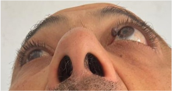
Figure 1: Clinical appearance at presentation showing left upper eyelid swelling, ptosis, and mild proptosis.
Radiological Findings
Contrast-enhanced facial computed tomography revealed a superolateral left intraorbital soft-tissue lesion measuring 34 × 23 mm and extending over approximately 25 mm. The process involved both the intraconal and extraconal compartments and was responsible for grade I proptosis. Based on its location and soft-tissue appearance, an orbital lymphomatous process was considered the leading radiological diagnosis, and tissue confirmation was recommended.
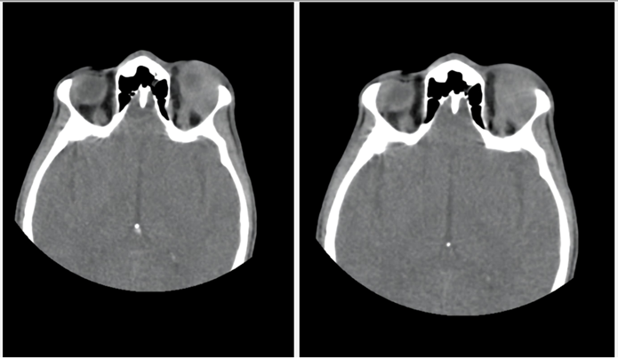
Figure 2: Contrast-enhanced computed tomography demonstrating a 34 × 23 mm superolateral left intraorbital lesion extending over 25 mm, involving the intraconal and extraconal compartments and producing grade I proptosis.
Surgical Biopsy and Histopathological Findings
Given the progressive clinical course and the suspicion of lymphoma, an incisional biopsy was undertaken through a lateral eyebrow-tail approach, providing direct access to the superolateral orbital compartment. Representative tissue fragments were submitted for routine histopathological examination and immunohistochemistry.
Microscopic examination showed a dense lymphoplasmacytic infiltrate containing numerous mature plasma cells. Immunohistochemical staining demonstrated CD138 positivity in numerous plasma cells. IgG4 immunostaining highlighted more than 20 IgG4-positive plasma cells per high-power field. CD20 stained B-cell lymphoid nodules, whereas CD5 and BCL2 highlighted the T-cell component. The Ki-67 proliferation index was low within the lymphoid infiltrate. CD10 and cyclin D1 were negative. Pancytokeratin AE1/AE3 stained only residual glandular structures. The overall morphological and immunohistochemical profile was considered compatible with orbital involvement by IgG4-related disease.
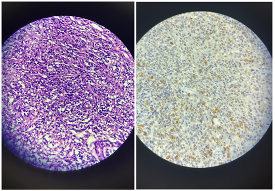
Figure 3: Histopathological findings. (A) Hematoxylin and eosin staining showing a dense lymphoplasmacytic infiltrate. (B) IgG4 immunohistochemistry highlighting numerous IgG4-positive plasma cells (>20 cells/high-power field).
Serum IgG4 concentration will be measured as part of the systemic work-up. A complementary pathological review, including assessment of the IgG4-positive/total IgG-positive plasma-cell ratio and evaluation for storiform fibrosis and obliterative phlebitis, is also planned. These investigations will provide a more complete clinicopathological and systemic correlation and help further support the diagnosis of IgG4-related orbital disease.
Systemic Assessment, Treatment, and Follow-Up
At the time of manuscript preparation, serum IgG4 testing and a systematic assessment for extraorbital involvement had not been performed. Because IgG4-RD may be multisystemic, the recommended post-biopsy work-up includes serum IgG subclasses, complete blood count, inflammatory markers, renal and hepatic profiles, complement levels, urinalysis, and targeted or whole-body imaging according to symptoms and local practice, with referral to internal medicine or rheumatology [1,4,6].
Discussion
IgG4-ROD is a recognized manifestation of IgG4-RD and may be the first or only clinically apparent site of disease. Although lacrimal gland enlargement is a classic pattern, involvement may extend to orbital fat, extraocular muscles, eyelids, the orbital apex, and trigeminal nerve branches [2-5]. Recent series confirm a broad spectrum of unilateral and bilateral presentations, with proptosis, eyelid swelling, ptosis, and motility restriction among the most common symptoms [2,4,5]. The present patient had a unilateral superolateral orbital mass with ptosis and impaired elevation, but no diplopia or reported visual loss.
The radiological distinction between IgG4-ROD and orbital lymphoma may be difficult. Both conditions can produce a homogeneous soft-tissue mass, lacrimal-region enlargement, extraocular muscle involvement, or diffuse orbital infiltration. Bilaterality, enlargement of infraorbital or supraorbital nerves, and multisite ocular adnexal involvement may support IgG4-ROD, but none is sufficiently specific to replace biopsy [2,3]. In this case, the unilateral intraconal and extraconal lesion was interpreted as possibly lymphomatous, an appropriate concern given the patient's age and the progressive mass effect.
Histopathology remains central to diagnosis. The characteristic morphological triad of systemic IgG4-RD consists of a dense lymphoplasmacytic infiltrate, storiform fibrosis, and obliterative phlebitis, accompanied by increased IgG4-positive plasma cells and an elevated IgG4-positive/IgG-positive ratio [8,9]. However, the full triad may be incomplete in orbital specimens, and quantitative thresholds vary according to tissue type and diagnostic framework. Current criteria therefore emphasize integration of clinical and radiological findings, serum IgG4, histological morphology, immunostaining, and exclusion of alternative diagnoses [3,7-9].
In the present specimen, the dense plasmacytic infiltrate, numerous CD138-positive plasma cells, more than 20 IgG4-positive cells per high-power field, and low proliferative activity supported IgG4-related disease. Nevertheless, the absence of a reported IgG4/IgG ratio, serum IgG4 level, and explicit assessment of storiform fibrosis and obliterative phlebitis limits formal categorization. This limitation should be stated rather than overcome by assuming unavailable data. Moreover, IgG4-positive plasma cells can occur in other inflammatory conditions and in some lymphomas. The 2023 revised ophthalmic criteria particularly stress the need to exclude MALT lymphoma and other lymphoid malignancies [3].
The differential diagnosis of a superolateral orbital mass includes lymphoproliferative disorders, idiopathic orbital inflammation, lacrimal gland epithelial tumors, sarcoidosis, granulomatosis with polyangiitis, thyroid eye disease, infection, and metastatic disease. Lymphoma is especially important because it may coexist with or histologically resemble IgG4-ROD. A low Ki-67 index and the absence of a specific immunophenotypic pattern in the available panel favored a non-high-grade lymphoid process, but definitive exclusion of lymphoma may require assessment of light-chain restriction, clonality studies, and hematopathological review when morphology or clinical evolution remains suspicious [2,3,9].
Once the diagnosis is established, systemic evaluation is essential. In a 2025 nationwide multicenter study, extraorbital involvement was identified in more than half of adequately evaluated patients with definite IgG4-ROD, reinforcing the need to assess the pancreas, biliary tract, salivary glands, kidneys, lungs, lymph nodes, retroperitoneum, and large vessels according to the clinical context [4]. Serum IgG4 is useful but is neither perfectly sensitive nor specific; a normal value does not exclude tissue-proven disease [1,7,8].
Systemic glucocorticoids remain the usual first-line induction treatment for active symptomatic IgG4-ROD, particularly when vision, ocular motility, or other organs are threatened. The initial response is often rapid, but relapse during or after tapering is common [1,2,10,11]. Steroid-sparing immunomodulators may be considered in relapsing or steroid-dependent disease, although evidence for conventional agents is heterogeneous. B-cell depletion with rituximab has shown substantial activity in refractory, relapsing, or organ-threatening IgG4-RD and IgG4-ROD [1,10-12]. Surgery is primarily diagnostic, although selected patients may benefit from debulking; recent data suggest an association between debulking and remission, but surgery does not replace systemic assessment or medical treatment when active multisystem disease is present [4].
Long-term surveillance is required because clinical improvement does not eliminate the risk of relapse, fibrotic sequelae, optic neuropathy, or unrecognized systemic involvement. Follow-up should combine ophthalmic examination, assessment of visual acuity and ocular motility, serial imaging when indicated, and monitoring of systemic disease activity. In the next phase of management, we plan to complete the systemic work-up, initiate an appropriate treatment according to the findings, and ensure regular clinical and radiological follow-up.
Conclusion
IgG4-related orbital disease may present as a unilateral superolateral orbital mass that is clinically and radiologically indistinguishable from lymphoma. A carefully planned biopsy is decisive, but the interpretation of increased IgG4-positive plasma cells must remain clinicopathological and must include exclusion of lymphoid malignancy. Serum IgG4 testing and systemic staging are required after an orbital diagnosis because clinically silent extraorbital involvement is common. This case illustrates the diagnostic value of the lateral eyebrow-tail approach for tissue sampling and the importance of multidisciplinary follow-up.
References
- Wallace ZS, Katz G, Hernandez-Barco YG, Baker MC. Current and future advances in practice: IgG4-related disease. Rheumatol Adv Pract, 2024; 8(2): rkae020. doi: 10.1093/rap/rkae020.
- Lai KKH, Ang TWX, Cheuk W, et al. Advances in understanding and management of IgG4-related ophthalmic disease. Asia Pac J Ophthalmol (Phila), 2024; 13(5): 100101. doi: 10.1016/j.apjo.2024.100101.
- Takahira M, Goto H, Azumi A. The 2023 revised diagnostic criteria for IgG4-related ophthalmic disease. Jpn J Ophthalmol, 2024; 68(4): 293-301. doi: 10.1007/s10384-024-01072-4.
- Ko JS, Lee MJ, Khwarg SI, et al. Immunoglobulin G4-related ophthalmic disease: a nationwide multicenter study in Korea. Ophthalmology, 2025; 132(9): 995-1004. doi: 10.1016/j.ophtha.2025.04.031.
- Kim D, Jeong S, Lew H. Unraveling the clinical features and outcomes of IgG4-related ophthalmic disease. J Clin Med, 2024; 13(13): 3780. doi: 10.3390/jcm13133780.
- Lai KKH, Aljufairi FMAA, Sebastian JU, et al. Systemic involvement in immunoglobulin G4-related ophthalmic disease. Ocul Immunol Inflamm, 2024; 32(8): 1852-1858. doi: 10.1080/09273948.2023.2280709.
- Wallace ZS, Naden RP, Chari S, et al. The 2019 American College of Rheumatology/European League Against Rheumatism classification criteria for IgG4-related disease. Arthritis Rheumatol, 2020; 72(1): 7-19. doi: 10.1002/art.41120.
- Umehara H, Okazaki K, Kawa S, et al. The 2020 revised comprehensive diagnostic criteria for IgG4-related disease. Mod Rheumatol, 2021; 31(3): 529-533. doi: 10.1080/14397595.2020.1859710.
- Deshpande V, Zen Y, Chan JKC, et al. Consensus statement on the pathology of IgG4-related disease. Mod Pathol, 2012; 25(9): 1181-1192. doi: 10.1038/modpathol.2012.72.
- Detiger SE, Karim AF, Verdijk RM, van Hagen PM, van Laar JAM, Paridaens D. The treatment outcomes in IgG4-related orbital disease: a systematic review of the literature. Acta Ophthalmol, 2019; 97(5): 451-459. doi: 10.1111/aos.14048.
- Khosroshahi A, Wallace ZS, Crowe JL, et al. International consensus guidance statement on the management and treatment of IgG4-related disease. Arthritis Rheumatol, 2015; 67(7): 1688-1699. doi: 10.1002/art.39132.
- Wu A, Andrew NH, Tsirbas A, Tan P, Gajdatsy A, Selva D. Rituximab for the treatment of IgG4-related orbital disease: experience from five cases. Eye (Lond), 2015; 29(1): 122-128. doi: 10.1038/eye.2014.251.
- Rattunde AL, Dittami GM, Sobolewska B, et al. IgG4-related orbital disease versus idiopathic orbital inflammation: clinical features, diagnostics, and management. Biomedicines, 2025; 13(9): 2311. doi: 10.3390/biomedicines13092311.