
Email id: info@ijclinmedcasereports.com
Giant Renal Cysts: Uro-CT Findings
Francesco Messina*, Grazia Calabrese, Carmela Tebala and Lorena Turano
Unit of Radiology - Riuniti Hospital, Azienda Ospedaliera Grande Ospedale Metropolitano (G.O.M.) “Bianchi-Melacrino-Morelli” Reggio Calabria, Italy
Received Date: 22/05/2026; Published Date: 14/09/2026
*Corresponding author: Francesco Messina, MD, Unit of Radiology - Riuniti Hospital, Azienda Ospedaliera Grande Ospedale Metropolitano, Bianchi–Melacrino, Morelli‖ Via Giuseppe Melacrino n.21, 89124 Reggio Calabria, Italy
Abstract
Renal Cysts (RC) are acquired lesions of the kidneys that occur commonly in the renal cortex, although the etiology is yet to be fully established. Most benign renal cysts are asymptomatic and require no treatment; they are commonly an incidental finding at imaging. RC can cause a variety of clinical symptoms when they are sufficiently large. However, cases of huge renal cysts have been rarely reported. Giant RC are clinically significant because they can cause physical complications and require targeted interventions. We report two cases of our experience of giant RC, that had big diameters at Computed Tomography (CT), and also a clinical symptomology with abdominal plain.
Keywords: Cysts Kidney Renal; CT-urography
Background
At birth, the kidneys may appear normal, but with time they can develop cysts. Clinical presentation of RC is variable and includes: dull flank pain of variable severity and time course (most common); abdominal or flank masses; haematuria; hypertension up to renal failure in the most advanced cases. Macroscopically the kidneys can demonstrate a variable size of the cysts (from a few millimeters to many centimeters). Besides monitoring renal function by standard measurements, the diagnosis and follow-up of patients with RC is based largely on radiologic investigations that are performed with Ultrasonography (US), CT and Magnetic Resonance (MRI), with the aim of evaluating renal cysts morphology and volume. The aim of this report is to highlight the role of CT-urography in the diagnosis and characterization of giant renal cysts, describing two cases of our experience of giant RC. Giant RC (generally larger than 8 cm) are clinically significant because, unlike common simple cysts, they can cause physical complications and require targeted interventions, sometimes even emergency ones.
Imaging technique (Uro-CT)
In both cases, we subsequently performed a CT-urography, using the Uro-CT scan protocol, that was made by:
- basal phase (without contrast);
- cortico-medullary phase (25-40 seconds after contrast administration);
- nephrographic phase (80-120 seconds) shows enhancement of the renal parenchyma;
- excretory phase (5-15 minutes) visualizes the urinary excretory tree (ureters and bladder).
The relative images were evaluated with an appropriate work-stations using MPR (axial, sagittal and coronal), cMIP, MIP thin and thick, and VRT reconstructions. The images were reconstructed as 1.2-mm-slices.
Case 1
We describe a 67-year-old woman with a giant renal cyst that had progressively increased in size. She arrived to the Emergency Department of our Hospital with persistent right and posterior abdominal flank pain (over a period of two months), without haematuria. The abdomen was painless on palpation and percussion. Routine hematology, biochemistry, and serum tumor markers were normal. The patient suffered from arterial hypertension (on drug therapy); she had a GFR of 75 mL/min, and had not other significant comorbidities. Previous renal tests showed no further significant alterations. No previous abdominal operations for the patient, and no fever.
An US was inconclusive for the presence of excessive intestinal gas. So an emergency abdominal unenhanced contrast CT was initially made, with a 128multislices Computed Tomography scan, and the images so obtained were analyzed with a slice-thickness of 1.2 mm and MPR reconstructions (axial, sagittal, and coronal). CT had shown the presence (Figure 1, a-b) of a giant renal cyst (about 13 cms) identified in the renal cortex of the right kidney, with hypodense appearance and exophytic development.
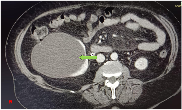
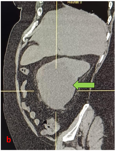
Figure 1 (a-b): Computed Tomography with axial (a) and sagittal (b) MPR reconstructions. CT had shown the presence of a giant renal cyst of about 13 cms (green arrow), in the renal cortex of the right kidney with hypodense appearance and exophytic development. CT had also shown the compression of the right ureter by the giant renal cyst.
Afterwards, a CT-urography was performed, using the Uro-CT scan protocol (as we previously described), that had confirmed (Figure 2, a-c) the presence of a giant renal cyst (13 cms) in the renal cortex of the right kidney, with hypodense appearance, exophytic development, without any contrast enhancement in the various phases of the CT study. CT-urography had also shown the presence of compression on the proximal section of the right ureter, and a consequent dilation of its proximal section, on the right.
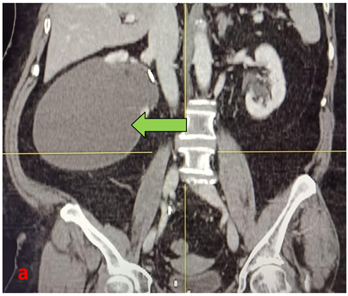
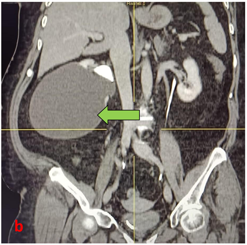
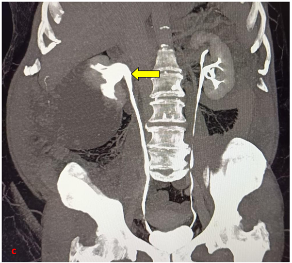
Figure 2 (a-c): CT-urography with Uro-CT scan protocol. CT coronal (a,b) MPR coronal reconstructions, and MIP coronal (c ) , had confirmed the presence of a giant renal cyst (13 cms) in the renal cortex of the right kidney (green arrows), with hypodense appearance, exophytic development, without any contrast enhancement in the various phases of the CT study. CT-urography had also shown the presence of a compression on the proximal section of the right ureter, and a consequent dilation of its proximal section, on the right (yellow arrow).
The patient is now monitored clinically and with laboratory tests, and will undergo periodic check-up based on her general clinical conditions.
Case 2
We describe the case of a 40-year-old man, that arrived to the Emergency Department of our Hospital with persistent right and posterior abdominal flank pain, with micro-haematuria. Routine hematology, biochemistry, and serum tumor markers were normal. No previous abdominal operations for the patient, and no fever.
An US was preliminarily made, but it was not-diagnostic because of the presence of intestinal gas. So an emergency abdominal CT was made (with a 128multislices Computed Tomography scan), at first as unenhanced and then a CT-urography with Uro-CT scan protocol (as we previously described), that had shown the presence (Figure 3, a-d) of a giant unilateral renal cyst (about 10 cms) identified in the renal cortex of the right kidney, with hypodense appearance and exophytic development.
The suggestive feature of this case two is that we found that the giant renal cyst, in its exophytic development, comes into contact at the same time with the lower margin of the right hepatic lobe, and compresses the proximal section of the right ureter; we particularly describe it in our report at Hospital.
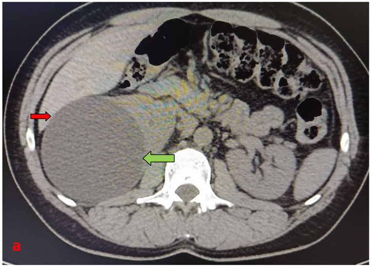
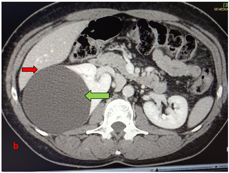
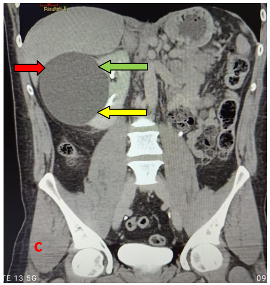
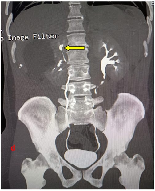
Figure 3 (a-d): CT-urography with Uro-CT scan protocol. Basal phase (a); axial reconstruction. Basal phase (a); cortico-medullary phase (b) ; nephrographic phase (c) and excretory phase (d) with a MIP coronal reconstruction. CT had shown the presence of a giant renal cyst (10 cms) in the renal cortex of the right kidney (green arrow), with hypodense appearance, exophytic development, without any contrast enhancement in the various phases of the CT study. CT-urography had also shown the presence of a compression at the same time with the lower margin of the right hepatic lobe (red arrows), and a compression of the proximal section of the right ureter (yellow arrows).
The patient, also in consideration of his symptoms, had understood and agreed to undergo surgery, and is now awaiting scheduled surgery to remove this giant renal cyst. However, he is now monitored clinically and with laboratory tests, monitoring overall his general clinical conditions.
Discussion
RC occur commonly in the renal cortex, with an exophytic development, although the etiology is yet to be fully established. RC is one of the most common benign lesions of the kidneys, and occurring with a frequency greater than 50% over the age of 50 years [1-3]. Most are simple RC and are usually detected incidentally on imaging. They are occasionally large enough to be palpable on routine clinical examination or by the patient himself. The origin of RC is in renal tubules, exactly from the diverticulum of the distal convoluted tubule [4]. Proliferation of tubule epithelial cells modulated by endocrine, paracrine, and autocrine factors is a major element in the pathogenesis of RC diseases [5]. However, majority of the RC are < 3 cm in size [6]. They may occur well within a kidney or on its surface, usually oval or round in shape, they have a smooth outline lined by a single layer of flattened epithelium [7]. The pathogenesis of cysts presenting at a later stage is less clear. If developmental, they may represent abnormalities of earlier generations of uriniferous tubules persisting as cystic collections. If acquired, they may represent renal tubular obstruction secondary to focal ischaemia and inflammation, factors that can produce epithelial cysts experimentally [8]. Giant RC are fluid-filled sacs that can form in the kidneys. Although they are often benign and asymptomatic, large cysts can cause symptoms such as flank pain, infection, or bleeding.
Furthermore, the clinical significance of giant renal cysts presents a varied range of symptoms, including: severe pain (due to distension of the renal capsule, resulting in dull, chronic pain in the flank); hypertensive compression (compression of the renal artery may cause secondary arterial hypertension); hydronephrosis (compression of the calyceal system or ureter can obstruct urinary flow, leading to parenchymal distress); risk of cyst rupture or hemorrhage (large cysts are exposed to external trauma, with a risk of spontaneous rupture or intracystic bleeding).
The International reference classification for the RC is the —Bosniak classification‖ (updated in 2019), which assesses the risk of malignancy based on morphological characteristics on contrast-enhanced CT, and cystic lesions are divided into five categories based on their visible characteristics (presence of septa, wall thickness, calcifications and how they react to contrast enhancement):
- Bosniak type I cysts are so-called simple benign cysts, with thin walls and no septa, calcifications, or solid components. Their contents have the same density as water.
- Bosniak type II cysts are minimally complicated cysts: they may have thin septa or fine calcifications; the content may be slightly denser than water but without contrast medium uptake. These are also benign cysts that do not require any special measures. In some situations —when the diagnosis is based only on US examination— may request a more accurate examination (such as a CT scan with contrast medium or an MRI). Once the diagnosis has been confirmed, no further investigations are necessary.
- Bosniak type IIF cysts differ from category II in that they have a greater number of septa with possible thickening, minimal calcifications, and/or slight contrast uptake. In 5-10% of cases, these cysts may be malignant, and therefore follow-up monitoring is recommended for the next 5 years after the initial diagnosis.
- Bosniak type III cysts are indeterminate cystic formations that often have thick walls or septa and take up contrast medium. They are malignant in 60-80% of cases and, in the absence of reassuring medical history data such as previous trauma or kidney infection, usually require histological data obtained through transcutaneous biopsy or exploratory surgery.
- Bosniak type IV cysts are highly suspicious for malignant neoplasia. These cysts contain obvious solid areas that can take up contrast medium. In these situations, surgery to remove the lesion should always be considered.
In our case, we observed giant RC of type 1 (according to Bosniak classification). In addition, in our long experience, in particular with CT scans, we have rarely encountered giant simple cysts which are not part of the findings of polycystic kidneys (which have a completely different approach) [9]. The RC that we have described in our case report are simple RC and represent the largest ones we have ever described in our radiological activity (13 cms).
CT-urography is a specialized CT application dedicated to the study of the urinary tract, capable of simultaneously analyzing the kidneys, ureters, and bladder (all-in-one study‖). UroCT scan is indicated for: diagnosis and staging of urinary tract tumors; evaluation of renal vascular abnormalities and congenital malformations; diagnosis of inflammatory/infectivous diseases; characterization of renal cysts; valuation of renal trauma.
Diagnosis of RC is made through imaging tests, such as US and CT. Simple cysts don’t require treatment, but giant cysts that cause symptoms (or have complex characteristics) may require regular monitoring and, in some selected cases, a surgical treatment (such as percutaneous or laparoscopic removal).
Conclusion
In our opinion and based on our experience, we think about the importance of UroCT scan in the diagnosis of nephro-urinary diseases, and in particular, of the Renal Cysts. In fact, CT-urography is the gold standard for evaluating RC for at least three reasons: assessment of their density (expressed in Hounsfield Units, HU); study of their contrast kinetics; anatomical precision (to demonstrate their relationship with surrounding structures). CT-urography can be considered as an —all-in-one study‖. It allows for the acquisition of high-quality images, in axial, sagittal, coronal, and three-dimensional (3D) multiplanar reconstructions (MPR). The entire abdomen cavity can be visualized during the same examination, and an accurate assessment of the number, size and extent of renal cysts is usually well demonstrated. So, CT-urography had an importance for the first diagnosis of RC, for the clinical management of patients and for the subsequent correct use of imaging in check-up.
Conflicts of interest: The authors certify that there is no conflict of interest with any financial organization regarding the material discussed in the manuscript.
Patient Consent Statement: The patient confirmed the consense for publication of our case report.
References
- Pedersen JF, Emamian SA, Nielsen MB. Simple renal cyst: relations to age and arterial blood pressure. Br J Radiol, 1993; 66: 581–584.
- Tada S, Yamagishi J, Kobayashi H, Hata Y, Kobari T. The incidence of simple renal cyst by Computed Tomography. Clin Radiol, 1983; 34: 437–439.
- Laucks SP Jr, Mc Lachlan MSF. Ageing and simple cysts of the kidney. Br J Radiol, 1981; 54: 12.
- Baert I, Steg Is the diverticulum of the distal and collecting tubules a preliminary stage of the simple cyst in the adult? J Urol, 1977; 118: 707–710.
- Gagnadoux Cystic kidney disease. Rev Prat, 1997; 47: 1536-1540.
- Tada S, Yamagishi J, Kobayashi H, et al. The incidence of simple renal cyst by computed tomography. Clin Radiol, 1983; 150: 207.
- Sandeep S, Vikas K, Harminder P, Dinesh G. A case report: an unusual cause of giant renal mass. J Indian Acad Clin Med, 2006; 7(3): 252–254.
- Siegel MJ, McAlister WH. Simple cysts of the kidney in J Urol, 1980; 123: 75–78.
- Messina F, Loria F, Loria G, Frosina L, Frosina F, Morano E. Sonography and contrast enhancement MDCT in the evaluation of complicated Autosomal Dominant Polycystic Kidney Disease. Peertechz - Archives of Renal Diseases and Management, 2015; 1(1): 003-007.