Email id: info@ijclinmedcasereports.com
Congenital Myasthenic Syndrome Linked to a Rare CHRNA1 Mutation: Clinical Insights from a Rare Case
Mouddane M1,*, Haddouali K1, Khattab H1, Sikkal A1, Bellakhdar1, El Otmani H1,2, El Moutawakil B1,2 and Rafai MA1,3
¹Department of Neurology, CHU Ibn Rochd, Casablanca, Morocco
2Laboratory of Genetics and Molecular Pathology, Faculty of Medicine and Pharmacy, Casablanca, Morocco
3Research Laboratory on Nervous System Diseases, Neurosensory Diseases and Disability, Faculty of Medicine and Pharmacy, Casablanca, Morocco
Received Date: 18/06/2026; Published Date: 09/09/2026
*Corresponding author: Mouddane Meryem, Department of Neurology, CHU Ibn Rochd, Casablanca, Morocco
Abstract
Background: Congenital Myasthenic Syndromes (CMS) are rare inherited disorders of neuromuscular transmission caused by defects in proteins of the neuromuscular junction. Mutations in CHRNA1, encoding the α-subunit of the nicotinic acetylcholine receptor, are an uncommon cause of postsynaptic CMS and have been associated with a broad clinical spectrum ranging from severe prenatal phenotypes to milder oculobulbar forms.
Case Presentation: We report the case of a 14-year-old boy born to consanguineous parents who presented with neonatal-onset ptosis and facial weakness. Symptoms began shortly after birth with fluctuating unilateral ptosis that rapidly progressed to bilateral involvement and later became persistent. During infancy, he developed hypophonia and limited oral aperture. Neurological examination revealed bilateral ptosis, ophthalmoplegia, facial diplegia, and hypophonia, while limb strength and deep tendon reflexes remained normal. Repetitive nerve stimulation demonstrated a significant decremental response consistent with a postsynaptic neuromuscular junction disorder. Autoimmune testing for acetylcholine receptor and MuSK antibodies was negative. Genetic analysis identified a homozygous pathogenic variant in CHRNA1, confirming the diagnosis of congenital myasthenic syndrome.
Discussion: CHRNA1-related CMS is exceptionally rare and exhibits marked phenotypic heterogeneity. The present case expands the clinical spectrum of this condition by illustrating a predominantly oculobulbar phenotype characterized by ptosis, ophthalmoplegia, facial weakness, and hypophonia without significant limb involvement. Recognition of this presentation is important because CMS may be misdiagnosed as autoimmune myasthenia gravis or other neuromuscular disorders, particularly in resource-limited settings.
Conclusion: This report represents, to our knowledge, the first genetically confirmed case of CHRNA1-related CMS reported from Africa. It highlights the importance of considering CMS in patients with early-onset ptosis and negative autoimmune markers and underscores the pivotal role of genetic testing in establishing the diagnosis and guiding targeted treatment strategies.
Introduction
Congenital Myasthenic Syndromes (CMS) are a heterogeneous group of inherited disorders of neuromuscular transmission characterized by fatigable muscle weakness and variable clinical severity [1]. CMS are classified as presynaptic, synaptic, or postsynaptic, according to the location of the primary defect within the neuromuscular junction. To date, over 35 genes have been associated with these disorders, with CHAT, COLQ, RAPSN, CHRNE, DOK7, and GFPT1 representing the most frequently affected [2].
In contrast, mutations in CHRNA1, encoding the alpha subunit of the nicotinic acetylcholine receptor, represent a rare cause of postsynaptic CMS and may result in either slow-channel or fast-channel syndromes [3,4]. To date, only a few case reports of CMS associated with CHRNA1 mutations have been documented.
We present, to our knowledge, the first reported case from Africa, contributing to the broader characterization of its clinical and molecular features.
Case Report
A 14-year-old male, born to third-degree consanguineous parents, was referred for evaluation of early-onset ptosis and facial weakness. The perinatal period was notable for neonatal distress requiring ILAM. Motor development was mildly delayed, with independent ambulation achieved at 18 months. There was no family history of neuromuscular disorders.
The patient’s symptoms manifested shortly after birth, initially as unilateral left-sided ptosis, progressing to bilateral involvement within the first month. The ptosis was initially fluctuating, more pronounced in the evening, and later became persistent. At one year of age, he developed a constant inability to fully open the mouth (oral aperture limitation) accompanied by hypophonia.
Neurological examination revealed bilateral ophthalmoplegia, symmetric ptosis, bilateral facial paresis, and hypophonia (Figure 1). Limb strength was preserved, and deep tendon reflexes were normal. No sensory deficits were observed.
Electrophysiological assessment demonstrated a significant decremental response on repetitive nerve stimulation across multiple nerve-muscle pairs, indicative of a postsynaptic neuromuscular junction defect. Serum testing for anti-acetylcholine receptor (AChR) and anti-MuSK antibodies was negative. Genetic testing confirmed a homozygous mutation in the CHRNA1 gene, consistent with a postsynaptic congenital myasthenic syndrome.
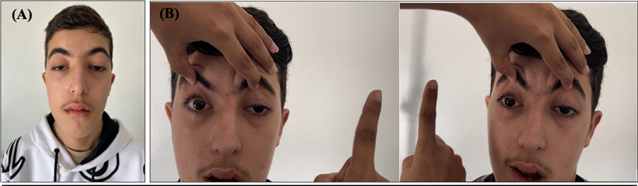
Figure 1: Representative clinical images of the patient. (A) Bilateral ptosis and facial diplegia. (B) Ophthalmoplegia, demonstrated by impaired eye movements during finger-pursuit examination.
Discussion
Mutations in the CHRNA1 gene, encoding the α-subunit of the postsynaptic nicotinic Acetylcholine Receptor (AChR), represent a rare cause of Congenital Myasthenic Syndromes (CMS). These mutations impair neuromuscular transmission primarily by reducing the number or function of AChRs at the postsynaptic membrane [5]. In addition, alternative splicing of CHRNA1, generating P3A− and P3A+ isoforms, plays a pathogenic role, as disease-associated variants may shift this balance toward the less functional isoform, further compromising receptor availability [6].
Clinically, CHRNA1-related CMS may present as either slow-channel or fast-channel syndromes, with autosomal dominant or recessive inheritance depending on the underlying mechanism. Only a limited number of cases have been reported to date, and the phenotypic spectrum is highly heterogeneous. Presentations range from severe prenatal manifestations [7], including fetal akinesia, contractures, and pterygia, to milder postnatal forms characterized by ptosis, ophthalmoplegia, facial and bulbar weakness, and variable limb involvement. Additional features such as arthrogryposis or fatal multiple pterygium syndrome have also been described [8].
The present case is consistent with a postsynaptic CMS due to a CHRNA1 mutation, characterized by early-onset fluctuating ptosis progressing to fixed ophthalmoplegia, facial weakness, and hypophonia. This relatively restricted phenotype aligns with previously reported milder forms and highlights the clinical variability associated with CHRNA1 variants.
From a therapeutic perspective, acetylcholinesterase inhibitors, particularly pyridostigmine, remain the first-line treatment and are generally associated with clinical improvement. Other agents such as fluoxetine or quinidine may be considered in slow-channel syndromes, whereas 3,4-diaminopyridine (3,4-DAP) appears less consistently effective [2]. Emerging therapeutic strategies, including antisense oligonucleotides (AONs), offer promising perspectives by targeting the underlying splicing defect and restoring the balance between CHRNA1 isoforms [6]. Although still experimental, these approaches may represent future disease-modifying therapies, particularly in genetically confirmed cases.
Conclusion
This report highlights a rare case of CHRNA1-related congenital myasthenic syndrome, expanding the phenotypic spectrum with a relatively mild, predominantly oculobulbar presentation. It underscores the importance of considering CMS in early-onset ptosis with negative autoimmune markers and emphasizes the key role of genetic testing for accurate diagnosis. Early recognition is essential, as targeted therapies such as pyridostigmine can significantly improve clinical outcomes, while emerging treatments may offer future disease-modifying options.
Acknowledgments: The authors would like to thank the Department of Neurology, CHU Ibn Rochd, for its support in the management and evaluation of this patient.
Conflicts of Interest: The authors declare that they have no conflicts of interest.
Funding: No funding was received for the preparation of this case report.
Consent for Publication: Written informed consent for publication of this case report and the accompanying clinical images was obtained from the patient's legal guardians. All identifying information has been removed, and every effort has been made to protect the patient's privacy and confidentiality.
References
- Beeson D. Congenital myasthenic syndromes. Handb Clin Neurol, 2024; 203: p. 69‑88.
- Finsterer J. Congenital myasthenic syndromes. Orphanet J Rare Dis, 2019; 14 (no 1): p. 57.
- Rodríguez Cruz PM, et al., A novel phenotype of AChR-deficiency syndrome with predominant facial and distal weakness resulting from the inclusion of an evolutionary alternatively-spliced exon in CHRNA1. Neuromuscul Disord, 2023; 33 (no 2): p. 161‑168.
- Ohno K, Ito M, Ohkawara B. Review of 40 genes causing congenital myasthenic syndromes. J Hum Genet, 2025.
- Gooneratne IK, et al., Slow-Channel Congenital Myasthenic Syndrome due to a Novel Mutation in the Acetylcholine Receptor Alpha Subunit in a South Asian: A Case Report. J Neuromuscul Dis, 2021; 8 (no 1): p. 163‑167.
- Tei S, Ishii HT, Mitsuhashi H, Ishiura S. Antisense oligonucleotide-mediated exon skipping of CHRNA1 pre-mRNA as potential therapy for Congenital Myasthenic Syndromes. Biochem Biophys Res Commun, 2015; 461 (no 3): p. 481‑486.
- Chiramel JJ, PKC, Thirumalesh PV. Congenital myasthenia syndrome due to CHRNA1 mutation resulting in respiratory failure. International Journal of Contemporary Pediatrics, 2025; 12 (no 2): p. 325‑327.
- Irumudomon O, Ghosh PS. Clinical Reasoning: A child with arthrogryposis: Congenital myasthenic syndrome-CHRNA1 mutation. Neurology, 2018; 91 (no 10): p. e995‑e998.