Preemptive Intraluminal Negative Pressure Therapy (PINPT) for Duodenal Resection Using a Double Lumen Open-Pore Film Drain (dOFD) – First Report of a New Method for Anastomotic Prophylaxis in Duodenum
Gunnar Loske1,*, Johannes Müller1, Martin Zeile2, Eckard Martens3 and Christian Theodor Müller4
1Department for General, Abdominal, Thoracic and Vascular Surgery, Katholisches Marienkrankenhaus Hamburg gGmbH, Hamburg, Germany
2Institute for Diagnostic and Interventional Radiology, Katholisches Marienkrankenhaus Hamburg gGmbH, Germany
3Department for Medical Oncology and Hematology, Gastroenterology and Infectious Diseases, Katholisches Marienkrankenhaus Hamburg gGmbH, Germany
4Department of Surgery, Wilhelmsburger Krankenhaus Groß-Sand, Germany
Received Date: 03/02/2025; Published Date: 08/04/2025
*Corresponding author: Dr. Gunnar Loske, med, Department for General, Abdominal, Thoracic and Vascular Surgery, Katholisches Marienkrankenhaus Hamburg gGmbH, Alfredstrasse 9, 22087 Hamburg, Germany
Abstract
Duodenal suture insufficiency after surgical resection is difficult to treat. The method of active drainage of digestive secretions for anastomotic prophylaxis with simultaneous enteral nutrition is already used as a new safety concept in oesophageal surgery. It can also be used in duodenal surgery.
A thin Double lumen Open pore Film Drain (dOFD) is used for PINPT. The dOFD is constructed with a thin open-pore drainage film and a drain. The film consists out of two multi-perforated membranes separated by a small gap. When negative pressure is applied to the film, fluids can be aspirated over the entire surface of the space and through the numerous pores. By wrapping the film around the drain, small lumen Open pore Film Drainages (OFD) can be created.
After duodenal resection, the dOFD is placed intraluminal covering the anastomotic region. Vacuum is applied with an electronic negative pressure device. During wound healing duodenal secretions are kept away from the anastomosis. At the same time, the patient can be fed via an integrated feeding tube.
The innovative method of PINPT for anastomotic prophylaxis is demonstrated in a case report of duodenal resection for a large adenoma. PINPT lasted for 7 days. The anastomosis healed without complications.
Keywords: Drainage; Endoscopic vacuum therapy; Leakage; Anastomosis; Insufficiency; Prophylaxis
Abbreviations: iT-integrated feeding tube; DE-Drainage Element; dOFD-Double lumen Open-pore Film Drain; ENPT-Endoscopic Negative Pressure Therapy; OFD-Open pore Film Drainage; PINPT-Pre-emptive Intraluminal Negative Pressure Therapy; PARD-Preemptive Active Reflux Drainage
Background
Duodenal suture insufficiency following surgical resection and other surgical or interventional procedures on the duodenum is difficult to treat. Endoscopic negative pressure therapy (ENPT) has become an important endoscopic treatment for oesophageal anastomotic defects [1]. This innovative technique can also be used successfully for duodenal leaks [2,3].
In recent years, ENPT has also been used preemptively in oesophagectomy as new safety concept for esophageal surgery. The aim of preemptive use is to reduce the rate of anastomotic insufficiency and thus increase patient safety in this high-risk procedure [4,5].
A therapeutic variant of ENPT in oesophagectomy is known as Preemptive Active Reflux Drainage (PARD) [5]. The therapeutic principle is to actively keep the reflux of digestive secretions away from the intrathoracic oesophageal anastomosis during the vulnerable early phase of anastomotic healing. To achieve this, a special double-lumen open-pore film drain (dOFD) is placed intraluminally in the gastric tube. A permanent negative pressure is applied to the dOFD. As the reflux secretions are aspirated, the gastric tube and anastomosis are decompressed. At the same time, enteral nutrition is provided via an integrated feeding tube. A 100% healing rate of the anastomosis has been achieved in oesophagectomy with PARD [6].
The method of active drainage of digestive secretions for anastomotic prophylaxis with simultaneous enteral nutrition can also be used in duodenal surgery. We present a case report of preemptive intraluminal negative pressure therapy (PINPT) in the duodenum.
Material and Method
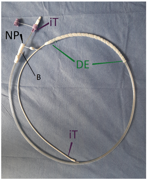
A dOFD is used for PINPT. To create a dOFD, the distal end of the gastric channel of a triluminal tube (Freka®Trelumina, CH/Fr 16/9, 150 cm, Fresenius, Germany) is covered with a 25 cm long, 3-4 cm wide strip of a thin, transparent, open-pore, double-layered film (DF) (Suprasorb® CNP drainage film, Lohmann & Rauscher International GmbH, Germany) [2,5-8] (Figure 1). Negative pressure can be applied to the film-covered drainage element (DE). Enteral nutrition can be provided simultaneously via the integrated feeding tube (iT).
The DF was originally developed for use in the open abdomen for peritonitis. The DF consists of two multi-perforated membranes separated by a small gap. When negative pressure is applied to the DF, fluids can be aspirated over the entire surface of the space and through the numerous pores. By wrapping the DF around a drain, small lumen open pore film drainages (OFD) can be created for ENPT. These are only 4-6 mm in diameter. OFDs can be placed transnasally using the same technique as a nasogastric tube (NGT) [5-8].
For PINPT, the dOFD is inserted into the duodenum. The film-coated portion is placed in the duodenal lumen to cover the anastomotic area. The iT is placed in the jejunum distal to the anastomosis. A continuous negative pressure of 125 mmHg is applied. This will permanently aspirate the duodenal secretions and keep them away from the anastomosis. Digestive secretions should have little or no effect at this vulnerable point. Feeding can take place through the iT at the same time as negative pressure is applied.
Results, Case Report with Clinical Application of PINPT
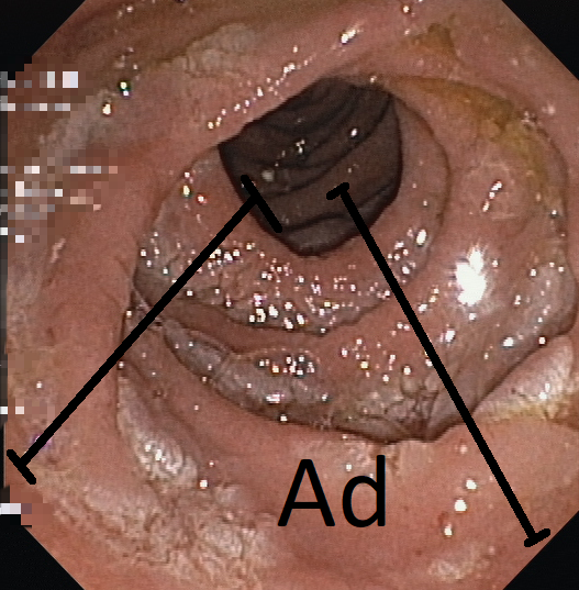
An 83-year-old woman was diagnosed with a circular adenoma of significant size a few centimetres distal to the greater duodenal papilla (Figure 1).

Figure 1: A large circular adenoma (Ad) was diagnosed in the duodenum, located a few centimeters distally from the major duodenal papilla.
Due to its size, surgical resection of the adenoma was recommended following a multidisciplinary consultation. To promote anastomotic healing, we introduced PINPT using a dOFD for the first time (Figure 2).

Figure 2: The figure shows a double lumen open-pore drainage (dOFD) with an integrated intestinal feeding tube (iT) used for pre-emptive intraluminale negative pressure therapy. The dOFD is constructed with a triluminal tube (Freka®Trelumina, Fresenius). For the 25 cm long drainage element (DE) the gastric channel is wrapped with the open-pore drainage film (Suprasorb® CNP Drainage film, Lohmann & Rauscher). The ventilation channel is blocked with a clamp (B). Negative pressure (NP) is applied to the gastric channel.
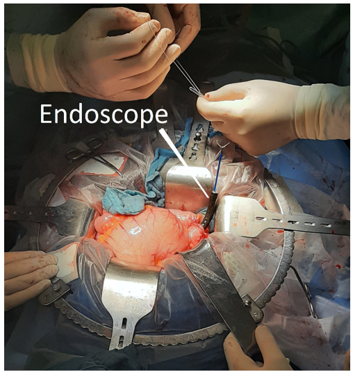
First, surgical resection of the duodenal segment was performed after exposure of the duodenum through the open abdomen, aided by intraoperative endoscopy to localise the adenoma. After the posterior wall of the jejuno-duodenotomy was anastomosed, a dOFD was placed transnasally using an endoscopic surgical rendezvous pull-through technique. The film-coated drainage element was placed at the level of the anastomosis (Figure 3)(Video). Available at: https://ijclinmedcasereports.com/pdf/Video123.mp4

Figure 3: View of the anastomosis of the duodenum (Duod) after performing the posterior wall of the anastomosis. The dOFd has been inserted with a pullthrough technic. The film covered drainagelement of the OFD can be seen through the open wound edges of the anterior wall of the anastomosis. The feeding tube was guided by the surgeon into the jejunum.

Video: The video image shows the preparation for the rendezvous maneuver. The video demonstrates a schematic clip of the principle of action of the PINPD, the construction of a dOFD, the pullthrough insertion technic of the dOFD and the fluoroscopic contrast and the endoscopy of the anastomosis.
Available at: https://ijclinmedcasereports.com/pdf/Video123.mp4
The feeding tube was digitally inserted into the jejunum to complete the positioning of the dOFD. The anterior wall was closed to complete the anastomosis. Negative pressure of 125 mmHg was applied using an electronic pump (ACTIV.A.C; KCI, San Antonio, Texas, USA). This collapsed the small bowel lumen and allowed permanent aspiration of biliary and pancreatic duodenal fluids, thus preventing digestive secretions from entering the anastomosis during the initial vulnerable period of anastomotic healing. Nutrition was provided via the integrated feeding tube (Figure 4).

Figure 4: The schematic figure demonstrates the pre-emptive intraluminal negative pressure therapy in the duodenum. After the surgical procedure on the duodenum, a dOFD is inserted transnasally into the duodenum. The film wrapped drainageelement (DE) covers the anastomotic region (A). On the film coated DE negative pressure can be applied to drain the duodenal secretions continuously (green arrows). Simultaneously, the patient can be fed via the integrated feeding tube (iT).
PINPT was performed for 7 days. Sufficiency of the anastomosis was demonstrated by fluoroscopic contrast injection through the feeding tube during removal of the dOFD (Figure 5) (video). Available at: https://ijclinmedcasereports.com/pdf/Video123.mp4

Figure 5: After removal of the dOFd. dOFD is still connected with the negative pressure pump (NPP). Drainageelement (DE), integrated jejunal feeding tube iT, blocked ventilation channel (B).
Oral feeding was started with a soft diet and anastomotic healing was checked endoscopically on days 1, 3 and 7 after the end of PINPT. The video demonstrates a schematic clip of the principle of action of the PINPD, the construction of a dOFD, the pullthrough insertion technic of the dOFD and the fluoroscopic contrast and the endoscopy of the anastomosis
Current Status
Preemptive ENPT is a further development of ENPT. A first meta-analysis has already described several indications [9].
Using a case report, we demonstrate that ENPT can also be used preemptively in the duodenum. We used a dOFD, which we have been using since 2017 for preemptive active reflux drainage (PARD) in esophagectomy [5,6]. We are now introducing PINPT, a new method for prophylaxis of anastomotic healing in the duodenum.
Conflict of Interest: Gunnar Loske is a consultant for Lohmann & Rauscher GmbH & Co.KG.His patents for negative pressure therapy have been transferred to Lohmann & Rauscher GmbH & Co.KG. A conflict of interest exists due to a financial interest in several negative pressure therapy products currently being launched.
Johannes Müller, Martin Zeile, Eckard Martens and Christian Theodor Müller declare no conflict of interest.
Funding: For this work no funds were received from any organization.
Acknowledgment: We like to thank Selda Schwarzhaupt and the nursing staff of the interdisciplinary endoscopic unit and the surgical department of the Marienkrankenhaus Hamburg for their excellent technical assistance. We gratefully acknowledge the support of the Department of Anesthesiology and Intensive Care Medicine.
References
- Pattynama LMD, Pouw RE, Henegouwen MIVB, et al. Endoscopic vacuum therapy for anastomotic leakage after upper gastrointestinal surgery. Endoscopy, 2023; 55(11): 1019-1025.
- Loske G, Rucktaeschel F, Schorsch T, et al. Endoscopic Negative Pressure Therapy (ENPT) for duodenal leakage - novel repair technique using open-pore film (OFD) and polyurethane-foam drainages (OPD). Endosc Int Open, 2019; 7(11): E1424-E1431.
- Wichmann D, Jansen KT, Onken, et al. Endoscopic negative pressure therapy as stand-alone treatment for perforated duodenal diverticulum: presentation of two cases. BMC Gastroenterol, 2021; 21(1): 436.
- Müller PC, Morell B, Vetter D, Raptis DA, Kapp JR, Gubler C, et al. Preemptive Endoluminal Vacuum Therapy to Reduce Morbidity After Minimally Invasive Ivor Lewis Esophagectomy: Including a Novel Grading System for Postoperative Endoscopic Assessment of GI-Anastomoses. Ann Surg, 2021; 274(5): 751-757.
- Loske G, Müller J, Schulze W, et al. Pre-emptive Active Drainage of Reflux (PARD) in Ivor-Lewis oesophagectomy with negative pressure and simultaneous enteral nutrition using a double-lumen open-pore film drain (dOFD). Surg Endosc, 2022; 36(3): 2208-2216.
- Loske G, Müller J, Schulze W, et al. Endoscopic negative pressure treatment: From management of complications to pre-emptive active reflux drainage in abdominothoracic esophageal resection-A new safety concept for esophageal surgery]. Chirurgie (Heidelb), 2023. doi: 10.1007/s00104-023-01970-2.
- Kouladouros K, Wichmann D, Loske G. The role of open-pore film drainage systems in endoscopic vacuum therapy: current status and review of the literature. Visc Med, 2023.
- Loske G, Schorsch T, Rucktaeschel F, et al. Open-pore Film Drainage (OFD): a new multipurpose tool for endoscopic negative pressure therapy (ENPT). Endosc Int Open, 2018; 6: E865-E871.
- Adamenko O, Ferrari C, Seewald S, et al. Prophylactic endoluminal vacuum therapy after major gastrointestinal surgery: a systematic review. Updates Surg, 2022; 74(4): 1177-1186.

