An Interesting Rare Case of Dual Sigmoid Volvulus
Khadija Kamal, Kamal Benzidane*, Lamiaa Mourafi, Abdelhak.Ettaoussi, Abdessamad Majd, Mounir Bouali, Abdellillah Elbakouri and Khalid El Hattabi
Hassan II University of Casablanca, Morocco Institution: Department of Visceral Surgical Emergency, Ibn Rochd University Hospital, Casablanca, Morocco
Received Date: 08/02/2025; Published Date: 14/03/2025
*Corresponding author: Kamal Benzidane, Hassan II University of Casablanca, Morocco, Institution: Department of Visceral Surgical, Ibn Rochd University Hospital, Casablanca, Morocco
Abstract
Colon volvulus is a significant cause of intestinal obstruction, predominantly affecting the sigmoid colon, while the transverse colon is rarely involved. A dual colonic volvulus, involving both sigmoid and transverse segments, is an uncommon presentation. Clinical presentation often includes occlusive syndrome and asymmetric abdominal distension. CT scans offer higher sensitivity, particularly with a double whirl sign. Management strategies for dual sigmoid colon volvulus prioritize surgical intervention in emergencies. Left hemicolectomy with delayed anastomosis is preferred to minimize complications.
Keywords: Dual colon volvulus; Colon volvulus; Intestinal obstruction; Surgical management
Introduction
Volvulus, is a word derived from a Latin word “volvere” which means a twist [1]. The sigmoid volvulus is a condition in which a part of the sigmoid colon wraps around itself and its own mesentery, causing a large bowel obstruction by strangulation may lead to ischemia and then necrosis [2,3].
The first case of sigmoid volvulus was described in 1836 by von Rokitansky [2]. It is the third leading cause of colonic obstruction worldwide, accounting for 3–5% of all acute bowel obstructions [4].
It remains particularly common in the geographic region known as the “Volvulus Belt” encompassing Africa, South America, Russia, the Middle East, and Eastern Europe determining [3,4].
The most commun site on colonic volvulus is the sigmoid colon by 60–75%, followed by the cecum in 25–40% of cases and the transverse colon in only 4% [4,5]. The dual location of the volvulus represents a rare situation and a major surgical emergency due to the high risk of gangrene and septic shock [6].
Due to the rarity of this clinical entity and the treatment options that are poorly codified, we present a case of a patient with double volvulus of the sigmoid colon.
Case Report
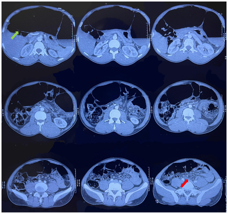
A 35-year-old patient, with no significant medical history, presented to the emergency department with an obstructive syndrome characterized by the cessation of bowel movements and gas, bilious and food vomiting, and pain with abdominal distension. This condition had been evolving for 3 days, along with a decline in the general state of health. The clinical examination revealed a conscious patient who was hemodynamically and respiratory stable, with the presence of an asymmetrical, tympanic, and non-tender distended abdomen. The hernial orifices were free, and the rectal exam revealed an empty ampulla. An abdominal CT scan, performed, showed colonic obstruction due to sigmoid volvulus, with distension of the sigmoid colon exhibiting a "coffee bean" sign, measuring 124 mm in diameter, along with the visualization of the whirlpool sign. There were no signs of digestive ischemia.

Figure 1: CT scan image showing the colonic sigmoid distension (green arrow) with the whirlpool sign (red arrow).
A hydro-electrolytic panel was done to check for any abnormalities and returned without issues.
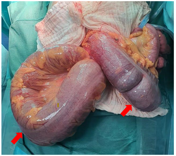
The patient was urgently admitted to the operating room. The intraoperative exploration revealed the presence of a dual volvulus of the sigmoid colon, without signs of ischemic damage and no involvement of the small bowel.

Figure 2: Per operative image showing the double volvulus of the sigmoid colon.
The procedure performed was a segmental left colonic resection, removing the two volvulated loops, followed by a double-barrel colostomy. Postoperative recovery was uncomplicated, with a functional stoma on postoperative day 1 and discharge on postoperative day 2. The pathological examination revealed no tumor lesions, and the restoration of colonic continuity was carried out 2 months after the initial procedure.
Discussion
Colon volvulus is a common cause of intestinal obstruction. The sigmoid colon is the segment most susceptible to this pathology; however, the transverse colon is the least affected segment. A colonic volvulus with dual localization, constitutes a rare entity of obstruction [7].
Colon volvulus has several factors that can be grouped into three categories:
- Congenital factors: intestinal malrotations, Hirschsprung’s disease, the lack of fixation of the right colon or splenic angle, shortening of the mesocolon [8,9].
- Anatomical factors leading to abnormal mobility of the colon: dolichocolon, associated with or without the megacolon or Chilaiditi syndrome, chronic inflammatory disease [10].
- Physiological factors: chronic constipation and/or overuse of laxatives leading to motility disturbances [11].
The etiological factors of colon volvulus are relatively the same regardless of the site. However, these different factors are often associated and have a synergistic effect in the increased mobility of the colon that predisposing it to the volvulus [12].
The two main problems in the colonic volvulus are the bowel obstuction and the vacular occlusion. The first one lead to the distention of the twisted loop due to mechanical obstruction and bacterial fermentation. On second place, the vascular occlusion and the colonic distention by decresing capillary perfusion due to the intra colonic pressure, lead to ichemia that lead to bacterial translocation and toxemia and to the colonic gangrene [13-18].
Clinically, the patient presents an occlusive syndrome, often accompanied by other symptoms such as vomiting and rectal bleeding. On abdominal examination, the patient presents an asymmetric abdominal distension on inspection and Von Wahl sign in palpation. The presence of rectal melanotic stool, rebound tenderness or muscular defense generally is a sign of complication like gangrene, perforation and peritonitis [19,20].
Plain abdominal radiography has been found diagnostic in 57%-90% of patients by showing various characteristic signs, notably omega or horseshoe sign, bird beak sign, inverted U sign, northern exposure sign, coffee bean sign. But all this signs are not found if there is a simultaneous volvulus [21-24].
The CT scan has better sensitivity and specificity in the diagnosis by showing the whirl sign. The presence of two concomitant whirl sign may suggest a simultaneous volvulus [25,26]. However the preoperative diagnosis of the simultaneous volvulus remains challenging and often discovered during the surgery [27,28].
The endoscopic derotation is the gold standard for sigmoid volvulus if there is no sign of necrosis or perforation, in second plan a colectomy. However, the first surgical approach is the most common due to the financial problem or lack of endoscopic equipment and it consist of a segmentary resection and colostomy or anastomosis but in emergency on an unprepared colon exposes to a lot of morbidities (fistula, sepsis) [25,29,30].
On the dual volvulus of the left colon, the surgical approch consist of left hemicolectomy with anastomosis if there is no necrosis and the patient is hemodynamically stable. Therefor delayed anastomosis seems to be the best option by reducing the risk of anastomotic leakage and morbidity [31-33].
Conclusion
In conclusion, colonic volvulus is a common cause of occlusion, but when it involves two segments, it becomes a rare entity that presents significant challenges in both diagnosis and treatment.
References
- Lianos G, Ignatiadou E, Lianou E, Anastasiadi Z, Fatouros M. Simultaneous volvulus of the transverse and sigmoid colon: case report. G Chir, 2012; 33(10): 324-326.
- Raveenthiran R, Madiba TE, Atamanalp SS, De U. Volvulus of the sigmoid colon. Colorectal Dis, 2010; 12: 1-17.
- Gingold D, Murrell Z. Management of colonic volvulus, Clin. Colon RectalSurg, 2012; 25(4): 236–244.
- Perrot L, Fohlen A, Alves A, Lubrano J. Management of the colonic volvulusin. J. Visc. Surg, 2016; 153(3): 183–192.
- Valsdottir E, Marks JH. Volvulus: small bowel and colon, Clin. Colon RectalSurg, 2008; 21(2): 91–93.
- Ndong A, et al. Synchronous sigmoid and transverse volvulus: A case report andqualitative systematic review. International Journal of Surgery Case Reports, 2020; 75: 297–301.
- Hellinger MD, Steinhagen RM, Colonic volvulus, in: Wolff BW, Fleshman JW, Beck DE, et al. The ASCRS Textbookof Colon and Rectal Surgery, 2nd ed., Springer, New York, 2008; pp. 291–294.
- Hoseini A, Eshragi Samani R, Parsamoin H, Jafari H. Synchronic volvulus ofsigmoid and transverse colon: a rare case of large bowel obstruction, Ann.Colorectal Res. 2014; 2(1): 1–2.
- Ballantyne GH. Review of Sigmoid Volvulus: History and Treatment Outcomes. Diseases of the Colon & Rectum, 1982; 25: 494-501. https://doi.org/10.1007/BF02553666.
- Hellinger MD, Steinhagen RM, Colonic volvulus, in: Wolff BG, Fleshman, Beck DE, Pemberton JH, et al. The ASCRS Textbookof Colon and Rectal Surgery, 2nd ed., Springer, New York, 2008; pp. 291–294.
- McBrearty A, Harris A, Gidwani A. Transverse-sigmoid colon knot: a rarecause of bowel obstruction, Ulster Med. J, 2011; 80(2): 107–108.
- Ndong A, et al. Synchronous sigmoid and transverse volvulus: A case report andqualitative systematic review. International Journal of Surgery Case Reports, 2020; 75: 297–301.
- Raveenthiran R, Madiba TE, Atamanalp SS, De U. Volvulus of the sigmoid colon. Colorectal Dis, 2010; 12: 1-17.
- Shepherd JJ. The epidemiology and clinical presentation of sigmoid volvulus. Br J Surg, 1969; 56: 353-359.
- Arigbabu AO, Badejo OA, Akinola DO. Colonoscopy in the emergency treatment of colonic volvulus in Nigeria. Dis Colon Rectum 1985,; 28: 795-798.
- Sinha RS. A clinical appraisal of volvulus of the pelvic colon with special reference to aetiology and treatment. Br J Surg, 1969; 56: 838-840.
- Gibney EJ. Volvulus of the sigmoid colon. Surg Gynecol Obstet, 1991; 173: 243-255.
- Madiba TE, Ramdial PK, Dada MA, Mokoena TR. Histological evidence of hypertrophy and ischemia in sigmoid volvulus among Africans. East Afr Med J, 1999; 76: 381-384.
- Bak MP, Boley SJ. Sigmoid volvulus in elderly patients. Am J Surg, 1986; 151: 71-75.
- Pahlman L, Enblad P, Rudberg C, Krog M. Volvulus of the colon. Acta Chir Scand, 1989; 155: 53-56.
- Mbengue A, Ndiaye A, Maher S, Schmutz G, Ranchoup Y, Blum A, et al. Imagerie des occlusions intestinales hautes de l’adulte, Feuillets Radiol, 2016; 56(5): 265–296.
- Avots-Avotins KV, Waugh DE. Colon volvulus and geriatric patient. Surg Clin North Am, 1982; 62: 249-260.
- Javors BR, Baker SR, Miller JA. The northern exposure sign: a newly described fi nding in the sigmoid volvulus. AJR, 1999; 173: 571-574.
- Feldman D. The coff ee bean sign. Radiology, 2000; 216: 178-179.
- Katsanos K, Ignatiadou E, Markouizos G, Doukas M, Siafakas M, Fatouros M, et al., Non-toxic megacolon due to transverse and sigmoid colon volvulus in a patient with ulcerative colitis, J. Crohns Colitis, 2009; 3(1): 38–41.
- McBrearty A, Harris A, Gidwani A. Transverse-sigmoid colon knot: a rare cause of bowel obstruction, Ulster Med. J, 2011; 80(2): 107–108.
- Wisler JR, Stawicki SP. Interesting clinical image: colonic double twist, OPUS 12 Sci, 2009; 3: 58–59.
- Lianos G, Ignatiadou E, Lianou E, Anastasiadi Z, Fatouros M. Simultaneous volvulus of the transverse and sigmoid colon: case report, Il G Chir, 2012; 33(10): 324–326.
- Oren D, Atamanalp SS, Aydinli B, et al. An algorithm for the management of sigmoid colon volvulus and the safety of primary resection: experience with 827 cases. Dis Colon Rectum, 2007; 50: 489-497.
- Madiba TE, Thomson SR. The management of sigmoid volvulus. J R Coll Surg Edinb, 2000; 45: 74-80.
- Lianos G, Ignantiadou E, Lianou E, et al. Simultaneous Volvulus of the Transverse and Sigmoid Colon: Case Report. G. Chir, 2012; 33: 324-326.
- Ndong A, et al. International Journal of Surgery Case Reports, 2020; 75: 297–301.
- Touré CT, Dieng M, Mbaye M, Sanou A, Ngom G, Ndiaye A, et al., Résultats de la colectomie en urgence dans le traitement du volvulus du côlon au centre hospitalier universitaire (CHU) de Dakar, Ann. Chir, 2003; 128(2): 98–101.

