Laparoscopic Non-Tension Hernioplastic of Lower Lumbar Hernia
Igor Černi*
Department of General and Abdominal Surgery, General and Teaching Hospital Celje, Slovenia
Received Date: 07/12/2024; Published Date: 09/01/2025
*Corresponding author: Igor Černi, MD, MS, Department of General and Abdominal Surgery, General and Teaching Hospital Celje, Slovenia
Background
Lumbar or petit hernia is a rare condition, traditional open operative treatment with reparation of hernia opening and surrounding tissue is difficult, painful, and with uncertain outcome.
Because of their rarity and complex anatomical location, they can pose a formidable challenge to surgeons. The challenges start with diagnosis and continue to the selection of treatment [1,2]. In this report, our aim is to present case report of Petit’s hernia.
Petit’s hernia is described as herniation of retroperitoneal fat through the aponeurosis of the internal abdominal oblique muscle between the erector spinae muscles in the inferior lumbar triangle. The neck of this hernia is usually large, and therefore, it has a lower risk of strangulation than other hernias [1]. Grynfeltt hernia is described as herniation of retroperitoneal fat through the aponeurosis of the transversalis muscle between the erector spinae muscles and internal oblique muscles in the superior lumbar triangle [2].
Lumbar hernias are rare lesions that are usually observed after trauma or surgery.
Symptoms and presentation of lumbar hernias can vary. They are frequently asymptomatic or may cause back pain in the sciatic nerve distribution area with or without a palpable mass. According to Light, it is the most probable diagnosis in young women and athletes with back pain [7].
Patient and Treatment
This is an attempt to present a 51-year-old female patient with painful lump in the left lumbar region suspended over the cervical ridge edge in standing position. Hernia proved to be reponible, and confirmed by ultrasound. (Figures 1, 2).
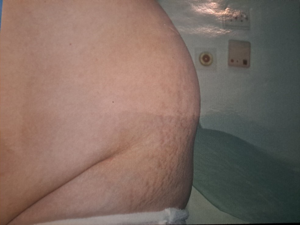
Figure 1: Lower lumbar hernia.
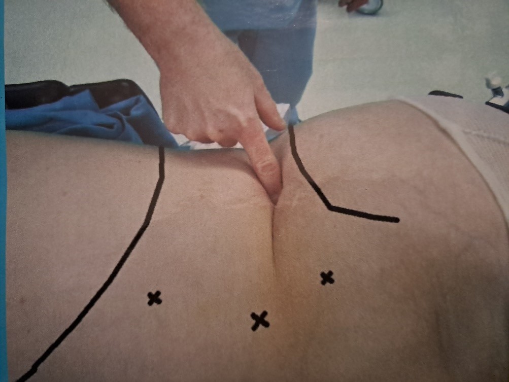
Figure 2: Port placement.
Laparoscopy was performed in general anaesthesia, accessed the retroperitoneal area, de-prepared hernial sack, and closed the muscular defect with PTFE screen. Operative surgery was concluded with the reparation of peritoneum (Figures 3, 4, 5).
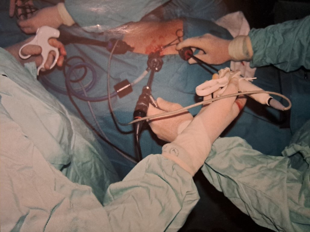
Figure 3: Laparoscopy.
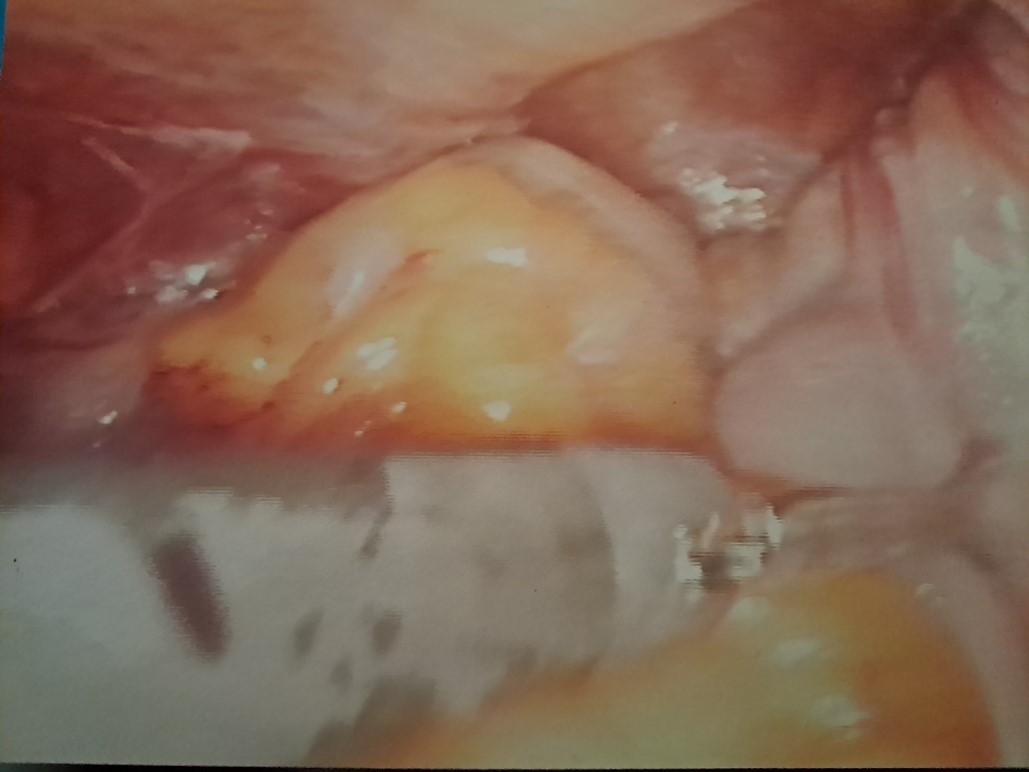
Figure 4: Hernia sac.
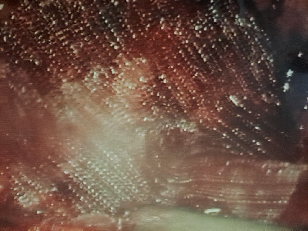
Figure 5: Mesh placement.
After the surgery the patient exhibited no difficulties, got out of bed on the very day of the surgery, and left the hospital two days later. Subsequent control indicated no problems (Figure 6).
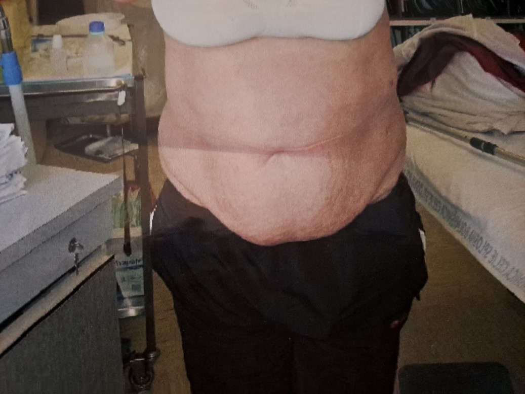
Figure 6: Patient after the operation.
Conclusion
Lumbar hernias are rare clinical entities and need suspicion to be diagnosed. Imaging studies, particularly UZ or CT, are useful in defining the anatomy and contents. Reconstruction can be achieved with synthetic mesh repair; this can be accomplished by either open or endoscopic methods with minor complications.Laparoscopic approach to the lumbar hernia reparation is simple, safe and very comfortable for patient.
References
- Petit JL. Traite des maladies chirurgicales, et des operations qui leur convenient. Vol 2. T.F. Didot; Paris: 1774, pp. 256–258.
- Grynfeltt J. Quelque mots sur la hernie lombaire. Montpellier Med, 1866; 16: 329.
- Suarez S, Hernandez JD. Laparoscopic repair of a lumbar hernia: report of a case and extensive review of the literature. Surg Endosc, 2013; 27: 3421–3429. doi: 10.1007/s00464-013-2884-9. https://doi.org/10.1007/s00464-013-2884-9.
- Saylam B, Duzgun AP, Ozer MV, Coskun F. Acute traumatic abdominal wall hernia after blunt trauma: A case report. Ulus Cerrahi Derg, 2011; 27: 43–45. https://doi.org/10.5097/1300-0705.UCD.502-10.00.
- Moreno-Egea A, Baena EG, Calle MC, Martínez JA, Albasini JL. Controversies in the current management of lumbar hernias. Arch Surg, 2007; 142: 82–88. doi: 10.1001/archsurg.142.1.82. https://doi.org/10.1001/archsurg.142.1.82.
- Mismar A, Al-Ardah M, Albsoul N, Younes N. Underlay mesh repair for spontaneous lumbar hernia. Int J Surg Case Rep, 2013; 4: 534–536. doi: 10.1016/j.ijscr.2013.03.005. https://doi.org/10.1016/j.ijscr.2013.03.005.
- Light HG. Hernia of the inferior lumbar space. A cause of back pain. Arch Surg, 1983; 118: 1077–1080. doi: 10.1001/archsurg.1983.01390090061014; https://doi.org/10.1001/archsurg.1983.01390090061014.
- Bigolin AV, Rodrigues AP, Trevisan CG, Geist AB, Coral RV, Rinaldi N, et al. Petit lumbar hernia-a double-layer technique for tension-free repair. Int Surg, 2014; 99: 556–559. doi: 10.9738/INTSURG-D-13-00135.1. https://doi.org/10.9738/INTSURG-D-13-00135.1.

