Uncommon Pancreatic Metastasis 10 Years After Nephrectomy in Clear Cell Renal Carcinoma
Joud Boutaleb1,*, Sarah Loubaris1, Amine Lalaoui Salim2, Ouijdane Zamani1, Rachida Saouab1 and Jamal El Fenni1
1Radiology Department Mohammed Vth military hospital, Rabat, Morocco
2Urology department, CHU- IBN SINA, Rabat, Morocco
Received Date: 30/11/2024; Published Date: 07/01/2025
*Corresponding author: Joud Boutaleb, Radiology Department Mohammed Vth military hospital, Rabat, Morocco
Introduction
Adult kidney cancer represents approximately 3% of all malignancies and ranks third among urological cancers, following prostate and bladder cancers. While the most common sites for metastasis include the lungs, bones, liver, and brain, pancreatic metastases are relatively rare. This report presents a case of pancreatic metastasis from renal cell carcinoma (RCC), which is an uncommon occurrence [1].
Case Report
A 56-year-old female patient with a history of clear-cell Renal Carcinoma (RCC) treated 10 years earlier presented with progressive weight loss and epigastric pain over the past week. A contrast-enhanced CT scan was performed to further evaluate the symptoms.
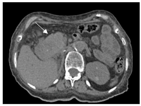
Imaging revealed a large, rounded mass with lobulated contours located in the head of the pancreas. The mass demonstrated significant enhancement following contrast administration (Figure 1, 2), measuring 67 x 51 x 37 mm (height x width x anterior-posterior). Notably, the mass caused dilation of both the extra- and intra-hepatic bile ducts, as well as the pancreatic duct (Wirsung’s duct), with a corresponding atrophy of the downstream pancreas. Additionally, a necrotic, heterogeneous mass was observed in the left adrenal gland.
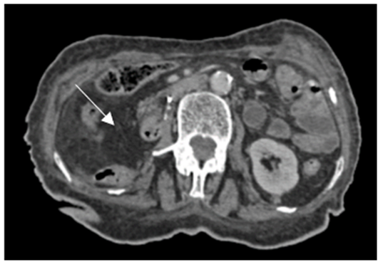
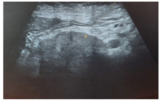
The right nephrectomy bed was unremarkable, with the contralateral kidney appearing normal (Figure 3). Ultrasound and biopsy were performed, confirming the metachronous nature of the pancreatic mass as originating from the prior renal carcinoma (Figure 4, 5). Subsequently, the patient underwent surgical resection of the pancreatic mass with curative intent.

Figure 1: Axial CT scan showing a mass in the head of the pancreas (C-phase) with lobulated contours.

Figure 2: (A) Axial CT scan revealing a hypervascular mass in the pancreas; (B) Coronal CT section demonstrating the hypervascular mass in the pancreatic head.

Figure 3: Axial CT scan illustrating the empty right nephrectomy bed.

Figure 4: Ultrasound showing a heterogeneous, hypoechoic mass in the head of the pancreas.

Figure 5: Histopathological image of the clear-cell adenocarcinoma in the pancreatic tissue.
Discussion
Clear cell renal carcinoma typically metastasizes to the lungs, bones, and liver. Pancreatic metastases from RCC are rare, with a latency period of several years following nephrectomy, typically ranging from 10 to 27 years [2-3]. The spread to the pancreas may occur through lymphatic pathways, connecting the pancreas to the renal arteries [4], or via venous routes, particularly through portocaval shunts associated with renal tumors [5].
While many patients with pancreatic metastasis from RCC remain asymptomatic, common clinical presentations include abdominal pain (often epigastric), jaundice, weight loss, palpable masses, and, in some cases, duodenal invasion leading to gastrointestinal hemorrhage [6].
Radiologically, pancreatic metastases from RCC can present as hypodense lesions with well-defined borders and intense, heterogeneous enhancement after contrast administration [7]. These lesions may also contain hypodense areas, which is a characteristic finding on CT imaging. On ultrasound, such lesions are typically hypoechoic, although cystic forms can occasionally be observed [7]. The differential diagnosis includes pancreatic neuroendocrine tumors, which require careful consideration in imaging [8].
The definitive diagnosis is usually made through histopathological examination following surgical resection or biopsy, as was performed in this case [9,10]. Surgical management is the treatment of choice for solitary pancreatic metastases from RCC. Common procedures include cephalic duodenopancreatectomy or left-sided pancreatectomy. In cases of multiple pancreatic metastases, subtotal pancreatectomy may be necessary [11].
The prognosis following surgical resection of pancreatic metastases from RCC is generally favorable, with reported 5-year survival rates ranging from 34% to 88%.
Conclusion
Pancreatic metastases from clear cell renal carcinoma are rare but may manifest several years after nephrectomy. Surgical resection remains the most effective treatment modality, offering the potential for improved survival and quality of life.
Author contributions: All authors contributed equally to this work.
Conflict of Interest: The authors declare that they have no conflicts with this manuscript.
Ethics approval: Our institution does not require ethical approval for reporting individual cases or case series.
Informed consent: Written informed consent was obtained from a legally authorized representative(s) for anonymized patient information to be published in this article.
References
- Méjean A, Lebret T. Management of renal cancer metastases. Atypical metastatic sites of renal cancer. Prog Urol, 2008; 18(7). doi: 10.1016/S1166-7087(08)74561-5.
- Robbins EG, Franceschi D, Barkin JS. Solitary metastatic tumors to the pancreas: a case report and review of the literature. Am J Gastroenterol, 1996; 91: 2414–2417.
- Fullarton GM, Burgoyne M. Gallbladder and pancreatic metastases from bilateral renal carcinoma presenting with hemobilia and anemia. Urology, 1991; 38:184–186.
- Nagakawa T, Konishi I, Ueno K, Ohta T, Kayahara M. A clinical study on lymphatic flow in carcinoma of the pancreatic head area, peripancreatic regional lymph node grouping. Hepatogastroenterology, 1993; 40: 457–462.
- Saitoh H, Yoshida K, Uchijima Y, Kobayashi N, Sawata J, Nakame Y. Possible metastatic routes via portacaval shunts in renal adenocarcinoma with liver metastases. Urology, 1991; 37: 598–601.
- P Roland CF, Van Heerden JA. Nonpancreatic primary tumors with metastasis to the pancreas. Surg Gynecol Obstet, 1989; 168: 345–347.
- Ng CS, Loyer EM, Lyer RB, David CL, Dubrow RA, Charnsangavej C. Metastases to the pancreas from renal cell carcinoma: findings on three-phase contrast enhanced helical CT. Am J Roentgenol, 1999; 172: 1555–1559.
- Ward EM, Stephens DH, Sheedy PF. Computed tomographic characteristics of pancreatic carcinoma: an analysis of 100 cases. Radiographics, 1983; 3: 547–565.
- Faure JP, Richer JP, Irani J, et al. Renal cancer and late pancreatic metastases. Report of 3 cases and review of the literature. Prog Urol, 1998; 8: 404-407.
- Machado NO, Chopra P. Pancreatic Metastasis from renal carcinoma managed by whipple resection pattern, Surgical Management and Outcome. A Case Report and Literature Review of Metastatic JOP. J Pancreas, 2009; 10: 413-418.
- Paparel P, Cotton F, Voiglio E, et al. A case of late pancreatic metastasis from clear cell renal carcinoma. Prog Urol, 2004; 14: 403-405.

