Emphysematous Cholecystitis
Soukaina Bahha*, Asmae Guennouni, Salma EL Aouadi, Chaimae Abourak, Fatima-Zahra Laamrani and Laila Jroundi
Radiology Department, Ibn Sina Hospital, Rabat, Morocco
Received Date: 28/11/2024; Published Date: 06/01/2025
*Corresponding author: Soukaina BAHHA, Radiology Department, Ibn Sina Hospital, Rabat, Rabat, Morocco
ORCID: https://orcid.org/0009-0007-9343-0999
Abstract
Emphysematous Cholecystitis (EC) is a rare but severe form of acute cholecystitis characterized by the presence of gas within the gallbladder wall and/or lumen. It is primarily caused by infection with gas-producing bacteria, most commonly Clostridium species, Escherichia coli, and Klebsiella spp. EC is more common in elderly males and is strongly associated with underlying conditions such as diabetes mellitus, immunocompromised states, and biliary obstruction. The clinical presentation can range from mild abdominal pain to septic shock, making early diagnosis challenging. Imaging, particularly abdominal CT and MRI, is crucial for identifying gas within the gallbladder and assessing complications like abscesses or perforation. Prompt initiation of broad-spectrum antibiotics and urgent cholecystectomy are essential for improving outcomes. Given the high risk of severe infection and complications, early and aggressive management is critical, especially in high-risk patients. With appropriate treatment, many patients recover fully, although delays in diagnosis and treatment can result in high morbidity and mortality.
Keywords: Emphysematous cholecystitis; CT scan; Imaging; Cholecystostomy
Introduction
Emphysematous Cholecystitis (EC) is a rare but serious form of acute cholecystitis characterized by the presence of gas within the gallbladder wall and/or lumen. This condition is often associated with a higher risk of complications and a more severe clinical course compared to typical acute cholecystitis. The gas is primarily caused by bacterial infection, typically involving gas-producing organisms such as Clostridium species, Escherichia coli, or Klebsiella spp. Due to its distinctive imaging features and the associated clinical severity, emphysematous cholecystitis requires timely diagnosis and prompt treatment to prevent life-threatening complications.
Case Report
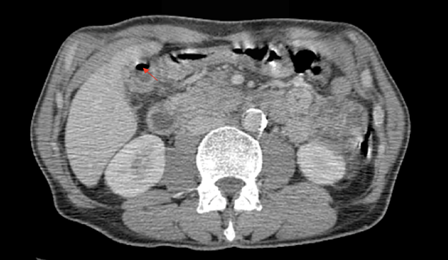
The patient is a 54-year-old man with a history of type 2 diabetes, who presents to the emergency department with right upper quadrant abdominal pain lasting for 4 days, accompanied by fever and vomiting. His laboratory results show a white blood cell count of 14,000, predominantly neutrophils, a CRP of 150, and abnormal liver function tests with moderate hepatocellular damage. A CT scan was performed, revealing a gallbladder wall thickened to 5mm, containing air, The patient was administered intravenous piperacillin-tazobactam (4g) as empirical antibiotic therapy and subsequently underwent an urgent cholecystectomy, which proceeded without any complications.

Figure: An abdominal contrast-enhanced axial CT scan shows a gallbladder with a thickened wall measuring 5mm, containing air (Red arrow), and associated with perivesicular fluid collection.
Discussion
Emphysematous cholecystitis was first described by Stoltz A. in 1901[1]. The symptoms of emphysematous cholecystitis are often challenging to differentiate from those of uncomplicated acute cholecystitis, other acute right upper abdominal conditions, or amoebic liver abscess, making diagnosis difficult. The presentation can vary widely, ranging from mild abdominal pain to sepsis and shock, depending on the severity and progression of the disease. Emphysematous cholecystitis is more common in males than females, with a male-to-female ratio of 7:3. Additionally, approximately 40% of patients affected by this condition have diabetes mellitus [2].
The diagnosis of emphysematous cholecystitis (EC) relies on the detection of gas in the gallbladder lumen, wall, and sometimes the bile ducts. In radiographic imaging, intraluminal gas appears as one or more round bubbles or a pear-shaped area of lucency in the right upper quadrant on a supine X-ray. On erect or decubitus films, an air-fluid level within the gallbladder may also be visible. CT imaging is the most sensitive modality for identifying intraluminal or intramural gas in the gallbladder. It is also helpful in assessing complications such as abscess formation and pericholecystic inflammatory changes [3].
Magnetic Resonance Imaging (MRI) can offer detailed insights into intramural necrosis and intraluminal gas. Gas within the gallbladder lumen and wall typically appears as floating, single void areas in the upper portions of the gallbladder [4].
Cholecystectomy is the definitive treatment for emphysematous cholecystitis. Emphysematous cholecystitis generally carries with it a poorer prognosis, and the mortality rate in emphysematous cholecystitis as high as 25%, compared with 4% in acute cholecystitis [5].
Conclusion
Emphysematous cholecystitis is a life-threatening condition requiring prompt diagnosis and intervention. Early imaging, broad-spectrum antibiotics, and urgent surgery are essential for better outcomes. Due to its association with severe infection and high mortality, aggressive management is crucial, especially in high-risk patients. Timely treatment can lead to full recovery and prevent serious complications.
References
- Lallemand B, De Keuleneer R, Maassarani F. Emphysematous cholecystitis, Acta Chir. , 2003; 103(no 2): p. 230‑232. doi: 10.1080/00015458.2003.11679413.
- Miyahara H, Shida D, Matsunaga H, Takahama Y, Miyamoto S. Emphysematous cholecystitis with massive gas in the abdominal cavity. World J. Gastroenterol. WJG, 2013; 19(no4): p. 604. doi: 10.3748/wjg.v19.i4.604.
- Khare S, Pujahari AK. A Rare Case of Emphysematous Cholecystitis. Clin. Diagn. Res. JCDR, 2015; 9(no 9): p. PD13. doi: 10.7860/JCDR/2015/10972.6463.
- Watanabe Y, et al. MR imaging of acute biliary disorders. Rev. Publ. Radiol. Soc. N. Am. Inc, 2007; 27(no2): p. 477‑495. doi: 10.1148/rg.272055148.
- Smith EA, Dillman JR, Elsayes KM, Menias CO, Bude RO. Cross-sectional imaging of acute and chronic gallbladder inflammatory disease. AJR Am. J. Roentgenol., 2009; 192(no1): p. 188‑196. doi: 10.2214/AJR.07.3803.

