Retrocaval Ureter, About 2 Cases
Ali Hannaoui, Anas Tmiri*, Adil Kbirou, Amine Moataz, Mohamed Dakir, Adil Debbagh and Rachid Aboutaieb
Department of Urology, University hospital center Ibn Rochd, Casablanca, Morocco
Received Date: 02/12/2024; Published Date: 31/12/2024
*Corresponding author: Anas Tmiri, Department of Urology, University hospital center Ibn Rochd, Casablanca, Morocco; Faculty of Medicine and Pharmacy, Hassan II University, Casablanca, Morocco
Abstract
Urinary tract malformations are multiple. Often these anomalies lead to chronic obstructions of the urinary tract and the formation of stones. Retrocaval ureter is a rare congenital malformation, characterized by a spiral trajectory of the right ureter around the inferior vena cava. It is an anomaly of the development of the inferior vena cava and not of the right superior excretory tract. The diagnosis is radiological and its treatment consists of a resection with anterization and reanastomosis of the retrocaval ureter by open surgery or laparoscopically.
We report two cases of retrocaval ureter treated by two different surgical techniques.
Introduction
Retrocaval ureter is a rare congenital malformation in which the ureter spirals around the inferior vena cava. This abnormality results from an irregularity in the embryological development of the inferior vena cava, not the ureter itself [1,2].
It affects about 0.9 per 1000 births and is three times more common in men than in women. This malformation can lead to obstruction of the ureter and cause hydronephrosis. Diagnosis is radiological, and treatment generally consists of surgical management1 2.
We describe two cases of retrocaval ureter, treated by two different surgical techniques.
Cases Presentation
Case 1:
A 19-year-old male presented to the outpatient department of urology at our hospital with intermittent right lower back pain for the past three months. His medical history was remarkable. There was no hematuria or other urinary tract symptoms. General physical examination revealed mild tenderness in the right flank. Other systems were normal.
Biologically, renal function was 7 mg/l, CRP negative and CBEU was sterile; other biological parameters were normal.
Abdominal ultrasound showed right hydronephrosis. The initial Uro-CT objectified: ureterohydronephrosis upstream of lumbar ureteral stenosis. In view of the hyperalgesic nature of the lumbar pain, the patient underwent a right ureteral endoprosthesis.
A second Uro-CT objectified: a discrete pyelocalic and right ureteral dilatation with an initial retrocaval and interaortocaval course of the ureter, resuming its normal course at the iliac and pelvic level (Figure 1).

Figure 1: Uro-CT objectified a discrete pyelocalic and right ureteral dilatation with an initial retrocaval and interaortocaval course of the ureter.
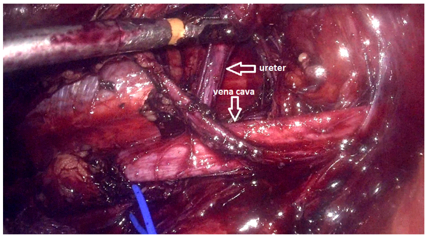
Based on these results, we decided to operate on the patient by transperitoneal laparoscopy. After locating the pelvis and the right ureter, which was dilated, the segment of the retrocaval ureter was located lower than the pyelo-ureteral junction. We sectioned the pathological segment (∼3 cm) of the ureter (Figure 2).
We then performed a uretero-ureteral anastomosis with insertion of a double-j stent (Figure 3).

Figure 2: Segment of the retrocaval ureter.

Figure 3: Uretero-ureteral anastomosis after sectioning the stenotic area.
Case 2:
36-year-old patient, without any particular pathological history, who presented with chronic right lower back pain without other accompanying signs, the clinical examination was normal
The preoperative assessment was unremarkable with correct renal function.
Abdominal ultrasound was performed, revealing right ureterohydronephrosis with no detectable obstacle (Figure 4).

Figure 4: Abdominal ultrasound objectified right ureterohydronephrosis.
Uro-CT revealed a retrocaval ureter responsible for major ureterohydronephrosis (Figure 5).

Figure 5: Uro-CT showing major ureterohydronephrosis following a retrocaval ureter.
Intravenous urography revealed a disparity in caliber between the upper lumbar ureter and the retrocaval portion (Figure 6).

Figure 6: Intravenous urography revealed a disparity in the caliber of the ureter at its lumbar portion.
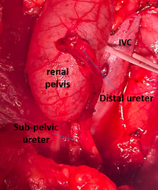
The patient underwent open surgery for ureteral resection with anteriorization and reanastomosis, a junction syndrome was associated for which a resection with creation of a new junction was performed with placement of a double J catheter (Figures 7, 8, 9, 10).

Figure 7: Retrocaval ureteral.

Figure 8: After resection of the retrocaval portion and the presence of pyeloureteral junction syndrome.

Figure 9: The pyeloureteral junction after pyeloplasty.

Figure 10: The double J catheter in place.
The post-operative follow-up was simple with good clinical and paraclinical progress.
Discussion
Retrocaval (circumcaval or postcaval) ureter is a rare congenital anomaly of the relationship of the inferior vena cava and the ureter where the infrarenal segment of the inferior vena cava is anterior to the embryologically normal ureter. Retrocaval ureter is usually associated with some degree of ureteral obstruction. It manifests mainly by right flank pain, urinary tract infection and stone formation. The demonstrated incidence of retrocaval ureter is 0.9—2 per 1000 cases, being 2.8 to 4 times more frequent in the male population. Given the increasing number of complications, it is treated, in most cases, by surgical intervention [3].
The first radiological paraclinical examination prescribed in the presence of painful symptoms of the right flank should be renal ultrasound followed by CT scan. In the past, excretory urography, retrograde ureteropyelography according to Chevassu and cavography were considered to be the main diagnostic methods for retrocaval ureter. The result of excretory urography that suggests the existence of retrocaval ureter is a deviation of the lumbar ureter with hydronephrosis. Since the median deviation of the lumbar ureter can be caused by retroperitoneal fibrosis, retroperitoneal mass and previous surgical interventions, it was necessary to confirm the diagnosis by retrograde ureteropyelography according to Chevassu and by cavography of the inferior vena cavav [3].
There are two types of retrocaval ureter, types 1 and 2. Type 1 is more common; According to the experience of some authors, type 1 occurs in 93 to 94% of cases; The course of the ureter is normal up to L3 height, Thereafter takes a superomedial direction forming a fishhook or inverted J appearance, The ureter then passes behind the inferior vena cava, goes around it and appears on its medial edge progressing towards the anterior position relative to the right iliac artery and entering the bladder orthotopically. Pronounced hydronephrosis occurs in approximately 40 to 50% of patients. Type 2 is found in 6-7% of cases. The renal pelvis and the initial segment of the ureter occupy an almost horizontal portion, The location of the median deviation of the ureter is higher than that of type 1, The curvature that the ureter forms when passing behind the inferior vena cava is slight and thus takes the shape of a sickle. Hydronephrosis is less pronounced [3-5].
Retrocaval ureter is most commonly treated surgically, depending on the complications and their progression. Several surgical techniques have been described including ureteral resection with anterization and reanastomosis; resection and reanastomosis; resection and reanastomosis of the inferior vena cava; the vena cava supporter; nephrectomy; laparoscopic surgery. There is a growing number of authors who describe the practice of laparoscopic reconstructive techniques including the "automatic suture device". Many of them believe that laparoscopic surgery should be one of the therapies of choice in the surgical treatment of retrocaval ureter. It has advantages over conventional open surgery: it is minimally invasive, the convalescence period is shorter and the cosmetic effect is better [3-5].
Conclusion
Retrocaval ureter is a rare congenital malformation that can lead to ureteral obstruction and hydronephrosis. The diagnosis is radiological, the initial methods are abdominal ultrasound and uro-CT. Inferior vena cava cavography is only applicable in exceptional cases. Although classical open surgical treatment has satisfactory results, laparoscopic surgery offers many advantages including a less invasive approach and good functional results.
References
- Tembely A, Diarra A, Berthé H, Diakité ML, Ouattara K. Uretere Retrocave: Deux Nouvelles Observations à L’hopital Du Point G A Bamako. African Journal of Urology, 2014; 20(2): 104-107. doi: 10.1016/j.afju.2013.11.007
- Cornu J-N, Sèbe P. Uretère rétrocave. EMC Urologie, 18-158-A-10, 2011.
- Hadzi-Djokic J, et al. Uretère rétrocave : à propos de 16 cas - ScienceDirect, 2024.
- Masson E. Uretère rétrocave. EM-Consulte, 2024.
- Kakanou A, Nchimi A, Ghuysen MS, Khamis J, Khuc T. Uretère rétrocave chez un enfant de dix ans. Annales de Chirurgie, 2001; 126(2): 156-158. doi: 10.1016/S0003-3944(00)00481-8.

