An Unusual Case of Giant Cell Tumor in a Patient with Pyknodysostosis
Navin Patil1,*, Shanti Gurung2, Ram Bhutani3, Marie Affana4, Noodee Al Khinalie5, Karim Slim5, William O Olotu5, Afolabi Deji Gabriel5 and Kristel Ferrol5
1Professor and Dean of Basic Sciences, All Saints University School of Medicine, Dominica.
2Associate Professor and Course Director, All Saints University School of Medicine, Dominica
3MD, Internal Medicine, Sinai Hospital, Baltimore, USA
4Assistant Professor and Assistant Dean of Student Affairs, All Saints University School of Medicine, Dominica
5MD student, All Saints University School of Medicine, Dominica
Received Date: 13/11/2024; Published Date: 04/12/2024
*Corresponding author: Dr. Navin Patil, Professor and Dean of Basic Sciences, All Saints University School of Medicine, Dominica
Abstract
Aim: Pyknodysostosis is a rare autosomal recessive disorder, with an incidence of 1.7 per million births. This lysosomal storage disorder occurs due to a defect in the gene that codes for cathepsin K, which is necessary for the normal functioning of osteoclasts. Co-existence of Pyknodysostosis and giant cell tumor is unknown and not described in the medical literature. We hereby report such a development.
Case presentation: A 26-year-old female patient presented with a swelling in the occipital region accompanied by a dull, aching pain. She reported no history of trauma, loss of consciousness, or seizures. Imaging studies, including MRI and CT scans, suggested a diagnosis of Pyknodysostosis. The swelling was subsequently excised through surgical intervention. Histopathological analysis confirmed the presence of a Grade II Giant Cell Tumor (GCT) of the bone, classified according to the Netherland grading system.
Conclusion: To the best of our knowledge, this is a very rare presentation of pyknodysostosis and giant cell tumor in the same patient. The emphasis is mainly on the early diagnosis as it has an important role in the general health of such patients and prevention of complications.
Keywords: Pyknodysostosis; Giant cell tumor; Cathepsin K
Introduction
The term Pyknodysostosis meaning ‘dense defective bone’ was first described in 1962 by Maroteaux and Lamy. Pyknodysostosis is a rare autosomal recessive disorder occurring due to mutated gene located on chromosome 1q21 and characterized by short limbs, short stature, delayed closure of sutures, wormian bones, and hyperostosis development of secondary neoplasm in a patient with pyknodysostosis is a rare phenomenon [1]. Giant cell tumor is a bone tumor with multinucleated giant cells. It usually occurs in long bones and articular surfaces, affects the metaphysis in skeletally immature patients. However it’s relation with Pyknodysostosis is unknown.
Development of GCT on Pyknodysostosis is unknown and not described in any literature and so we hereby present a case report of such development with a review of the literature.
Case Report
A 26 year old female with stunted growth presented to our hospital with complaints of swelling over the occipital region since 2011.This was associated with dull aching pain. However, there was no previous history of trauma or loss of consciousness or seizures. Local examination of the swelling revealed an 8.0 x 8.0 cm spherical swelling in the occipital region, which was soft and not fluctuant. Skin over the swelling was thinned out and old operative scar was present. Patient was admitted and advised MRI and CT scan. MRI of the brain revealed extra calvarial enhancing lesion in the subgaleal plane of the left occipital region with no extension of lesion into underlying bone or intracranial cavity. CT Brain showed resorption of the skull bone around the occipital region up to the vertex. MRI and CT findings were suggestive of Pyknodysostosis and the swelling was surgically excised at a local hospital with a histopathological diagnosis (giant cell tumor of bone grade II Netherland classification) of Giant cell tumor (GCT). There was no history of similar swellings in the body.
Imaging
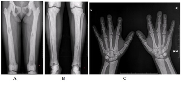
A) &B) Frontal radiographs of bilateral leg showing sclerosis of bone with ill-defined medullary lucencies
C) Hand radiograph showing short stubby fingers with diffuse sclerosis of bone
D) Foot, chest and
E) CT coronal sections of skull shows expansion of medullary cavity with diffuse sclerosis of bone.
F) Lateral skull X ray shows frontal bossing
Features are consistent with sclerosing bone disease –pyknodysostosis
(#) T2, flair, T1+c, T1,T1 SAG and CT sagittal& axial images showing extra calvarial enhancing lesion in the subgaleal plane of the left occipital region No extension of lesion into underlying bone/intracranial cavity
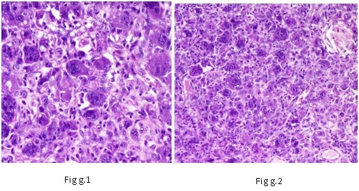
G) Histopathology:

A) & B) Frontal radiographs of bilateral leg showing sclerosis of bone with ill-defined medullary lucencies.
C)Hand radiograph showing short stubby fingers with diffuse sclerosis of bone.

D) Foot, chest and
E) CT coronal sections of skull shows expansion of medullary cavity with diffuse sclerosis of bone.
F) Lateral skull X ray shows frontal bossing.

Figure g.1: Autolyzed fibro collagenous tissue with muscle fibers.
Figure g.2: Tumor composed of multinucleated osteoclastic giant cells with mononuclear stromal cells with elongated pleomorphic vesicular nuclei and prominent nuclei infiltrating the fibro collagenous tissue. Also contains lymphoid aggregates with congested blood vessels, bony spicules and hemorrhage.
Discussion
Pyknodysostosis is a rare inherited disorder, with an incidence estimated to be 1.7 per million births [1]. A mutation in the gene that codes for the enzyme cathepsin K inhibits the normal functioning of osteoclasts. Cathepsin K is a lysosomal cysteine protease expressed in osteoclasts that is responsible for degrading collagen type 1. Dysfunction of osteoclasts causes impairment in bone resorption and remodeling. Hence, such affected bones are therefore abnormally dense and brittle and easily fracture. Sparing of the medullary cavity within the long bones is characteristic of the disorder, resulting in normal hematopoietic function [2]. The disorder is normally diagnosed at a young age owing to the characteristic phonotypical appearance with dwarfism and dysmorphic faces. Cognitive functioning and life expectancy of such patients are normal.
MRI findings of pyknodysostosis generally reveal normal cortical thickness in the calvaria, however there is increased trabecular bone within the medullary cavity. There is limited literature describing the CT findings, which includes hypoplastic sinuses, poor dentition, and thickening of the calvaria.
In our case study, the general features present were short stature, wrinkled skin, finger and nail abnormalities as well as acro-osteolysis with sclerosis of the terminal phalanges which is an essential pathognomonic feature. The radiological findings may show some degree of widening of the distal femur. The skull shows open anterior fontanel and sutures with small facial bones,
Nonpneumatised paranasal sinuses, and flattened mandibular angle. Terminal phalanges of the hands are partially or totally aplastic with loss of ungual tufts.
The differential diagnosis of pyknodysostosis is established with osteopetrosis, cleidocranial dysplasia and idiopathic acro-osteolysis [3].
GCT is a benign tumor composed of multinucleated giant cells that shows osteoclastic activity.
It usually develops in long bones, however unusual locations are also described in many case reports it can mimic other benign or malignant lesions at both radiological and histologic analysis GCT may occur in skull or pelvis secondary to Paget’s disease [4] Treatment in this condition is mainly supportive with emphasis on fracture prevention and management along with dental hygiene and regular checkups to prevent further complications.
Conclusion
Pyknodysostosis along with GCT is a rare phenomenon occurring in apolostotic PYNK. Hence requires a multispecialty approach. Early recognition of clinical features allows correct treatment planning and reduces the chance of complications in future thus ensuring a better quality of life to the patient.
Declaration:
Acknowledgments: Not applicable
Conflict of Interest: Nil
Funding: Not applicable
Availability of data and materials: Not applicable
Consent for publication: Written informed consent was obtained from the patient for publication of this case report and all the accompanying images.
References
- Barnard B, Hiddema W. Pyknodysostosis with the focus on clinical and radiographic findings: case report. SA J Radiol, 2012; 16: 74-76.
- Nirupama C, Sarasakavitha D, Palanivelu S, Guhan B. Pyknodysostosis: a case report of rare entity. J Indian Acad Oral Med Radiol, 2013; 25: 161-163.
- Mujawar Q, Naganoor R, Patil H, Thobbi AN, Ukkali S, Malagi N. Pyknodysostosis with unusual findings: a case report. Cases J, 2009; 2: 6544.
- Hunt NP, Cunningham SJ, Adnan N, Harris M. The dental, craniofacial, and biochemical features of pyknodysostosis: a report of three new cases. J Oral Maxillofac Surg, 1998; 56: 497-504.

