Ectopic Abutment of the Ureteral Meatus at the Level of the Prostatic Urethra Complicated by Acute Pyelonephritis
Mehdi Safieddine, Anas Tmiri*, Adil Kbirou, Amine Moataz, Mohamed Dakir, Adil Debbagh, Rachid Aboutaieb
Department of Urology, University hospital center Ibn Rochd, Casablanca, Morocco
Received Date: 14/11/2024; Published Date: 25/11/2024
*Corresponding author: Anas Tmiri, Department of Urology, University hospital center Ibn Rochd, Casablanca, Morocco; Faculty of Medicine and Pharmacy, Hassan II University, Casablanca, Morocco
Abstract
Congenital malformations of the ureter include several different obstructive and/or refluxing entities. Ectopy of the ureteral meatus is very rare since it is often asymptomatic and its true incidence is uncertain.
Acute pyelonephritis is a fairly common complication in these types of malformations which can be life-threatening, making it a medical-surgical emergency.
We report a case of acute pyelonephritis on ectopic ureteral meatus in a 39-year-old patient.
Keywords: Malformative uropathy; Ectopic abutment; The ureteral meatus; Acute pyelonephritis; Medical-surgical emergency
Introduction
Congenital malformations of the ureters are frequently associated with kidney malformations, but they can occur independently. Ectopy of the ureteral meatus is very rare since it is often asymptomatic and its true incidence is uncertain. Acute pyelonephritis is a fairly common complication in these types of malformations which can be life-threatening, making it a medical-surgical emergency [1-3].
We report a case of acute pyelonephritis in ectopic ureteral meatus.
Case Report
Patient aged 39, with no particular pathological history, who presents with an acute abdominal picture with suspicion of left renal colic accentuated 4 days before his admission.
On clinical examination, he presented with tenderness in the left lumbar fossa, hypogastrium and left flank with a fever of 38.3°C, with the presence of nitrites, leukocytes and hematuria on the urine dipstick.
On paraclinical assessment he presented a leukocytosis of 11,000/mm3, a CRP of 11 mg/l and acute renal failure with a creatinine of 110 mg/l.
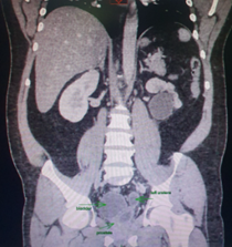
Abdominopelvic CT revealed major chronic uretero-hydronephrosis with atrophy and destruction of the left kidney (Figure 1 and 2).

Figure 1: Abdominopelvic CT in coronal section.

Figure 2: Abdominopelvic CT in frontal section.
The patient benefited from a double J catheter, the exploration of which revealed the arrival of the left ureteral meatus at the level of the prostatic urethra (Figure 3), with resection of the suspicious flat hyperemic bladder lesions.

Figure 3: Ectopic abutment of the ureteral meatus at the level of the prostatic urethra.
He was put on dual antibiotic therapy (Ceftriaxone and Amikacin), analgesic with good rehydration and a good clinical-biological evolution, a renal scintigraphy was prescribed upon discharge.
Discussion
Congenital malformations of the ureter include several different obstructive and/or refluxing entities. The term ectopic ureter is used to describe a ureter that terminates in the lateral wall of the bladder, lower along the trigone, at the level of the bladder neck, in the urethra in girls downstream of the sphincter, in the genital tract (prostate, seminal vesicle, uterus or vagina) or open externally [2,3].
The incidence of ectopic ureter remains uncertain because this malformation is often asymptomatic but it remains more common in women (2 to 12 times more common in women than in men) [3].
This malformation remains rare and can be described in the literature; we find a similar case of a 33-year-old man who presented with an ectopic opening of the left ureteral meatus at the level of the prostatic urethra above the veru montanum (Confirmed by the pre-operative urethro-cystoscope) complicated by uretero-renal reflux (Grade 3 on retrograde and voiding urethro-cystography) who consulted following chronic left low back pain distant from acute left pyelonephritis caused by Colibacille, evolution was favorable, on a small left kidney on renal ultrasound. DMSA renal scintigraphy confirmed a very poorly functional left kidney, with relative uptake of 11% versus 89% for the right kidney. A left nephroureterectomy by trans-peritoneal laparoscopy was performed with simple post-operative outcomes and good progress [3].
Another described case of abutment of the ureteral meatus at the vulvar level in a 4-month-old girl who presented a toxic state of dehydration, urea at 1.10 g/l and pyuria after resuscitation the urea level dropped to 0.20 g/l, an IVU was carried out showing a right kidney secreting normally with a silent left kidney, the cystogram showing a ureterocele. Faced with the persistence of the infection with pyuria and the discovery of a palpable left lumbar mass, a simple open nephrectomy was carried out secondly after a bladder size by which the operators objectified the presence of two ureters, one abuts at the trigonal level and the other at the vulvar level. The postoperative course was simple with good progress [4].
Conclusion
Severe obstructive or refluxing uropathy sometimes requires antibiotic prophylaxis to prevent pyelonephritis, but this remains the subject of debate. The indication for surgery is based on the function of the overlying kidney, urinary symptoms and the frequency of infectious episodes.
References
- Masson E. Ectopic ureteral outlets. EM-Consulte, 2024.
- Ureteral anomalies - Pediatrics. MSD Manual Professional Edition, 2024.
- André D, Hamie F, Chautard D, Colombel P, Soret JY. Outlet of a trifid ectopic ureter with uretero-renal reflux into the prostatic urethra. Progress in Urology, 2006.
- On a case of ectopic ureteral outlet | Urologia Internationalis | Karger Publishers, 2024.

