Rapid Intraoperative Tissue Expansion for Closure of Lumbosacral Meningomyelocele
Vijay Bhatia1, Deepanjali Kalra2, Manisha Singh2, Charmi Bodra2, Himadri Joshi2, Rajvee Gohil2, Jay Adeshra2 and Kushal Shah3
1Professor and Head of Department of Burns and Plastic Surgery, Smt NHL Municipal Medical College, Consultant, Sterling Hospital, Ahmedabad, Gujarat, 380059, India
2Residents, Smt. NHL Municipal medical college, Sardar Vallabhbhai Patel Institute of Medical Sciences and research, India
3Assitant Professor Department of Neurosurgery, Smt NHL Municipal Medical College, Ahmedabad, Gujarat, 380059, India
Received Date: 09/11/2024; Published Date: 22/11/2024
*Corresponding author: Vijay Bhatia, Professor and Head of Department of Burns and Plastic Surgery, Smt. NHL Municipal Medical College; Consultant, Sterling Hospital Ahmedabad, Gujarat, 380059, India
ORCID ID: 0000-0001-7563-6271
Abstract
Swellings due to incomplete closure of neural tube can anywhere over the spine however, most commonly located in lumbosacral region. Defects up to 5 cm can be closed primarily, however larger defects would require other reconstructive strategies. Here, we present a unique way of reconstructing the post myelomeningocele repair in a 28-day old neonate using rapid intraoperative tissue expansion technique. Size of the defect was large 10*8 cm where primary closure wasn’t possible. Post rapid intraoperative expansion two flaps were created which were sutured and the wound closed under no tension. Rapid Intraoperative tissue expansion is an excellent method for reconstruction of lumbosacral defects in children. Proper knowledge and following stringent protocols can yield best results. With the help of this technique various complications due to other reconstructive options like kyphosis, impaired muscular function, blood loss, wound necrosis and dehiscence can be avoided.
Introduction
Swellings due to incomplete closure of neural tube can anywhere over the spine however, most commonly located in lumbosacral region [1,2]. The most common birth defect of the nervous system is myelomeningocele [3-6]. It is most common form of open spinal dysraphism and forms the defect of the spinal cord, vertebrae and overlying skin. Defects up to 5 cm can be closed primarily, however larger defects would require other reconstructive strategies [7]. Reconstruction of huge defects post myelomeningocele excision has always been challenging. Newborn skin circulation is sensitive and by supplied by terminal vessels hence, undermining larger area of skin would result in harming vascularity resulting in ischemic necrosis and blood loss.
Tissue expansion was first introduced by Neuman in 1957 which was popularised and developed by Radovan. The modification of this process as Rapid Intraoperative Tissue Expansion (RITE) was introduced by Sasaki in 1987 (8,9). The stretching property and viscoelasticity are utilized to increase the surface area to reconstruct large defects without hindering vascularity of flaps. In literature, cases have been described in which reconstruction of post myelomeningocele defect has been done using the tissue expansion. However rarely rapid intraoperative tissue expansion has been used for this purpose. The advantage of rapid tissue expansion over the controlled tissue expansion is that only one stage, reduces the number of OPD visits and prevents the cosmetic deformity for the duration of the expansion process.
Here we present a unique way of reconstructing the defect post myelomeningocele repair in a neonate using rapid intraoperative tissue expansion technique.
Case
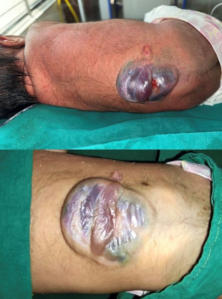
A newborn female, daughter of 24-year-old primigravida mother was delivered with elective cesarean section at 38 weeks and 4 days with birth weight 2.3 kg, was found to have swelling at lower lumbosacral region. Patient was admitted under pediatrician and neurosurgeon and plastic surgeons were called intraoperatively for closure of the defect. Patient was diagnosed with Arnold chiari malformation type 2 on antenatal scan at 8 months intrauterine and at birth presented with lumbosacral swelling. Patient had APGAR score of 8 at 1 min and 10 at 5 min. Patient had movements of all four limbs. After that she was shifted immediately to neonatal intensive care unit for observation. On second post birth day, patient was not maintaining saturation on room air, so NIV support was given and planned for MRI brain and spine. During MRI scan one episode of convulsion occurred and patient was intubated. Surgery was deferred till the patient was stabilized. MRI scan revealed herniation of cerebellar tonsil through foramen magnum and dilatation of bilateral lateral and 3rd ventricle with hydrocephalus. Herniation of nervous tissue and cerebrospinal fluid through spina bifida with sac of size 4.5 *4 cm at L2-L4 level which communicate with spinal cord via defect of 8.5 mm with posteriorly tethered spinal cord ends abnormally at the level of S1 vertebra.
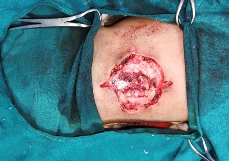
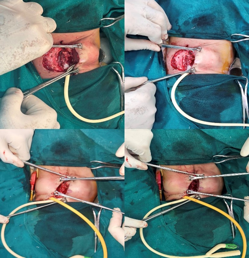
On post birth day 28, surgery was planned by neurosurgeon. During the operation it was found by the neurosurgeon that the dura of the spinal cord was adhered overlying skin. Elliptical skin incisions were taken and spinal cord was separated from the overlying skin. In this process there was tear in the dura for which the dural patch was used for repair and right-side medium pressure ventriculoperitoneal shunting was done. Post dural repair the skin and soft tissue defect size of 10*8 cm was created. Minimal undermining of the wound was done using blunt dissecting scissors and Foleys catheter of size 14 was used as a tissue expander, inserted in sterile manner into the wound through its edge bilaterally and was slowly inflated with saline till maximum capacity for 5 mins and deflated for 3 mins. This was repeated two times. Care was taken to maintain the catheter position all throughout the process in the wound so that it doesn’t get slipped out. Skin was expanded rapidly intraoperatively and two incisons were made to change the direction of suture line and convert into two rotation flaps. Tension free primary closure of wound was done in two layers with monocryl 3-0 and ethilon 3-0.

Figure 1: Preoperative picture showing sac size.

Figure 2: Intraoperative picture of defect size 10*8 cm after repair of meningomyelocele.

Figure 3: Technique of intraoperative rapid tissue expansion.

Figure 4: Intraoperative picture after primary closure of defect.

Figure 5: Healthy suture line on post operative day 2.

Figure 6: Flaps healed well and suture line healthy at postoperative day 10.

Figure 7: Postoperative day 21 suture removed and flaps healed well.
Results
Regular dressings were done on alternate day starting from post operative day 2. Suture-line was healthy. Flaps healed well. Patient was discharged on post operative day 10. Suture removal done on day 21. Patient was followed for one month. Wound healed well with acceptable functional and aesthetic outcomes.
Discussion
Major objectives of surgical treatment of meningomyelocele are 1) watertight closure of dura 2) long term soft tissue coverage. Small to moderate defects upto 5 cm can be closed primarily. However, defects more than 5 cm or greater than half the width of the back can be requires regional reconstruction procedures.
Large defects requiring reconstructive modalities have been described in literature and newer techniques have also come up. Various reconstructive modalities for large lumbosacral defects are: Skin grafts: Not a suitable option for coverage as causes kyphosis of spine and cosmetic deformities requiring spinal surgeries later on [10]. Local flaps like transposition, rotation, bipedicled , VY advancement , keystone flap , musculocuataneous flaps , perforator based flaps etc .which may or may not require skin grafting requires extensive undermining of the skin flap which is not suitable in children as sensitive skin supplied by terminal vessels leading to skin ischaemia and necrosis . Highest incidence of problems has been associated with musculocutaneous flaps which would require extensive dissection and technically challenging [11,12]. Tissue expansion a novel method of reconstruction utilizing the viscoelastic properties of skin for expanding the skin. It is an excellent method of reconstruction however, multiple stages, long operative stay, cosmetic deformity and delayed closure of wound are various disadvantages of this technique.
RITE is simple and effective method of reconstruction of large defects. Tissue expansion can be used in all the parts of body and in all age groups [12,13]. There are various advantages of this technique like single staged procedure, easy closure, cost effective, decreases the length of stay, avoids the body disfigurement and multiple sittings. Actual amount of flap width increased by immediate intraoperative technique is 15-20% reported by Hofmann and Baker in forehead flaps [14].
Advantages of using this technique has overpowered the other modalities of reconstruction and made it more preferable compare to other modalities. Rapid Tissue expansion works on the phenomenon of mechanical creep described by Gibson and Gibson et al (15) based on relative dehydration of tissue, random positioning of collagen fibers, micro fragmentation of elastic fibers and migration of adjacent tissues into the field.
Here, we present a case report of newborn, in whom post excision of myelomeningocele defect have been reconstructed using Rapid Intraoperative Tissue Expansion technique (RITE) which would have otherwise reconstructed using skin grafting or local flap. In children lower back region there is less capacity of the skin to return back to original size during the post operative period which we utilised for our technique.
Fewer studies have been published for using tissue expansion for expanding skin and closure of defect have been described in literature. However rapid intraoperative tissue expansion with foleys for reconstruction of lumbosacral defects have been nowhere described.
Conclusion
Rapid Intraoperative tissue expansion is an excellent method for reconstruction of lumbosacral defects in children. Proper knowledge and following stringent protocols can yield best results. With the help of this technique various complications due to other reconstructive options like kyphosis, impared muscular function, blood loss, wound necrosis and dehiscence can be avoided.
Author Contributions:
Vijay Bhatia is the chief plastic surgeon in our study who has performed the reconstruction.
Deepanjali Kalra wrote the manuscript.
Manisha.Singh, Charmi Bodra, Himadri Joshi, Rajvee Gohil and Jay Adeshra has assisted in operation.
Kushah Shah is the chief neurosgeon in the operation
Conflicts of Interest: The authors declare that there are no relevant conflicts of interest.
Sources of Funding: None
Acknowledgements: None
IRB Approval: As this study is case report IRB approval is not needed.
References
- Gürer B, Kertmen H, Akturk UD, Kalan M, Sekerci Z. Use of the bovine pericardial patch and fibrin sealant in meningomyelocele closure. Acta neurochirurgica, 2014; 156: 1345-1350.
- Schoellhammer L, Gudmundsdottir G, Rasmussen MM, Sandager P, Heje M, Damsgaard TE. Repair of myelomeningocele using autologous amnion graft and local flaps. A report of two cases. JPRAS open, 2018; 17: 9-14.
- Mutaf M, Bekerecioglu M, Erkutlu I, Bulut Ö. A new technique for closure of large meningomyelocele defects. Annals of plastic surgery, 2007; 59(5): 538-543.
- Bozkurt C, Akın S, Doğan Ş, Özdamar E, Aytaç S, Aksoy K, Erol O. Using the sac membrane to close the flap donor site in large meningomyeloceles. British journal of plastic surgery, 2004; 57(3): 273-277.
- Moldenhauer JS, Adzick NS. Fetal surgery for myelomeningocele: After the Management of Myelomeningocele Study (MOMS). InSeminars in fetal and neonatal medicine. WB Saunders, 2017; 22(6): pp. 360-366.
- Elbabaa SK, Gildehaus AM, Pierson MJ, Albers JA, Vlastos EJ. First 60 fetal in-utero myelomeningocele repairs at Saint Louis Fetal Care Institute in the post-MOMS trial era: hydrocephalus treatment outcomes (endoscopic third ventriculostomy versus ventriculo-peritoneal shunt). Child's Nervous System, 2017; 33: 1157-1168.
- Danish SF, Samdani AF, Storm PB, Sutton L. Use of allogeneic skin graft for the closure of large meningomyeloceles: technical case report. Operative Neurosurgery, 2006; 58(4): ONS-E376.
- Neumann CG. The expansion of an area of skin by progressive distention of a subcutaneous balloon: use of the method for securing skin for subtotal reconstruction of the ear. Plastic and reconstructive surgery, 1957; 19(2): 124-130.
- Sasaki GH. Intraoperative sustained limited expansion (ISLE) as an immediate reconstructive technique. Clinics in Plastic Surgery, 1987; 14(3): 563-573.
- Mustoe TA, Gifford GH, Lach E. Rapid tissue expansion in the treatment of myelomeningocele. Annals of plastic surgery, 1988; 21(1): 70-73.
- Mowatt DJ, Thomson DN, Dunaway DJ. Tissue expansion for the delayed closure of large myelomeningoceles. Journal of Neurosurgery: Pediatrics, 2005; 103(6): 544-548.
- Shurtleff DB, Lemire RJ. Epidemiology, etiologic factors, and prenatal diagnosis of open spinal dysraphism. Neurosurgery Clinics of North America, 1995; 6(2): 183-93.
- Arnell K. Primary and secondary tissue expansion gives high quality skin and subcutaneous coverage in children with a large myelomeningocele and kyphosis. Acta Neurochirurgica, 2006; 148: 293-297.
- Hoffman HT, Baker SR. Nasal reconstruction with the rapidly expanded forehead flap. The Laryngoscope, 1989; 99(10): 1096-1098.
- Gibson T, Kenedi RM, Craik JE. The mobile micro-architecture of dermal collagen: a bio-engineering study. Journal of British Surgery, 1965; 52(10): 764-770.

