Intrathroid Ectopic Parathyroid Glands about a Case
Amine Bachar, Malik Baallal Zarhouni*, El Azhari Ilias, Zakaria Essaidi, Taoufiq Al-Abbassi and Fatima Zahra Bensardi
Department of Visceral Surgery wing 1, Ibn Rochd University Hospital, Hassane II University, Faculty of Medicine and Pharmacy (FMPC), Morocco
Received Date: 15/10/2024; Published Date: 08/11/2024
*Corresponding author: Zarhouni Baallal Malik, Department of Visceral Surgery, Ibn Rochd University Hospital, Hassane II University, Faculty of Medicine and Pharmacy (FMPC), Casablanca, Morocco
Abstract
The ectopic localization of the parathyroid glands in intrathyoidinn is very rare and may be a cause of postoperative hypocalcemia, especially if all the parathyroid glands are ectopic. We report the observation of a 54-year-old patient with no particular pathological history operated for a multiheteronodular goiter with 2 right lobar nodules classified EU tirads 5 with clinical and biological eutyroidy, the patient benefited from a total thyroidectomy. The postoperative follow-up was simple. The histopathological examination of the excision piece found an invasive thyroid carcinoma of the right midlobar papillary type and also an ectopic parathyroid level of the left lobe surrounded by thyroid parenchyma, it is hyperplastic and without capsule, the patient subsequently presented hypocalcemia they always followed under calcium supplementation.
Introduction
The variations in the position of the parathyroid glands are well known. They can be located in an area extended from the level of the submandibular glands to that of the pericardium, within the carotid axes [1]. Rarely they can be included in the thyroid gland. We report an observation where it is discovered by the anatomo-pathological examination of the thyroidectomy specimen.
Observation
This is a 54-year-old patient with no particular pathological history presented for an anterior cervical swelling evolving for 6 months, not painful, without compressive signs or signs of dystoroidia, the clinical examination objectified a low anterior cervical swelling without inflammatory signs in the non-painful regard, soft on palpation and mobile on swallowing
Cervical ultrasound objectified a multiheteronodular goiter with the 2 largest nodules located in the right superior lobar and classified as tirads 5.
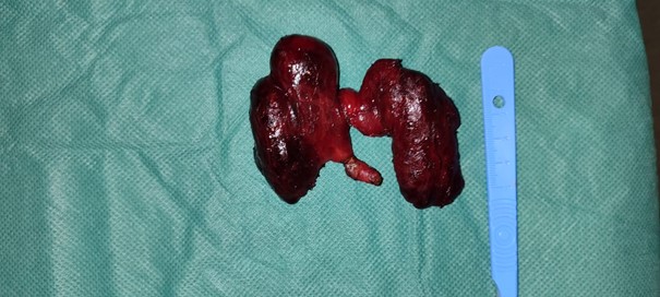
The patient's thyroid test was normal. The patient underwent a total thyroidectomy. The postoperative follow-up was simple, in particular no dysphonia or dyspnea nor clinical signs of hypocalcemia, the patient was declared discharged on D2 postoperatively. The histopathological examination of the excision piece found an invasive thyroid carcinoma of the right medielobar papillary type and also an ectopic parathyroid level of the left lobe surrounded by thyroid parenchyma, it is hyperplastic and without capsule, the phosphocalcium profile of the patient subsequently was disturbed with signs of hypocalcemia the patient was put on oral calcium supplementation 500mg per day; The patient is currently still being followed up with a 2-month follow-up with regular phosphocalcic tests to determine whether her hypocalcemia is transient or definitive.

Figure: Surgical specimen-thyroid.
Comment
Intrathyroid localization of parathyroids is rare. AKERSTOM et al found only 3 cases out of 503 autopsies, but in the series of cervical explorations for parathyroid adenoma, its incidence is higher varying from 1.4% to 3.2% [2]. This intrathyroid localization can be explained by embryology. The upper parathyroids come from the 4th gill pouch, they attach to the dorsal aspect of the thyroid gland as it descends from the ventral wall of the pharynx. The parathyroid can be trapped in the thyroid parenchyma when the lateral lobe fuses with the median lobe, and an intrathyroid upper parathyroid will be left [2].
The 3rd brachial pouch gives the thymus and the lower parathyroid. This attracts the lower parathyroid with it during its descent. This explains why the lower parathyroid can be very low: thoracic; and the presence of thymic tissue with the ectopic lower parathyroid [2]. The lower parathyroid can be imprisoned in the same way as the upper one [2].
Histologically, to affirm that a parathyroid is intrathyroid, it must be surrounded by the thyroid parenchyma and must not have a capsule [3]. And this is the case for our patient
This eliminates the parathyroids that are located in a fold of the capsule or in the thyroid capsule and in both cases the parathyroid has its own capsule in addition to the thyroid capsule [2,3].
These variations in the location of the parathyroids explain the difficulty encountered in the search for parathyroid adenomas. This research can be aided by ultrasound, transesophageal endoscopic ultrasound, scintigraphy, computed tomography, magnetic resonance imaging, ultrasound-guided cervical puncture and intraoperative ultrasound [2]. But the exact role of all these means of investigation is not yet well established and are for the most part indicated only after the first exploratory cervicotomy [2].
Conclusion
This intrathyroid localization of the parathyroids is a very rare cause of definitive or transient postoperative hypocalcemias that must be distinguished from involuntary parathroidectomy that are linked to known risk factors (duration of the operation, recurrent mediastinal evidment and malignancy).
References
- Dubost CL, Tourneur R, Bay M. Parathyroid glands: anatomy, histology, surgical exploration. E.M.C. (Paris) Glandes endocrines, 1968; 10011: A10.
- Feliciano DV. Parathyroid pathology in an intrathyroidal position. * Surgery "B" Department, ** Pathological Anatomy Department. American joumal of surgery, 1992; 164(1°5): 496-500.
- Hyperfunctioning intrathyroid parathyroid gland: a potential cause of failure in paratyroide surgery J R. soc med, 1981; 74: 49-52.

