Atypical Emphysematous Cystitis Complicated by Asymptomatic Bladder Perforation
Mohamed Bakhri1,2,*, Jaafar Marrakchi Benjaafar1,2, Rhyan Ouaddane Alami1,2, Mustapha Ahssaini1,2, Soufiane Mellas1,2, Jallal Eddine El Ammari1,2, Mohammed Fadl Tazi1,2 and Mohammed Jama El Fassi1,2
1Department of Urology, Hassan II University Hospital, Morocco
2Faculty of Medicine, Pharmacy and Dentistry of Fez, Sidi Mohammed Ben Abdellah University,
Morocco
Received Date: 15/09/2024; Published Date: 01/11/2024
*Corresponding author: Mohamed Bakhri, Department of Urology, Hassan II University Hospital, Morocco; Faculty of Medicine, Pharmacy and Dentistry of Fez, Sidi Mohammed Ben Abdellah University,
Morocco
Abstract
Emphysematous cystitis is a severe and potentially life-threatening bladder inflammation, characterized by gas production primarily due to infection by E. coli and Klebsiella pneumoniae. It is most commonly observed in elderly diabetic women with poorly controlled diabetes. Clinically, it presents a highly variable clinical picture from one patient to another, with diagnosis confirmed through radiology. Treatment involves prolonged antibiotic therapy and bladder drainage. The prognosis depends on the timeliness of diagnosis and treatment.
Keywords: Emphysematous; Cystitis; Perforation; Bladder
Introduction
Emphysematous cystitis is an acute, rare, and severe form of cystitis, primarily found in elderly diabetic women, characterized by the presence of gas in the bladder. The clinical presentation varies from patient to patient and can range from being asymptomatic to septic shock. Diagnostic confirmation is primarily achieved through imaging, especially CT scans. We present the case of a male patient, diabetic and hypertensive, who developed emphysematous cystitis complicated by bladder perforation.
Case Presentation
Mr. BD, an 82-year-old male, presented to the emergency department with a chronic cough associated with diffuse, intermittent abdominal pain, which had been ongoing for a week and was more pronounced in the hypogastric region, all while maintaining a generally stable condition. His medical history includes poorly controlled type 2 diabetes for 25 years, hypertension managed with dual therapy, and recurrent urinary tract infections, which were symptomatically treated by a private general practitioner without significant improvement.
Clinical examination revealed a hemodynamically stable patient (BP: 140/90 mmHg, HR: 86 bpm), with stable respiratory function (SaO2 99%, RR 14 breaths per minute), slightly febrile at 38.4°C, with a soft abdomen and preserved urine output. Biologically, there was an inflammatory syndrome with leukocytes at 16,000, CRP at 230 mg/L, and a urine culture positive for Klebsiella Pneumoniae (KP).
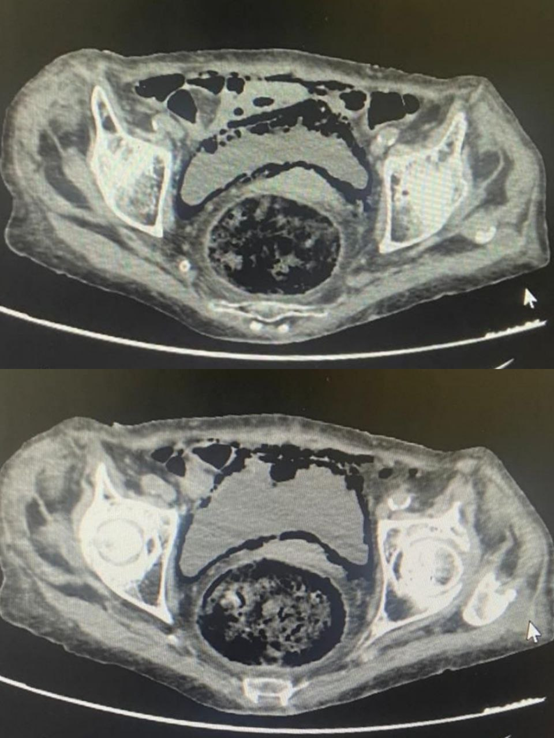
An abdominal-pelvic CT scan without contrast injection revealed gas images within the bladder wall, consistent with emphysematous cystitis, along with additional gas images in the perivesical area, suggesting bladder perforation (Figure 1).

Figure 1: Axial slice of an abdominal CT scan showing the presence of gas in the bladder wall, consistent with emphysematous cystitis, along with perivesical gas images suggesting bladder perforation.
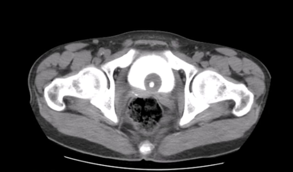
The patient underwent bladder catheterization, which confirmed preserved urine output. He was initially placed on dual antibiotic therapy, which was later adjusted according to the antibiogram. A markedly favorable evolution was observed: clinically, the patient was no longer febrile, with the disappearance of abdominal tenderness; radiologically, a follow-up CT urogram showed no contrast extravasation and the disappearance of the perivesical gas image (Figure 2); and biologically, there was improvement.

Figure 2: Axial slice of a late-phase follow-up CT urogram showing no contrast extravasation and resolution of the perivesical gas image.
Discussion
Emphysematous cystitis is a rare condition that complicates urinary tract infections. The average age of patients with this condition reported in the literature is 70 years old [1]. The main predisposing factors include poorly controlled diabetes, urinary stasis (neurogenic bladder, prostatic hypertrophy), malnutrition, immunosuppression, and chronic urinary tract infection. Diabetes remains the primary predisposing factor, implicated in 60% of cases [2,3]. The clinical presentation of emphysematous cystitis is often nonspecific, with pain occurring in 80% of cases and bladder irritative symptoms in 50%. It is asymptomatic in 7% of cases [4]. In elderly patients, clinical examination is often altered. Fever is inconsistent, despite the presence of an advanced infectious condition, and abdominal guarding is often absent [5].
The most commonly involved pathogens are Gram-negative bacilli, with Escherichia coli being implicated in 60% of cases. Other reported pathogens include Klebsiella pneumoniae, Proteus mirabilis or vulgaris, Aerobacter aerogenes, and Candida albicans [7]. Anaerobic bacteria are exceptionally rare in this pathology. Gas formation results from the fermentation of glucose into formic acid, followed by the production of carbon dioxide and hydrogen, under the combined influence of acidic urinary pH, glycosuria, and certain bacteria [8]. Abdominopelvic CT scanning is the reference imaging technique: it allows for a positive diagnosis (presence of air in the bladder lumen and/or wall), evaluation of the extent of gas collections, and the detection of associated renal involvement [6]. It also helps rule out differential diagnoses such as primary pneumaturia and communication with hollow organs like vesicodigestive or vesicovaginal fistulas [4].
Treatment is most often medical, involving broad-spectrum intravenous dual antibiotic therapy, initially combined with bladder drainage via an indwelling catheter. The duration of treatment is not well-defined and depends on the clinical response [6,1]. Generally, it ranges from 3 to 6 weeks. Surgical treatment may be necessary in cases of unfavorable evolution with necrotizing involvement, leading to partial or total cystectomy.
Conclusion
Emphysematous cystitis is a condition rarely encountered by urologists. Diabetes remains the primary predisposing factor. Abdominopelvic CT scanning is the reference imaging technique, and management involves bladder drainage combined with dual antibiotic therapy.
Compliance with ethical standards
Disclosure of conflict of interest: No conflict of interest to be disclosed.
Statement of informed consent: Informed consent was obtained from all individual participants included in the study.
References:
- Thomas AA, Lane BR, Thomas AZ, et al. Emphysematous cystitis: a review of 135 cases. BJU Int, 2007; 100: 17-20.
- Toyota N, Ogawa D, Ishii K, Hirata K, Wada J, Shikata K, et al. Emphysematouscystitis in a patient with type 2 diabetes mellitus. Acta Med Okayama, 2011; 65(2): 129–133.
- Perlemoine C, Neau D, Ragnaud JM, Gin H, Sahnoun A, Pariente JL, et al. Emphysematous cystitis. Diabetes Metab, 2004; 30(4): 377–379.
- Grupper M, Kravtsov A, Potasman I. Emphysematous cystitis: illustrative case report and review of literature. Medicine, 2007; 86(1): 47–53.
- Wroblewski M, Mikulowski P. Peritonitis in geriatric patients. Age Ageing, 1991; 20: 90-94.
- Grayson DE, Abbott RM, Levy AD, Sherman PM. Emphysematous infections of the abdomen and pelvis: a pictorial review. Radiographics, 2002; 22: 543–561.
- Paparel P, Cognet F, Cercueil JP, Krause D, Michel F. Stratégie diagnostique et thérapeutique dans la pyélonéphrite emphysémateuse. Ann Med Interne, 2003; 154: 259-262.
- Huang JJ, Chen KW, Ruaan MK. Mixed acid fermentation of glucose as a mecanism of emphysematous urinary tract infection. J Urol, 1991; 146: 148-151.

