Ultrasonographic Aspects of Holoprosencephaly in the Context of a Polymalformative Syndrome: A Report of Two Cases
Medemagh Malak*, Toumi Dhekra, Chalbi Nadia, Cheikh Mohamed Chayma, Ben Farhat Imen, Amorri Safa, Lazreg Hanan, Bergaoui Haifa and Faleh Raja
Department of Gynecology and Obstetrics, Fattouma Bourguiba University Hospital, Monastir, Tunisia
Received Date: 12/09/2024; Published Date: 01/11/2024
*Corresponding author: Malak Medemagh, Department of Gynecology and Obstetrics, Fattouma Bourguiba University Hospital, Monastir, Tunisia
ORCID number of STC: 0000-0003-4005-209
Abstract
Holoprosencephaly is a rare malformation encountered in newborns. It is caracterised by the absent or incomplete division of the prosencephalon.
The first case, was diagnosed by ultrasonogram at 16 weeks of gestation with alobar holoprosencephaly, hypotelorism and single umbilical artery.
The second case was also diagnosed during the first-trimester ultrasound at 15 weeks of gestation, which showed incomplete separation of the brain’s lobes, with the brain remaining largely fused. Both cases indicated a severe developmental abnormality, suggesting a diagnosis of holoprosencephaly.
Keywords: Holoprosencephaly; Brain; Fetal malformation
Introduction
Holoprosencephaly (HPE) is a complex brain malformation resulting from an early defect in the median cleavage of the embryonic brain, occurring in the prosencephalon between the 18th and 28th day of gestation, affecting both the brain and the face. It is a rare fetal pathology with significant etiological heterogeneity, occurring in about 1 in 15,000 to 16,000 births [1].
Based on the degree of hemispheric individuation and decreasing severity, three forms have been defined: alobar or complete, semi-lobar, and lobar or incomplete. The prenatal diagnosis mainly relies on ultrasound [2].
The aim of our work is to analyze the etiopathogenic aspects and prenatal diagnostic modalities of HPE.
Case Reports
Case 1: The patient is a 38-year-old woman with no significant medical history and no known consanguinity. She is currently pregnant for the second time (G2P1AO). Her first pregnancy was uneventful and resulted in a cesarean delivery in 2019 due to chorioamnionitis.
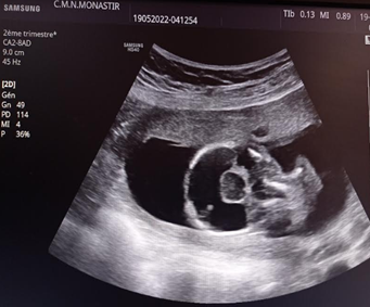
At the 16-week of her current pregnancy, her primary care physician detected a severe brain malformation during a routine first-trimester ultrasound. The diagnosis was holoprosencephaly (HPE), a condition where the brain fails to divide into two hemispheres. To further evaluate the situation, a detailed morphological ultrasound was conducted. This follow-up imaging confirmed the presence of a single fetus with a positive cardiac heartbeat. However, the ultrasound also revealed a complex syndrome involving multiple abnormalities: alobar holoprosencephaly, hypotelorism (a condition where the distance between the eyes is reduced), and a single umbilical artery (a deviation from the usual two-artery umbilical cord) (Figure1).

Figure 1: Alobar HPE : US image obtained at 16 weeks shows fused thalami and a monoventricle.

Figure 2 : Post-expulsion fetus.
Given the severity of the detected anomalies and the associated risk of poor outcomes, the decision was made to proceed with a medical termination of the pregnancy.
Case 2
The second patient is a 26-year-old woman, (G5P0A4). She has a history of four prior pregnancies, all of which ended in spontaneous abortions, and she previously underwent a medical termination of pregnancy in 2020 due to a diagnosis of holoprosencephaly, a serious and rare brain malformation. The patient also has a family history of second-degree consanguinity.
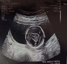
At 15 weeks of amenorrhea, during a routine first-trimester ultrasound, a single fetus was detected. The ultrasound revealed a significant midline anomaly in the fetus, specifically semilobar holoprosencephaly. This condition is characterized by incomplete separation of the brain’s lobes, where the brain remains largely fused, indicating a severe developmental abnormality. Additionally, the imaging showed a midline facial structure that might be indicative of a proboscis, a rare and severe congenital malformation involving an abnormal extension or projection of facial tissue.

Figure 3: Obstetric ultrasound, in axial, showing fusion of the thalami and cerebral hemispheres with a single ventricular cavity.
In response to these findings, an amniocentesis was conducted to further investigate and identify any potential chromosomal abnormalities that could be contributing to these severe malformations. The results of the genetic testing were crucial for understanding the full scope of the fetal condition.
Considering the severity of the detected anomalies and the associated high risk of poor outcomes for the fetus, including significant impact on quality of life and potential for severe disability, the medical team recommended and the patient consented to a medical termination of pregnancy.
Discussion
Holoprosencephaly (HPE) is a severe congenital brain malformation often associated with distinctive facial anomalies [3,4].
Several authors agree on the association between maternal age over 30 years and the occurrence of HPE. The concept of consanguinity has been reported in the literature, along with the implication of several genetic mutations [5]. In our patient, a second-degree consanguinity was noted.
It results from a failure in neuroectoderm induction during the third week of embryonic development, leading to a developmental anomaly in the prosencephalon. This results in a median hemispheric mass replacing the two cerebral hemispheres and an absence of median structures.
The classification system for HPE, established by DeMyer, categorizes the condition into three main types: alobar, where there is no separation between the cerebral hemispheres and a large single ventricle is present; semi-lobar, where only the anterior lobes remain fused while the parieto-occipital regions are separated by the interhemispheric fissure and the falx cerebri; and lobar, characterized by fusion in only the most rostral and inferior portions of the frontal lobes [6,7].
HPE is often associated with various facial anomalies, such as a median cleft, cyclopia, and nasal abnormalities. In 80% of cases, these predictive malformations reflect the severity of the brain abnormalities [5].
It is linked to a high rate of postnatal mortality. The overall estimated mortality rate for all HPE subtypes is 33% within the first 24 hours after birth and 58% within the first month. The survival rate after one year of age is approximately 29% [8].
The antenatal diagnosis of holoprosencephaly (HPE) is primarily based on conventional obstetrical ultrasound, supplemented by fetal MRI, which reveals cerebral anomalies such as a single ventricular cavity, absence or abnormal development of midline structures, thalamic fusion, and microcephaly. The extent of these abnormalities varies depending on the anatomical form of HPE. An ultrasound diagnosis can be made as early as the first trimester, enabling early pregnancy termination, particularly in cases of the alobar and semi-lobar forms [9].
The prognosis depends on the extent of brain fusion and malformations, as well as any accompanying health complications. Alobar and semilobar forms of HPE are typically fatal. Children born with lobar HPE can survive for several years but often face significant neurological issues and severe intellectual disability [10].
References:
- Ramakrishnan S, Das JM. Holoprosencephaly. In: StatPearls [Internet]. Treasure Island (FL): StatPearls Publishing, 2024.
- Kauvar EF, Muenke M. Holoprosencephaly: recommendations for diagnosis and management. Curr Opin Pediatr, 2010; 22(6): 687-695. doi: 10.1097/MOP.0b013e32833f56d5.
- Dwight RC, Minal T, Jill AH. The etiopathologies of holoprosencephaly; Drug Discov Today Dis Mech, 2005; pp. 529–537.
- Fedoua W, Mouna H, Hasana S, Boufettal H, Mahdaoui S, Samouh N. Holoprosencephaly (HPE) : case report and review of the literature. Int J Surg Case Rep, 2023; 110: 108723. doi: 10.1016/j.ijscr.2023.108723.
- Fatnassi R, Turki E, Belhaj J, Labidi I, et al. Holoprosencephaly: pathogenesis, phenotypic characteristics - About four cases. Morphologie, 2011; 95(310): 79–82.
- Leibovitz Z, Lerman-Sagie T, Haddad L. Fetal Brain Development: Regulating Processes and Related Malformations. Life, 2022; 12: 809. doi: 10.3390/life12060809.
- Moutard ML, Fallet-Blanco C. Pathologie neurologique malformative fœtale. EMC – Pédiatrie, 2004; 1(2): 210–231.
- Malta M, AlMutiri R, Martin CS, Srour M. Holoprosencephaly: Review of Embryology, Clinical Phenotypes, Etiology and Management. Children (Basel), 2023; 10(4): 647. doi: 10.3390/children10040647.
- Tongsong T, Wanapiraka C, Chanprapapha P, Siriangkul S. First trimester sonographic diagnosis of holoprosencephaly. J. Gynaecol. Obstet, 1999; 66: 165–169.
- Poenaru MO, Vilcea ID, Marin A. Holoprosencephaly: two case reports. Maedica (Bucur), 2012; 7(1): 58-62.

