Neurodermatitis Tumoral, Aspect Atypical of Affection Common
Julian Camilo Sánchez Guerrero*, Paulina Gabriela Pacheco Buñay, and Paulo Ricardo Martins Souza
Dermatology Service, Hospital da Santa Casa de Misericórdia de Porto Alegre, Porto Alegre, RS, Brazil; Department of Internal Medicine/Dermatology, Federal University of Health Sciences of Porto Alegre, Porto Alegre, RS, Brazil
Received Date: 25/08/2024; Published Date: 25/10/2024
*Corresponding author: Julian Camilo Sánchez Guerrero, Dermatology Service, Hospital da Santa Casa de Misericórdia de Porto Alegre, Porto Alegre, RS, Brazil; Department of Internal Medicine/Dermatology, Federal University of Health Sciences of Porto Alegre, Porto Alegre, RS, Brazil
Introduction
Neurodermatitis (or chronic lichen simplex) is defined as a form of skin thickening secondary to pruritus, a factor that precedes the appearance of lesions. Although its pathogenesis is debatable, it is a condition that is easy to diagnose. It may begin with small erythematous papules in the itchy areas, which later group together to form plaques. Over time and with continued pruritus, the area becomes thickened, causing accentuation of the skin lines. Darkening from the area and frequent, mainly in individuals with phototypes higher. The injuries can They can be solitary or multiple, and show a predilection for the posterolateral region of the neck, occipital region, upper back and lumbar region, anogenital region (scrotum, vulva), calves, ankles and the dorsal aspect of the hands, feet and forearms. Predisposing factors include anxiety, obsessive-compulsive disorder, and pruritus related to systemic diseases [1].
Its morphological/visual aspect generally allows its identification, through the accentuation of skin lines, or the thickening of the skin, which may or may not be associated with hyperchromia [1].
Treatment strategies include emollients, doxepin, capsaicin, immunomodulators (tacrolimus and pimecrolimus), topical (or occlusive) and intralesional corticosteroids in more severe cases. Psychotherapy, phototherapy, psychiatric medications, and the use of topical antipruritics as "substitutes" for scratching/scratching the skin may be useful, and botulinum toxin injections may help in patients who do not respond to conventional treatments. Surgical measures, such as cryosurgery or surgical excision, are options in persistent cases [4,5].
Case Report
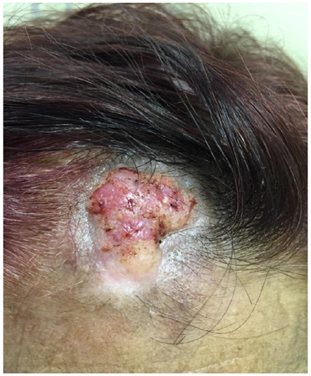
A 61-year-old woman was consulted for a pruritic lesion on her forehead that began 10 years ago after aneurysm surgery, with progressive growth. She admitted touching the lesion a lot, sometimes without feeling itchy, reporting that it worsened when she got nervous. On examination, she presented an infiltrated, elevated plaque with a fibrotic appearance, in the hair implantation line (Figure 1) and onychophagia (Figure 2) since childhood. The anatomopathological examination showed prurigoid dermatitis. Infiltration with triamcinolone + occlusive drenison was performed. After 6 months, there was slight improvement in the lesion and she still reports manipulating the lesion due to anxiety. A second anatomopathological examination showed skin with acanthopapillomatosis, hyperkeratosis and chronic eczema-like inflammation. The patient continued to undergo follow-up and injections with triamcinolone with unsatisfactory progress.
Discussion
By the term neurodermatitis, Brocq demonstrated the primary characteristic, the constancy and intensity of the pruritus that is pre-eruptive, having related the condition to the nervousness of the individuals. By the term lichenification (lichen facere), understanding that the pathogenesis of the lesion is the repeated trauma that, in predisposed subjects, ends up producing the characteristic changes in the appearance and structure of the skin [2].
In addition to the classic aspect, neurodermatitis can have some variations, such as purely papular, lichenoid papular, hyperkeratotic, or even a giant aspect when very extensive, with varying degrees of hyperchromia in any of these lesions. The areas most frequently affected are those that are easiest to reach with the hands. Deposits may occur of amyloid substance secondarily [3].
With a longer evolution time and/or more intense itching, neurodermatitis can evolve with an appearance hyperkeratotic, which occurs more easily in areas where the stratum corneum is thicker, such as knees, feet, and hands, but virtually any other area subject to constant trauma related to itching can assume this characteristic [4]. Although the hyperkeratotic aspect is frequent, we did not find in the literature a report of neurodermatitis with a tumoral appearance, as in this case.

Figure 1: Tumor lesion with fibrotic appearance and consistency, measuring 3.5 cm in the frontal hairline implantation line.

Figure 2: Traumatic onychodystrophy.
References
- Bologna JL, Schaffer JV, Cerroni Dermatology (4th ed.). Elsevier, 2018.
- Brocq Accurate elementary of dermatology. J.-B. Ballerina et son, 1906.
- Burns T, Breathnach S, Cox N, Griffiths Book of text of dermatology of Rook (8th ed.). Wiley- Blackwell, 2010.
- Charifa A, Badri T, Harris Lichen Simplex Chronicus. In: StatPearls [Internet]. Treasure Island (FL): StatPearls Publishing, 2024.
- Pruritus: therapies for localized pruritus. Disponible en.

