Unusual Cause of Urinary Retention
Sidki Kenza*, Madina Rabileh, Sara Ez Zaky, Omor Youssef and Latib Rachida
Department of Medicine, Mohamed V University Rabat, Morocco
Received Date: 18/08/2024; Published Date: 24/10/2024
*Corresponding author: Sidki Kenza, Department of Medicine, Mohamed V University Rabat, Morocco
Keywords : Carcinoma; Uretral; Urinary; Retention
History
A 34-year-old man presents to the emergency department with acute urinary retention and a sensation of suprapubic pain. His medical history, obtained during the clinical examination, revealed urological issues. The symptoms began two months prior with dysuria, pollakiuria, and three nocturnal voids with a weak urine stream, prompting a consultation with a urologist. The patient was started on antibiotic therapy after a positive cytobacteriological urine test, but there was only a temporary clinical improvement until his emergency visit. The patient also had a history of unprotected sexual intercourse and active smoking. Clinical examination showed a palpable bladder, induration of the bulbar urethra with bloody discharge, and inguinal lymphadenopathy. After placing a cystostomy catheter, a renal-bladder-prostate ultrasound was normal. However, a penile ultrasound and urethro-cystoscopy revealed a stenosing thickening of the bulbar urethra, and the histopathological study of the urethral biopsy confirmed urethral carcinoma. Based on all clinical, paraclinical, and histological findings, and after evaluating regional and distant tumor extension with MRI and CT scan, the patient underwent radical urethrectomy.
Diagnosis: Carcinoma of the bulbar segment of the male urethra.
Comments
Primary urethral carcinoma is considered a rare cancer, representing less than 1% of all malignant tumors, with a male predominance (sex ratio of 2.9). The peak incidence is in individuals older than 75 years, and it is negligible in those under 55 years, making this case somewhat unusual. Various predisposing factors have been reported, including urethral strictures, chronic irritation after multiple catheterizations, and chronic or recurrent urethritis due to sexually transmitted diseases (as seen in our patient). Urothelial carcinoma of the urethra is the predominant histological type (54-65%), followed by squamous cell carcinoma (16-22%) and adenocarcinoma (10-16%). Clinical examination should be thorough, including examination of the external genitalia with rectal examination and palpation of inguinal areas, sometimes under general anesthesia to assess the clinical stage.
Among the paraclinical exams for suspected urethral tumor, urine cytology is limited with sensitivity between 55 and 59%. Urethro-cystoscopy with biopsy provides histological proof, and radiological imaging aims to evaluate regional extension and detect distant metastases using CT and MRI; urography is now abandoned except for specific post-operative indications. The 1-year and 5-year relative survival rates for patients with urethral carcinoma in Europe are 71% and 54%, respectively. Predictive survival factors include patient age, Black race, tumor size and stage, histological type, and therapeutic modalities (extent of surgical treatment). Surgical excision remains the standard treatment for urethral carcinoma, with wide safety margins, improving functional outcomes and quality of life, often combined with preoperative cisplatin-based chemotherapy.


Figure 1:Penile ultrasound images: Axial section (A) and sagittal section (B) show heterogeneous hypoechoic tissue thickening of the penile urethra (asterisk) with diffuse infiltration and thickening of the penile tunics (arrow).


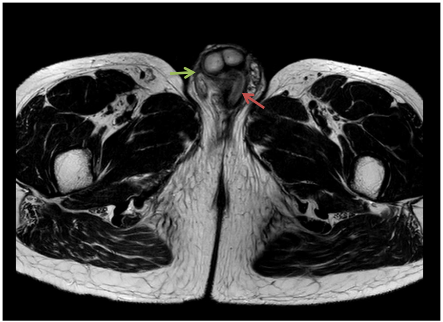
Figures 2: MRI images: Axial T2-weighted sequence (A) and coronal T2-weighted sequence (B); axial diffusion B1000 sequence (C); and axial T1 Fat Sat sequence with Gadolinium injection: shows intermediate T2 signal tissue thickening (pink arrow) of the penile urethra, with hyperintensity on diffusion B1000 and enhancement after Gadolinium injection. There is also infiltration of the penis (green arrow).

Figure 3: Microscopic image confirming urothelial carcinoma.
Conflict of Interest Statement: The authors declare no conflicts of interest.

