Arteria Lusoria: A Rare Aantomical Variant
Rabileh Madina*, Sidki Kenza, Insumbo Paulino, Lanjerie Safae, Wen-Yam Traoré, Saleheddine Tariq, Edderai Meryem and El Fenni Jamal
Department of Radiology, Mohammed V of the Military Hospital, Rabat, Morocco
Received Date: 01/08/2024; Published Date: 18/10/2024
*Corresponding author: Rabileh Madina, Department of Radiology, Mohammed V of the Military Hospital, Rabat, Morocco
Abstract
The arteria lusoria or right retro-oesophageal subclavian artery is the most common malformation of the aortic arch with a prevalence of 0.5-2.5%, can be associated with other congenital anomalies of the heart and great vessels. In most cases it is an asymptomatic conditions but It may be discovered in the face of symptoms of airway and/or oesophagea.
We are reporting the case of a patient who presented with a right axillary mass, which was confirmed to be an axillary PNET through biopsy. A cervico –thoraco abdominal pelvic CT scan conducted as part of the assessment for disease extension fortuitously revealed a right retroesophageal subclavian artery.
Keywords: Arc aortique; Arteria lusoria; Lymph node
Introduction
The term "arteria lusoria" refers to an anatomical variation of the arteries related to the supra-aortic trunks, specifically affecting the right subclavian artery. In this variation, the right subclavian artery originates directly from the aorta below the left subclavian artery and crosses the body's midline to reach the right subclavian area. This condition is the most common anomaly among the supra-aortic trunks. It takes an aberrant route, with an incidence of about 0.5% to 2% in the general population, and can be associated in 30% of cases with a bicarotid trunk [1]. It is most often asymptomatic and discovered incidentally.
Observation
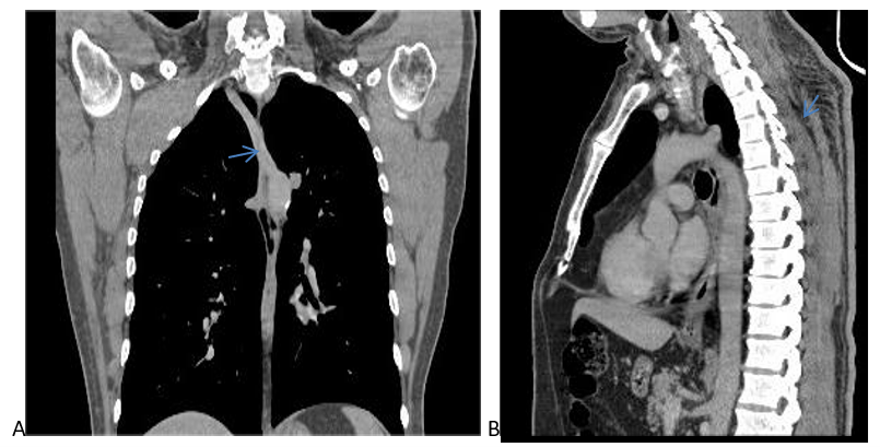
This concerns a 68-year-old patient with no notable prior medical history who presented with a large right axillary swelling, firm slighltyt movable compared to deep planes and skin, that has been developing for 8 months, accompanied by a deterioration in his general condition and long with tingling and numbness in the fingers of the hand. The anatomopathological analysis of the biopsy samples confirmed the diagnosis a Primitive Neuroendocrine Tumor (PNET) in the right axilla. She has been referred to our radiology department for a cervico-thoraco-abdominopelvic CT scan to clarify her anatomical relationships, especially as part of the assessment for disease extension. The analysis of media and parenchymal window cuts following the injection of iodine-based contrast agent has highlighted a voluminous double-component lesion process) and cyst portion, centered on the right axillary area and the pectoralis minor muscle, with an indistinct margin in the axillary region heterogeneously enhanced after injection. This process extends into the intercostoclavicular space and reaches the cervical base, respecting the thoracic cage without any underlying bone lysis additionally, multiple bilateral micronodular and parenchymal pulmonary lesions of random distribution, suggestive of secondary origins . The analysis of the computed tomography scans also highlighted the emergence of a vessel from the aortic arch that crosses the midline, travels posterior to the esophagus, and then proceeds forward and to the right towards the axillary region on the right side (Figure 1, 2), Its caliber was measured at 2.7 mm.
The patient was planned for salvage chemotherapy followed by radiotherapy sessions and The report, however, mentioned the presence of this anatomical variant of the supra-aortic trunks.

Figure 1: CT scan of the thorax in mediastinal window with injection of contrast product: (A: coronal section); (B: sagital section) , We note the direct origin of the right subclavian artery from the aorta (blue arrow).

Figure 2: CT of the mediastinal window with injection of contrast product (axial sections ) showing the course of vessel of the aortic arch, which crosses the median line, travels retroesophageally and finally directs forward and to the right to reach the level of the axillary region right (yellow arrow).
Discussion
Aberrant right subclavian artery, also known as arteria lusoria, is the most common malformation of the aortic arch, with an estimated incidence between 0.5 and 2% in the general population [1]. This vascular anomaly was described for the first time, to our knowledge, in 1735 by Hunauld [2].
Embryologically, the right subclavian artery develops through the remodeling of the fourth right aortic arch, the section of the dorsal aorta distal to this arch, and the sixth cervical intersegmental artery. The right dorsal aorta regresses beyond the sixth cervical intersegmental artery, and as the aorta grows caudally, anchored by its thoracic parietal branches, it separates. This detachment allows the right subclavian artery to be released from its connection to the thoracic aorta. The emergence of an arteria lusoria is a consequence of an interruption in this remodeling process There is abnormal degeneration of the fourth right aortic arch, and a lack of involution of the distal part of the right dorsal aorta. The aberrant right subclavian artery will therefore no longer be connected to the proximal part of the arch (ascending aorta) but will be attached to the descending aorta by the remainder of the right dorsal aorta. It then becomes the 4th and last branch of the aortic arch [4]. The pathophysiology of this malformation remains poorly understood and could involve a hemodynamic factor, in addition to a genetic and evolutionary basis [3].
The anatomical variation most frequently associated with the arteria lusoria is the origin of the left common carotid artery from the brachiocephalic arterial trunk, referred to as truncus bicaroticus. This occurs in about 30% of cases, as observed in our study [7]. Clinically, the arteria lusoria is often asymptomatic, as it does not form a complete ring around the esophagus or trachea. It is typically discovered incidentally during thoracic examinations for other conditions. However, it can become symptomatic primarily in three scenarios: firstly, when the esophagus and trachea are compressed between the arteria lusoria behind and the bicarotid trunk in front [5]; secondly, in the case of an aneurysm of this artery, which poses a significant complication. Lastly, with age, there may be atherosclerotic degeneration of the artery or the occurrence of fibro-muscular dysplasia [6]. The main clinical sign is dysphagia, referred to as lusorian dysphagia.
The diagnosis of arteria lusoria is established through imaging techniques. The first description of this anatomical variant was made by Kommerell, who utilized an esophageal transit X-ray with a radiopaque substance. As technology has advanced, so too have the methods of diagnosis. In this series, the diagnosis of arteria lusoria was confirmed through a CT scan following intravenous administration of a contrast agent. This method allowed for detailed visualization of the artery's origin, course, caliber, and other characteristics. Computed tomography is the preferred technique for incidental diagnoses, as it is a standard procedure commonly employed to investigate other thoracic lesions [8,9]. No treatment is required for an asymptomatic arteria lusoria. Treatment is only warranted if it leads to bothersome dysphagia or in cases of complications related to this artery, such as aneurysms, whether symptomatic or not.
Conclusion
Arteria lusoria is a rare vascular malformation, often asymptomatic, discovered incidentally. Diagnostic sensitivity of arteria lusoria is 100% on 64 multisplice computed tomography, allowing surgeons to be aware of this condition as it may complicate surgery to the mediastinum , and its diagnosis must lead the radiologist to look for cardiac and large vessel abnormalitie.
References
- Zapata H, Edwards JE, Titus JL. Aberrant right subclavian artery with left aortic arch: associated cardiac anomalies. Pediatr Cardiol, 1993; 14(3): 159-216.
- Hunauld PM. Variétés dans la distribution des vaisseaux. Hist Acad Roy Sci, 1735; VII:20.
- Louryan S, Vanmuylder N. Interprétation phylogénétique des anomalies des arcs aortiques et de la septation bulbaire Morphologie. EMC, 2014; 98(320): 18-26.
- Fanette Jeannon Arteria Lusoria: morphodensitometric study of 150 cases. Clinical applications. Life Sciences [q-bio], 2011; hal-01734018.
- Klinkhamer AC. Aberrant right subclavian artery: clinical and roentgenologic aspects. Am J Roentgenol Radium Ther Nucl Med, 1966; 97(2): 438-444.
- Gross RE, Ware PF. The surgical significance of aortic arch anomalies. Surg Gynecol Obstet, 1946; 83: 435-448.
- Myers PO, Fasel JHD, Kalangos A, Gailloud P. Arteria luso-ria: Developmental anatomy, clinical, radiological and surgical aspects. Ann Cardiol Angeiol (Paris), 2010; 59(3): 147-54.
- Kader Ndiaye, et al. Dyspneizing arteria lusoria: report of a case. Pan African Medical Journal, 2020; 37(318). doi: 10.11604/pamj.2020.37.318.23253.
- Nasser M, Petrocheli BB, Felippe TKS, et al. Aberrant right subclavian artery: case report and literature review. J Vasc Bras, 2023; 22: e20210151. https://doi.org/10.1590/1677-5449.20210151.

