Colloid Cyst: The Ticking Time Bomb within the Brain
Dr. Naser Mohamad Mansoor1, Dr. Ali Haider Ali2,*, Dr. Maryam Mahmood Ali2, Jainisha Thadhani3 and Dr. Marwa Al-Doseri4
1Consultant Emergency Medicine, Salmaniya Medical Complex, Kingdom of Bahrain
2Resident Emergency Medicine, Salmaniya Medical Complex, Kingdom of Bahrain
3Medical Student, Royal College of Surgeons of Ireland, Kingdom of Bahrain
4Resident Accident & Emergency, Salmaniya Medical Complex, kingdom of Bahrain
Received Date: 05/07/2024; Published Date: 10/10/2024
*Corresponding author: Ali Haider Ali, Resident Emergency Medicine, Salmaniya Medical Complex, Kingdom of Bahrain
Abstract
Colloid cysts are rare non-cancerous brain tumor, lined with an epithelium and containing a mucoid material. Colloid cysts are mainly occurring in the third ventricle nearby the foramen of Morno. Mostly, colloid cysts are asymptomatic and found incidentally on the brain images. However, as the colloid cysts grow, they can obstruct the flow of Cerebrospinal Fluid (CSF), which leads to hydrocephalus, brain herniation and sudden death. Colid cysts are identified as hyperdense mass on unenhanced Computer Tomography (CT) of the brain, whereas Magnetic Resonance Imaging (MRI) features are variable. Colloid cysts are life threating if misdiagnose or not treated properly. Treatment of the colloid cysts depend on the severity of the presentation. A life-saving procedure by inserting an External Ventricular Drain (EVD) should be done to relieve acute hydrocephalus. The common surgical management of colloid cysts include craniotomy with excision via a transcortical approach, interhemispheric transcallosal approach, or the endoscopic approach. In some cases, close observation is needed.
Here, we present two cases of colloid cyst, each with different presentations and variable courses of the disease, in order to emphasize the importance of promptly recognizing threatening signs and conducting clinical decisions. Founding the proper diagnostic approach early helps in making an accurate diagnosis and thereby enables appropriate management.
Keywords: Colloid Cyst; Neurosurgery; Headache; Surgery; Intra-Cranial Pathology
Introduction
Intracranial pathologies remain an interesting point of discussion within the medical field. With their unclear etiology and impact, such pathologies remain a huge area of debate within the neurosurgical community. One such pathology is known as a colloid cyst. Colloid, a benign epithelial growth usually contains gelatinous fluids [1]. Such fluids include mucin, blood, cholesterol, and particular ions, giving it its consistency [1,2]. Although can be found within any area of the cranium, it is usually located within the third ventricle or the foreman of Monro [1,2]. Due to its location, a spectrum of symptoms may appear which includes headaches up to sudden death [2]. Therefore, it is considered as a special and rare entity of mass growth within the neurosurgical field. In a report published in the year 2000, it was noted that 1.6% of cases within the neurosurgical department of Prince Albert Hospital in Australia were of colloid cysts. This case report will discuss two distinct presentations of colloid cysts, with an extensive literature review of the current literature.
Case 1
A 37-year-old South Asian male presented to the Emergency Department as a transfer from another hospital. The patient presented with a severe, tightening, headache for one day duration associated with multiple episodes of non-bilious, non-bloody vomiting. The patient underwent a Computed Tomography (CT) brain scan in the referring hospital which indicated dilated ventricles bilaterally with a hyperdense lesion within the third ventricle, noted as a possible colloid cyst. The patient at the time of the presentation was tired, making incomprehensible sounds associated with a decreased level of consciousness. A GCS 12 of out of 15 was calculated. Pupils were reactive and moving all four limbs with vital signs including BP 119/87, Heart Rate of 72 beats per minute, Respiratory Rate 15, Saturation 97%, and Temperature 37.4 degrees. CT was repeated and showed the same findings. While the patient was being referred to neurosurgery, the patient was intubated and sedated due to a decreased level of consciousness with a drop of saturation to 85% and blood pressure dropped to 86/48. On assessment by neurosurgery, the patient’s pupils were fixed and dilated with no response to pain and no corneal reflex. The patient was taken to the operation theater for bilateral external ventricle drain insertion. Surgery was done and during admission, the patient had a repeated CT showing Posterior Reversible Encephalopathy Syndrome (PRES). The patient collapsed with advanced cardiac life support protocol started. The patient expired after 20 minutes.
Case 2
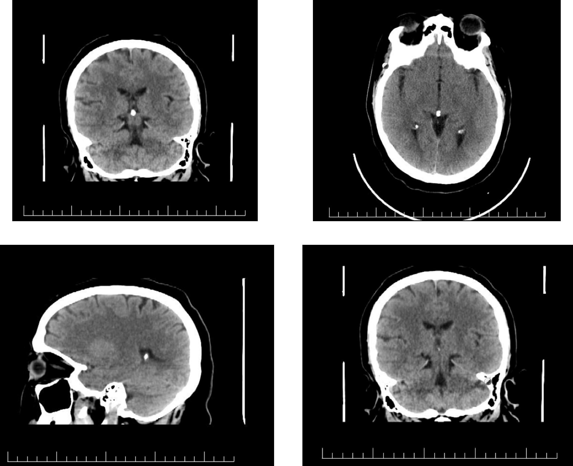
A 46-year-old Middle Eastern Female presented to the Emergency Department with complaints of severe headache associated with elevated blood pressure and one episode of vomiting. The patient is a known case of hypertension, diabetes mellitus type 2, hyperlipidemia, chronic kidney disease stage 4, and gastritis. The patient was noted to be in hypertensive emergency due to the symptoms with a blood pressure reading of 261/132. She was given captopril 25mg alongside amlodipine 5mg initially with CT brain arranged alongside basic laboratory investigations. After an hour and a half, the blood pressure reading was 213/141 and another dose of amlodipine 5mg was given. CT Brain was done and showed the presence of a colloid cyst. The patient was referred to Cardiology and Neurosurgery for elevated blood pressure and the incidental findings of the colloid cyst. Unfortunately, the patient signed leave against medical advice and left the hospital although educated about the seriousness of her findings. CT images can be seen in Figure One.

Figure 1: Initial CT Findings of Colloid Cyst.
Discussion
As previously mentioned, colloid cysts are benign epithelial growth associated with a gelatinous interior. The etiology of colloid cysts has been an area of debate. Most
Authors have agreed that it may have a congenital etiology, being remnants of certain ducts within the cerebrum [1,3]. Most argue that since it is commonly found within the third ventricle, it originates from the paraphysis element [3]. The paraphysis element is an embryonic duct located between the two hemispheres [3]. Yet, colloid cysts have been found in the frontal lobe and cerebrum, indicating that although congenital, they may arise from multiple embryonic origins [3]. In terms of epidemiology, colloid cysts are less than 2% of primary central nervous system tumors found [3,4], with about 99% of the tumors reported presenting within the third ventricle [2]. Therefore, it can be noted that the third ventricle is the most common location of colloid cysts in the cerebrum [2,4].
An interesting aspect concerning colloid cysts is the pathophysiology and its impact on the presentation. As mentioned previously, colloid cysts are usually found within the third ventricle, within the foreman of Monero. The foreman plays a major role in the outflow of Cerebrospinal Fluid (CSF) from the lateral ventricles into the third ventricle [3,5]. Therefore, due to the location of the cyst, it will act as a valve and may obstruct the inflow of CSF [3]. The backflow of CSF will enlarge the ventricles, leading to hydrocephalus [5]. This can be noted within the previously discussed case one. This has led to the presentation being noted, as an increase in fluids has led to compression symptoms upon presentation [5]. It is important to note that colloid cysts are usually slow-growing yet may lead to acute neurological deterioration such as sudden death [3,5]. These are noted in the two cases presented where a patient presented with an acute deterioration, while the second patient presented with acute onset of headache. As mentioned above, the clinical spectrum of presentation varies depending on the clinical route of growth. Symptoms include [4,5]:
- Asymptomatic, which is the usual presentation.
- Symptomatic, which usually occurs due to non-communicating hydrocephalus. These symptoms and signs include headache, ataxia, visual disturbances, altered mental state, urinary incontinence, lethargy, coma, increased reflexes, papilledema, nystagmus, inability to walk, and frontal release signs with chronic hydrocephalus.
Headaches are noted to be the most common symptomatic complaint [6]. These headaches have certain features, they are intermittent with increased severity and intensity [6]. Furthermore, they are short and mainly noted as frontal [6]. They are also usually relieved by lying down and are often associated with nausea and vomiting [6]. Such features may aid in the earlier diagnosis and requirement of further evaluation within the Emergency Department.
In terms of evaluation, imaging is the chosen modality. Either Computed Tomography (CT) or Magnetic Resonance imaging (MRI) of the brain are used [1,5]. CT is usually used due to its accessibility and time-saving prosperities [5]. Usually, a hyperdense mass is noted within the third ventricle associated with hydrocephalus, if chronic [5,6]. In severe cases, brain herniation may occur due to hydrocephalus [5]. MRI is usually the preferred method used for further evaluation, as an inpatient. They usually show hyperintense, isointense, or hypointense of MRI depending on the view used. These findings may correlate to histopathology and the viscosity of content [5]. In terms of management, it depends on the presentation. An acute presentation requires immediate management with external ventricular drains and removal of the cysts [7]. Surgical intervention may include craniotomy, endoscopic resection, transcortical, or stereotactic aspiration [7,8]. Endoscopic resection is the preferred method as it is less invasive with relatively high success rates [8]. In a publication reviewing about 105 cases, it was noted that about 90 patients had an endoscopic approach with minimal to no complication post-operatively [8]. Furthermore, the overall 5 years prognosis has been seen as 100% complete and successful resection with no complication nor reoccurrence [5,7-8]. Therefore, the overall prognosis of colloid cysts remains relatively positive, although the risk of sudden death and brain herniation, as seen in case one, remains plausible [8].
Conclusion
A colloid cyst is a benign and rare intracranial tumor. With an unclear etiology and serious effects of its pathophysiology, it may lead to serious complications such as sudden death and brain herniation. Therefore, prompt imaging such as brain CT must be done to exclude such diagnosis, especially with the presentation of red flags and rapid neurological deterioration. Management is usually noted as a surgical approach.
References
- Armao, Diane, et al. Colloid Cyst of the Third Ventricle: Imaging-Pathologic Correlation. American Journal of Neuroradiology, 2000; 21(8): pp. 1470–1477, www.ajnr.org/content/21/8/1470.short.
- Decq Philippe, et al. Endoscopic Management of Colloid Cysts. Neurosurgery, 1998; 42(6): p. 1288.
- Desai, Ketan I, et al. Surgical Management of Colloid Cyst of the Third Ventricle—a Study of 105 Cases. Surgical Neurology, 2002; 57(5): pp. 295–302, https://doi.org/10.1016/s0090-3019(02)00701-2.
- Humphries Roger L, et al. Colloid Cyst: A Case Report and Literature Review of a Rare but Deadly Condition. The Journal of Emergency Medicine, 2011; 40(1): pp. e5-9, https://doi.org/10.1016/j.jemermed.2007.11.110.
- Jeffree Rosalind L, Michael Besser. Colloid Cyst of the Third Ventricle: A Clinical Review of 39 Cases. Journal of Clinical Neuroscience, 2001; 8(4): pp. 328–31, https://doi.org/10.1054/jocn.2000.0800.
- Lagman Carlito, et al. Fatal Colloid Cysts: A Systematic Review. World Neurosurgery, 2017; 107: pp. 409–415, https://doi.org/10.1016/j.wneu.2017.07.183.
- Spears Roderick C. Colloid Cyst Headache. Current Pain and Headache Reports, 2004; 8(4): pp. 297–300. https://doi.org/10.1007/s11916-004-0011-2.
- Tenny Steven, William Thorell. Colloid Brain Cyst. Nih.gov, StatPearls Publishing, 2019.

