Association of a Volvulus of the Small Bowel and an Incomplete Jejunal Diaphragm in Newborns
Assia Mouad1,2,*, Fadoua Boughaleb1,2, Loubna Aqqaoui1,2,3, Houda Oubejja1,2,3,4,5 and Fouad Ettayebi1,2
1Paediatric Surgical Emergencies Department, Children’s Hospital of Rabat, Morocco
2Faculty of Medecine and Pharmacy, University Mohammed V, Rabat, Morocco
3Laboratory of Genetic and Biometry, Faculty of Science, University Ibn Tofail Kenitra, Morocco
4Laboratory of Epidemiology, Clinical Research and Biostatistics, Faculty of Medicine and Pharmacy, University Mohammed V, Rabat, Morocco
5SIM: Moroccan society of simulation in health care, Morocco
Received Date: 27/06/2024; Published Date: 09/10/2024
*Corresponding author: Assia Mouad, Paediatric Surgical Emergencies Department, Children's Hospital of Rabat, Ibn Sina University Hospital, Mohammed V Faculty of Medicine, Rabat, Morocco
Abstract
Intestinal malrotation, can result in volvulus and intestinal necrosis. Midgut volvulus, arising from malrotation, may be identified prenatally via ultrasound or MRI.
Congenital jejunal stenosis, a rare anomaly in newborns, along with different forms of intestinal atresias.
We present a unique case of small bowel volvulus linked to congenital incomplete jejunal diaphragm, diagnosed intraoperatively.
Keywords: Volvulus of the small bowel; Incomplete jejunal diaphragm; Newborn
Introduction
Anomalies in the development of the gastrointestinal tract, such as malrotation (more accurately described as malposition per Kluth et al.), lead to a constriction of the mesenteric root of the small intestine. This results in an unstable positioning of the gut, which is a significant factor in the occurrence of volvulus, posing a risk of extensive intestinal necrosis [1]. Complete midgut volvulus can subsequently result in irreversible intestinal necrosis, often necessitating extensive resection. Therefore, it is acknowledged as one of the three primary causes of short bowel syndrome in the pediatric population, alongside necrotizing enterocolitis and intestinal atresia [2,3].
We present a rare case of a volvulus of the small bowel associated with a congenital incomplete jejunal diaphragm, which was diagnosed in per-operatory.
Case Report
We report the case of a female newborn, admitted on day 3 of life for bilious vomiting.
On clinical examination, the newborn was hypotonic, with normally colored conjunctiva. The abdomen was flat.
The patient was placed in a warming table and underwent an oxygen therapy, fluid resuscitation and the placement of a nasogastric tube and a urinary catheter.
Radiological assessment included:
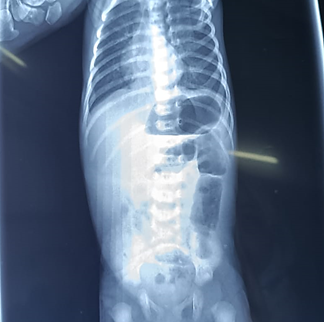
- Chest-abdominal X-ray in the upright position, which showed a double bubble appearance with aeration of the rest of the digestive tract (Figure 1).
- Abdominal Doppler ultrasound, which showed reversal of mesenteric flow with the superior mesenteric artery on the right and the vein on the left, and the presence of a Whirlpool Sign.

Figure 1: Chest-abdominal X-ray showing a double bubble appearance with aeration of the rest of the digestive tract.
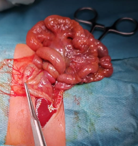
Following these assessments and after patient preparation, she was admitted to the operating room for small intestine volvulus. A right supraumbilical laparotomy revealed a volvulus of the small intestine with three twists but no intestinal necrosis (Figure 2).
Detorsion of the volvulus was performed, followed by release of Ladd's bands and mesenteric fusion between the first and last loops, and a precautionary appendectomy. The digestive tract was positioned in complete common mesentery before closure in layers.

Figure 2: Per operative view showing the volvulus of the small intestine with 3 twists.
The 39-day postoperative follow-up was marked by the appearance of an occlusive syndrome with bilious vomiting. The patient was readmitted to the operating room after preparation. Surgical exploration revealed loose non-occlusive adhesions throughout the small intestine and two tight occlusive adhesions adjacent to the first loop where two perforations and an incomplete jejunal diaphragm were discovered (Figure 3). Resection of the first loop followed by an end-to-end anastomosis between the duodenum and the second jejunal loop was performed.
The recovery was uneventful, with good clinical progress.

Figure 3: Surgical specimen: the 1st jejunal loop with 2 perforations and a jejunal diaphragm.
Discussion
Midgut volvulus arises due to intestinal malrotation and malfixation. The typical embryological bowel rotation, where the duodenojejunal and ileocolic segments of the bowel rotate 270° counterclockwise around the axis of the superior mesenteric artery, fails to take place [4]. Both the duodenojejunal junction (DJJ) and the ileocecal junction are improperly positioned, and the base of the small bowel mesentery is shortened, increasing the risk of volvulus occurring in the distal duodenum and proximal jejunum [5].
Midgut volvulus resulting from malrotation can potentially be identified on prenatal ultrasound through visualization of the "Whirlpool" or "snail sign" using color Doppler imaging [18]. While prenatal diagnosis of jejunoileal atresia (JIA) associated with volvulus is feasible, most cases also involve malrotation [19]. Antenatal MRI can provide precise delineation of both JIA and volvulus features [20]. However, in our case, prenatal diagnosis was not achieved. The small bowel volvulus was diagnosed through postnatal echodoppler, while the discovery of the jejunal diaphragm was only made during surgery.
Congenital jejunal stenosis is an exceedingly rare anomaly of the gastrointestinal (GI) tract in newborns. The prevalence rate of jejunoileal atresia and stenosis has been reported to range between 1 in 330 and 0.9 in 10,000 live births, with stenosis occurring in 7% of cases [6].
Conventionally, four types of intestinal (jejunoileal) atresias, each characterized by distinct anatomical features such as bowel continuity, length, mesentery defect, and vascular supply. Type I atresia is differentiated by intact bowel ends, continuous bowel wall and mesentery, and normal intestinal length. It includes a subtype with a perforated jejunal membrane obstructing the lumen, typically 2-3 mm thick with a central or eccentric aperture of 1-6 mm in diameter. Symptoms manifesting in childhood are inversely related to the size of the aperture, with smaller apertures presenting earlier. Due to its rarity, there are few reports on managing perforated jejunal diaphragm cases. Management typically involves surgical excision, with some cases utilizing intraoperative endoscopy for localization. Reported cases involve neonates to children, with procedures like web excision and enterotomy yielding successful outcomes [7-13].
Multiple authors have documented jejunoileal atresia (JIA) coupled with volvulus [15-17] . In the report of Shalini and al, the incidence of JIA with volvulus was 16.9% [14]. For instance, Komuro et al. reported a 27% occurrence of volvulus in 13 out of 48 patients with JIA [15].
Conclusion
Intestinal malrotation resulting in small bowel volvulus is one of the most common causes of neonatal obstruction. However, its association with incomplete jejunal diaphragm is rare. Diagnosis is often made intraoperatively.
References
- Kluth D, Hillen M, Lambrecht W. The principles of normal and abnormal hindgut development. J Pediatr Surg, 1995; 30: 1143-1147.
- Goulet O, Ruemmele F. Causes and management of intestinal failure in children. Gastroenterology, 2006; 130(2 Suppl 1): S16-28.
- Squires RH, Duggan C, Teitelbaum DH, Wales PW, Balint J, Venick R, et al. Pediatric Intestinal Failure Consortium. Natural history of pediatric intestinal failure: initial report from the Pediatric Intestinal Failure Consortium. J Pediatr, 2012; 161(4): 723-8.e2.
- Shah Mansi R, et al. "Volvulus of the entire small bowel with normal bowel fixation simulating malrotation and midgut volvulus." Pediatric radiology, 2015; 45: 1953-1956.
- Lampl B, Levin TL, Berdon WE, et al. Malrotation and midgut volvulus: a historical review and current controversies in diagnosis and management. Pediatr Radiol, 2009; 39: 359–366.
- Stollman TH, de Blaauw I, Wijnen MH, van der Staak FH, Rieu PN, Draaisma JM, et al. Decreased mortality but increased morbidity in neonates with jejunoileal atresia; a study of 114 cases over a 34-year period. J Pediatr Surg, 2009; 44: 217–221.
- Dave S, Gupta DK. Jejunoileal atresia. In: Gupta DK, editor. Textbook of Neonatal Surgery. 1st ed. New Delhi: Modern Publishers, 2000; p.181‑190.
- Grosfeld JL. Jejunoileal atresia and stenosis. In: O’Neill JA, Rowe MI, Grosfeld JL, Fonkalsrud EW, Coran AG, editors. Pediatric Surgery. 5th ed. St. Louis: Mosby, 1998; p. 1145‑1158.
- Thapa BR, Sahni A, Jethi SC, Rao KL, Mehta S. Jejunal diaphragm. Indian Pediatr, 1991; 28: 544‑546.
- Dalla Vecchia LK, Grosfeld JL, West KW, Rescorla FJ, Scherer LR, Engum SA. Intestinal atresia and stenosis: A 25‑year experience with 277 cases. Arch Surg, 1998; 133: 490‑496.
- De Backer T, Voet V, Vandenplas Y, Deconinck P. Simultaneous laparotomy and intraoperative endoscopy for the treatment of high jejunal membranous stenosis in a 1‑year‑old boy. Surg Laparosc Endosc, 1993; 3: 333‑336.
- Seltz LB. Case 1: A green case of failure to thrive. Paediatr Child Health, 2008; 13: 685‑687.
- Kothari PR, Kothari NP. Jejunal web with late presentation. Indian Pediatr, 2003; 40: 1109‑1110.
- Sinha Shalini, Yogesh Kumar Sarin. "Outcome of jejuno-ileal atresia associated with intraoperative finding of volvulus of small bowel." Journal of Neonatal Surgery, 2012; 1(3).
- Komuro H, Hori T, Amagai T, Hirai M, Yotsumoto K, Urita Y, et al. The etiologic role of intrauterine volvulus and intussusception in jejunoileal atresia. J Pediatr Surg, 2004; 39: 1812-1814.
- Dalla Vecchia LK, Grosfeld JL, West KW, West KW, Rescorla FJ, Scherer LR, et al. Intestinal atresia and stenosis: a 25-year experience with 277 cases. Arch Surg, 1998; 133: 490-496.
- Stollman TH, de Blaauw I, Wijnen MH, van der Staak FH, Rieu PN, Draaisma JM, et al. Decreased mortality but increased morbidity in neonates with jejunoileal atresia; a study of 114 cases over a 34-year period. J Pediatr Surg, 2009; 44: 217-221.
- Yoo SJ, Park KW, Cho SY, Sim JS, Hhan KS. Definitive diagnosis of intestinal volvulus in utero. Ultrasound Obstet Gynecol, 1999 ; 13: 200-203.
- Yu W, Ailu C, Bing W. Sonographic diagnosis of fetal intestinal volvulus with ileal atresia: A case report. J Clin Ultrasound, 2012. doi: 10.1002/jcu.21896.
- Simonovský V, Lisý J. Meconium pseudocyst secondary to ileal atresia complicated by volvulus: antenatal MR demonstration. Pediatr Radiol, 2007; 37: 305-309.

