Cardiac Metastases: An Unusual Site for Colon Cancer Metastases
Arpit Jain1, Varun Goyal2,*, Satyajeet Soni1, Satya Narayan1, Pallavi Redhu1, Pankaj Goyal1, Akanksha Jaju3, Nivedita Patnaik4 and Vineet Talwar1
1Department of Medical Oncology, Rajiv Gandhi Cancer Institute and Research Centre, Delhi, India
2Department of Medical Oncology, Sri Ram Cancer Center, MGMCH, Jaipur, India
3Department of Pathology, ESIC Hospital, Basaidarapur, Delhi, India
4Department of Pathology, BLK-Max Super Speciality Hospital, Delhi, India
Received Date: 18/06/2024; Published Date: 07/10/2024
*Corresponding author: Satyajeet Soni, Consultant, Department of Medical Oncology, Sri Ram Cancer Center, MGMCH,Jaipur, India
Abstract
Heart is an uncommon site of metastases for any primary. Carcinoma colon metastasize to heart is rarely occurring event and least rarely reported. To our knowledge, over the last 20 years only a few cases have been reported in the entire literature. We present a case of young lady known case of carcinoma colon presented with breathlessness found to have metastatic involvement of heart on further evaluation. Despite having progressive disease on follow up scan, our patient on treatment this metastatic lesion of heart responded well and symptoms improved.
Keywords: Cardiac metastases; Carcinoma colon; FOLFOX chemotherapy
Introduction
Secondary neoplasm of heart is uncommon and primary neoplasm is even lesser [1,2]. The most commonly involved primary tumors are carcinoma of the lung, breast, lymphoma, and malignant melanoma [3]. Metastatic cardiac involvement occurs most often in end stage of the malignant disease and associated with wide spread of the tumor, and generally diagnosed during autopsy. In colorectal cancer, the incidence of cardiac metastasis is highly variable ranging from 1.4%-7.2% in recent autopsy series [3,4]. To our knowledge, only a few cases have been reported of cardiac metastasis from colorectal cancer [5-8] and most of the cases diagnosed in autopsy, as most of the time patient has non-specific symptoms.
Case Presentation
A 41-year-old female a known case of Carcinoma sigmoid colon with liver metastases who underwent Total colectomy in July 2015 elsewhere and histopathology was suggestive of moderately differentiated adenocarcinoma {pT3N1M1} followed by she received six cycles of FOLFIRI based chemotherapy till end of 2015. In view of progression of disease including progressive liver lesion and new development of lung lesion she was treated with 18 cycles of FOLFOX till January 2017 with interim response evaluation and further she was continued on capecitabine.
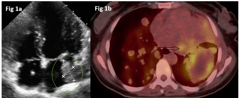
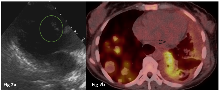
Further she was presented with complaints of cough with expectoration and breathlessness in April 2018. On evaluation chest X-ray was suggestive of multiple rounded radio opaque lesion scattered in bilateral lung parenchyma and a routine 2D echocardiography demonstrated left atrial (LA) enlargement with an irregular echogenic and inhomogeneous mass lesion (3 x 2 cm with area of 6 cm2) arising from lateral wall of Left atrium (Figure 1a). A PET-CT (May 2018) revealed metabolically active multiple pulmonary nodules, liver lesions and retroperitoneal lymph nodes, and a left atrial mass (Figure 1b). Serum CEA was 686 ng/ml and CT-guided biopsy from the liver lesion was suggestive of metastatic adenocarcinoma. On molecular testing it was MMR proficient and wild type KRAS/NRAS/BRAF. She was treated with FOLFIRI based chemotherapy and Panitumumab. After 6 cycles she had progressive disease on PET CT with subsequent reduction in the size of cardiac lesion size and her symptoms were also improved. She was started on FOLFOX + Bevacizumab-based chemotherapy and after 4th cycle, in view of hypersensitivity to Oxaliplatin chemotherapy regimen was changed FOLFIRI with Bevacizumab. After 10th cycle patient had again progression including liver lesion, lung lesion and bony disease with further reduction in size of cardiac metastases, however patient was asymptomatic for same. A follow-up ECHO showed regression of the cardiac disease (Figure 2a) with persistent and progression of disease on PET CT and absent cardiac activity as seen previously (Figure 2b). She was started on Regorafenib on progression but due to poor tolerance it was stopped and continued on best supportive care.

Figure 1a: 2D ECHO shows a moderate sized mass arising from lateral wall of Left atrium.
Figure 1b: Fused FDG PET/CT axial image shows metabolic activity in left atrium and metabolically active bilateral lung metastases.

Figure 2a: 2D ECHO shows significant decrease in size of mass almost negligible in comparison to previous mass.
Figure 2b: Fused FDG PET/CT axial image shows metabolically active bilateral lung metastases with regression of the left atrial mass.
Discussion
Colonic cancer metastasizes by hematogenous or lymphatic routes; lymph node, liver, lung and bones are sites commonly seen. Spleen, thyroid gland, spermatic cord, and skeletal muscles are unusual location and have been reported previously [9,10].
Cardiac metastasis from any malignancy is rare, but may be seen in carcinoma of the lung, breast, lymphoma, and malignant melanoma [3]. It may spread from primary neoplasm via lymphatic, hematogenous, or potentially by direct extension from an adjacent tissue.
The incidence of cardiac metastases reported in literature is highly variable, ranging from 2.3% and 18.3%.11 The actual incidence of cardiac metastatic disease may be underestimated because it is often be clinically silent and often missed during the initial evaluation of the primary malignancy as there presenting signs and symptoms may vary, depends on the location of tumor deposit. Potential manifestations are dyspnea and congestive heart failure, hypotension: malignant pericardial effusion, infarction and arrhythmias. However, in the majority of the reported cases, approximately 90% of the patients, cardiac metastasis is silent and diagnosed only on autopsy, whereas our patient had metastasis on progression of disease and found on routine 2D Echocardiography and PET Scan, it was located in left atrium and she was symptomatic due to cardiac lesion only as suggested on further follow up cardiac metastases responded well to treatment despite of continuous progression on other sites. Patient become asymptomatic and no any cardiac complication was noted.
Conclusion
In conclusion, cardiac metastasis in colonic cancer is rare; a chance finding of cardiac metastasis is likely to be increased by prompt investigation. Long-term survival can be achieved by early diagnosis and timely taken treatment decisions. Further studies and clinical trials are needed to establish treatment guidelines.
Declaration of patient consent: The authors certify that they have obtained all appropriate patient consent forms. In the form, the patient has given her consent for her clinical images and other clinical information to be reported in the journal. The patients understand that their names and initials will not be published and due efforts will be made to conceal their identity, but anonymity cannot be guaranteed.
Financial support and sponsorship: None
Conflicts of interest: None
References
- Lam KY, Dickens P, Chan AC. Tumors of the heart. A 20-year experience with a review of 12,485 consecutive autopsies. Arch Pathol Lab Med, 1993; 117(10): 1027-1031.
- Löffler H, Grille W. Classification of malignant cardiac tumors with respect to oncological treatment. Thorac Cardiovasc Surg, 1990; 38 Suppl 2: 173-175. doi: 10.1055/s-2007-1014062.
- Klatt EC, Heitz DR. Cardiac metastases. Cancer, 1990; 65(6): 1456-1459. doi: 10.1002/1097-0142(19900315)65:6<1456::aid-cncr2820650634>3.0.co;2-5.
- Scully RE, Mark EJ, McNeely WF, Ebeling SH, Phillips LD. Case records of the Massachusetts General Hospital. Weekly clinicopathological exercises. Case 20-1997. A 74-year-old man with progressive cough, dyspnea, and pleural thickening. N Engl J Med, 1997; 336(26): 1895-1903. doi: 10.1056/NEJM199706263362608.
- Teixeira H, Timóteo T, Marcão I. Metástases cardíacas de tumor do cólon [Cardiac metastases from a colonic tumor]. Acta Med Port, 1997; 10(4): 331-334.
- Lord RV, Tie H, Tran D, Thorburn CW. Cardiac metastasis from a rectal adenocarcinoma. Clin Cardiol, 1999; 22(11): 749. doi:10.1002/clc.4960221115.
- Oneglia C, Negri A, Bonora-Ottoni D, Gambarotti M, Bisleri G, Rusconi C, et al. Congestive heart failure secondary to right ventricular metastasis of colon cancer. A case report and review of the literature. Ital Heart J, 2005; 6(9): 778-781.
- Koizumi J, Agematsu K, Ohkado A, Shiikawa A, Uchida T. Solitary cardiac metastasis of rectal adenocarcinoma. Jpn J Thorac Cardiovasc Surg, 2003; 51(7): 330-332. doi: 10.1007/BF02719389.
- Bigot P, Goodman C, Hamy A, Teyssedou C, Arnaud JP. Isolated splenic metastasis from colorectal cancer: report of a case. J Gastrointest Surg, 2008; 12(5): 981-982. doi: 10.1007/s11605-007-0326-5.
- Choi PW, Kim CN, Kim HS, Lee JM, Heo TG, Park JH, et al. Skeletal muscle metastasis from colorectal cancer: report of a case. Journal of the Korean Society of Coloproctology, 2008; 24(6): 492. doi: 10.3393/jksc.2008.24.6.492.
- Bussani R, De-Giorgio F, Abbate A, Silvestri F. Cardiac metastases. J Clin Pathol, 2007; 60(1): 27-34. doi: 10.1136/jcp.2005.035105.

