Congenital Urethral Polyp: A Rare Cause of Lower Urinary Tract Obstruction
Mohamed Rami1,3, Badr Rouijel2,3,*, Hanae KhirAllah1,3, Asmae Abbassi1,3, Nawfal Fejjal1,3, Rachid Belkacem1,3 and Mohamed Amine Bouhafs1,3
1Pediatric Urology Department, Children's Hospital of Rabat, Morocco
2Pediatric Surgical Emergency Department, Children's Hospital of Rabat, Morocco
3Faculty of Medicine and Pharmacy of Rabat- Mohamed V University of Rabat, Morocco
Received Date: 30/03/2024; Published Date: 22/08/2024
*Corresponding author: Badr Rouijel, Pediatric Surgical Emergency Department, Children's Hospital of Rabat, Morocco; Faculty of Medicine and Pharmacy of Rabat- Mohamed V University of Rabat, Morocco
Abstract
Fibroepithelial polyps (FEP) of the lower urinary tract are relatively common in adults but rare in children. The clinical presentation is often nonspecific and varied : acute retention of urine, frequent urination and urination difficulties, urinary tract infection or hematuria. The diagnosis is made by ultrasonography, cystourethrography, and particularly urethrocystoscopy, which is currently the best method available for identification, histologic diagnosis and treatment of these polyps. In this papper, we present a case report of a boy of 16 months old with dysuria and intermittent acute urinary retention, who was diagnosed with an urethral fibroepithelial polyp.
Keywords: Fibroepithelial polyps; Acute urinary retention; Urethrocystoscopy; Transurethral resection
Introduction
Urethral fibroepithelial polyps (FEP) are considered as an infrequent benign neoplasms originating in mesenchymal tissue [1]. Urinary polyps can occur anywhere from the middle calices of the kidney to the anterior urethra. While they are less prevalent in the lower urinary tract compared to the upper urinary tract, they are often observed in children than adults, with a higher incidence among boys than girls [2]. Their occurrence is high during the first decade of an individual’s life. The etiology of this disease remains uncertain. However, in pediatric cases, congenital factors appear to be more common [3]. Although rare, urethral FEP should be highly considered in the differential diagnosis of lower urinary tract obstruction.
Case Presentation
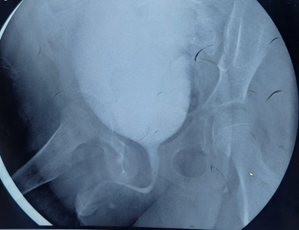
We report a case of a 16 months old boy, who came to consultation with a history of 4 months of dysuria and drip urination. He also presented intermittent acute urinary retention. He had no medical history including absence of urinary tract infection. The physical examination was normal. The ultrasound of the urinary tree revealed no abnormality as well as the retrograde urethrocystography which objectified a free urethra (Figure 1). Urinalysis, blood tests were within normal ranges.

Figure 1: Urethrocystography showing a free urethra.
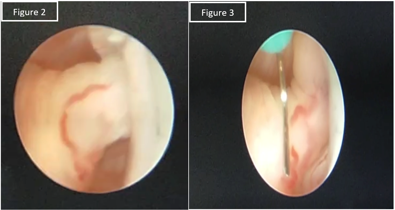
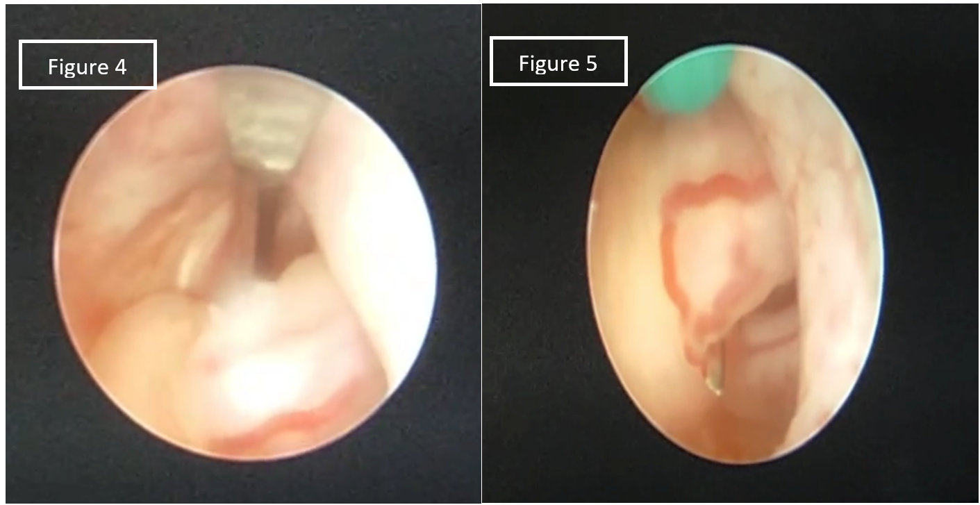
The patient underwent urethrocystoscopy, under general anesthesia, using a 9 Fr rigid pediatric cystoscope. Examination showed mild bladder trabeculation and a pedunculated polypoid mass of 5 mm beside the verumontanum, and located in the posterior urethral wall (Figure 2-3). Then, the base of the polyp was completely resected under cold blade, avoiding injury of the ejaculatory ducts (Figure 4-5). At the end of the procedure, an 8 Fr Foley catheter was placed.

Figure 2-3: Urethrocystoscopy revealing a pedunculated polyp of 5 mm beside the verumontanum.

Figure 4-5: Urethrocystoscopy showing the resection of the base of the polyp under cold blade.
The postoperative period was uneventful, the catheter was removed after 2 days and the patient was discharged the same day after spontaneous urination of clear urine.Anatomopathological examination of the excised polyp revealed a pink-tan, polypoid mass, measuring 5mm. Microscopic findings showed the tumor’s polypoid structure supported by a large dense hypervascularized fibroconjunctive axis, with normal urothelium and without evidence of malignancy. This finding confirmed the diagnosis of a urethral fibroepithelial polyp (Figure 6).

Figure 6 : Microscopic findings showingthe tumor’s polypoid structure supported by a large dense hypervascularized fibroconjunctive axis, with normal urothelium ( HE stain, x200 original magnification).After one year of follow-up, the patient was asymptomatic and no evidence of reccurence was reported.
Discussion
Fibroepithelial polyps (FEP) are a rare entity that can be encountered during childhood, as a pedunculated lesion mostly located in the posterior urethral wall [4].
The incidence is unknown because few series are reported, with fewer than 250 cases in the literature to date [4]. FEP are Found mainly in boys and exceptionally in girls, with a median age of 5.2 years [6]. The etiology is essentially congenital but various factors such as infective, irritative, traumatic, and obstructive causes have been proposed [3].
The lesion is usually single, but multiple polyps have been described [7]. Association with another abnormality of the urinary tree, in particular vesicoureteral reflux, is reported in 50% of cases [5].
The clinical triad of intermittent urinary retention, hematuria, and lower urinary tract symptoms has already been described by Akbarzadeh et al. in 2014, as being clearly suggestive of urethral polyps in children [5].
Ultrasonography is an adequate assessment tool when a diagnosis of FEP is likely, and can be considered to be the first-line morphological examination, which shows indirect signs of bladder outflow obstruction (hydronephrosis and large bladder with or without hypertrophy of the wall) and sometimes the presence of a pedunculated structure of moderate visible mobile echogenicity on decubitus images [8]. In case of doubt or no visible mass by ultrasonography, cystourethrography appears to be the second-line examination. It also has the advantage of excluding posterior urethral valves, which is the differential diagnosis in case of obstructive bladder symptoms in males [8]. The definitive diagnosis is endoscopic. Indeed, cystoscopy can be employed both for the diagnosis, particularly histopathological confirmation, as well as therapeutic purposes.
The standard treatment for polyps is transurethral resection, typically conducted using a resectoscope or forceps to effectively fulgurate the base of the polyp. The use of the HOLMIUM-YAG laser in endo-urology has been proven to perform ablation with finer precision than diathermic electrocoagulation to reduce damage to the external sphincter or ejaculatory ducts [6]. But there are no documented publications regarding its application in lower urinary tract locations in children. Laser therapy, particularly with Holmium, is preferred for ureteral polyps, with several polypectomies using this method reported in children [9].
Conclusion
While relatively uncommon, the diagnosis of urethral FEPs in pediatric patients should not be overlooked, particularly in cases presenting with intermittent urinary retention, hematuria, and lower urinary tract symptoms. Ultrasonography and cystourethrography can be used to diagnose, but urethrocystoscopy coupled with histologic findings is required for conclusive diagnosis. Transurethral polyp resection has been demonstrated as a safe and effective method for managing FEPs. They are considered as a benign tumors with an excellent prognosis since there is often no recurrence after a complete resection.
Conflict of Interest: All the authors declare that they do not have any conflict of interest.
Consent of Publication: Consent from parents has been taken.
Author’s Contribution: All the authors have contributed to the redaction of this manuscript.
References
- Liu XS, Kreiger PA, Gould SW, Hagerty JA. Congenital urethral polyps in the pediatric population. Can J Urol, 2013; 20(5): 6974–6977.
- Demircan M, Ceran C, Karaman A, Uguralp S, Mizrak B. Urethral polyps in children: a review of the literature and report of two cases. Int J Urol, 2006; 13(6): 841–843.
- Razzok A, Muhamad MS, Ismaeel A, Alyousef K, Oukan M. Posterior urethral polyp in a male child: a rare case report. omac131 Oxf Med Case Rep, 2022; 12: 2022. https://doi.org/10.1093/omcr/omac131.
- Rousseau S, et al. Management of lower urinary tract fibroepithelial polyps in children Journal of Pediatric Surgery, 2021: 56(2): 332-336.
- Akbarzadeh A, Khorramirouz R, Kajbafzadeh AM. Congenital urethral polyps in children: report of 18 patients and review of literature. J Pediatr Surg, 2014; 49(5): 835–839.
- Torino G, Brandigi E, Roberti A, Turra F, Palazzo G, Di Iorio G. Posterior Urethral Polyp Treated with Endoscopic Transurethral Resection: The Second Pediatric Case Managed with Holmium Laser Urology, 2020; 143: 238−240.
- Jain P, Shah H, Parelkar SV, Borwankar SS. Posterior urethral polyps and review of literature. Indian J Urol, 2007; 23: 206-207.
- Chiari ÆPG, Romano ÆF. Cassani et al Urethral polyp in a 1-month-old child Pediatr Radiol, 2005; 35: 691–693. DOI 10.1007/s00247-004-1397-z
- Li R, Lightfoot M, Alsyouf M, et al. Diagnosis and management of ureteral fibroepithelial polyps in children: a new treatment algorithm. J Pediatr Urol, 2015; 11: 22.e21 26.

