Laparoscopic Transperitoneal Surgery of the Retrocaval Ureter: A Case Report with Review of the Literature
Yassine Daghdagh, Anas Tmiri*, Adil Kbirou, Amine Moataz, Mohamed Dakir, Adil Debbagh, Rachid Aboutaieb
Urology Department, Ibn Rochd University Hospital Center, Casablanca, Morocco
Received Date: 14/03/2024; Published Date: 02/08/2024
*Corresponding author: Anas Tmiri, Urology Department, Ibn Rochd University Hospital Center, Casablanca, Morocco
Abstract
Retrocaval ureter or circumcaval ureter is considered a rare congenital anomaly, the cause of this malformation is an anomaly in the embryogenesis of the inferior vena cava. We present here the case of a 19-year-old man with a retrocaval ureter. Who was the first case successfully treated by laparoscopy in the Urology Department of the Ibn Rochd University Hospital.
Keywords: Laparoscopy; Retrocaval ureter
Introduction
The retrocave (or circumcave) ureter is a congenital malformation characterized by a spiral course of the right lumbar ureter around the Inferior Vena Cava (IVC).
The cause of this malformation is an anomaly in the embryogenesis of the inferior vena cava and not of the upper urinary tract. From an embryological point of view, this anomaly corresponds to a pre-ureteral IVC. This malformation may remain quiescent, or lead to ureteral obstruction, which may require surgical treatment [1].
We present here the case of a 19-year-old man with a retrocaval ureter. Who was the first case successfully treated by laparoscopy in the Urology Department of the Ibn Rochd University Hospital?
Case Presentation
A 19-year-old male presented to the outpatient department of urology at our hospital with intermittent right lower back pain for the past three months. His medical history was unremarkable. There was no hematuria or other urinary tract symptoms. General physical examination revealed mild tenderness in the right flank. Other systems were normal.
Biologically, renal function was 7 mg/l, CRP negative and CBEU was sterile; other biological parameters were normal.
Abdominal ultrasound showed right hydronephrosis. The initial Uro-CT objectified: ureterohydronephrosis upstream of lumbar ureteral stenosis. In view of the hyperalgesic nature of the lumbar pain, the patient underwent a right ureteral endoprosthesis.
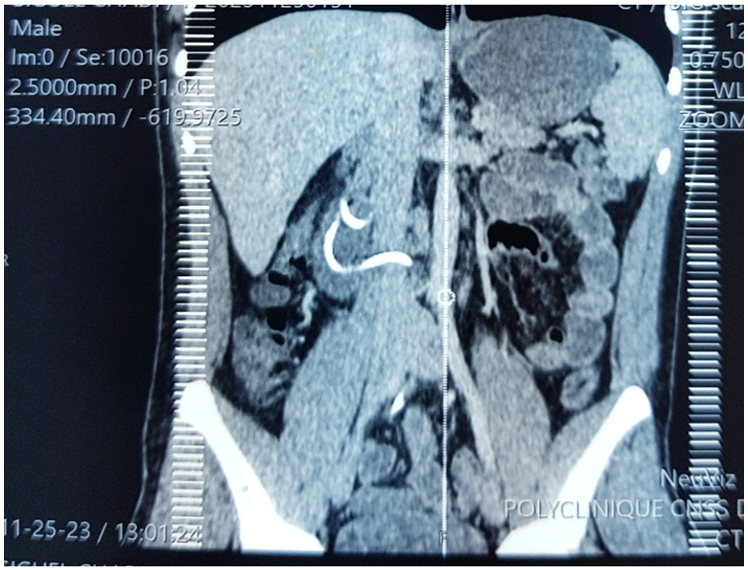
A second Uro-CT objectified: a discreet pyelocalic and right ureteral dilatation with an initial retrocaval and interaortocaval course of the ureter, resuming its normal course at the iliac and pelvic level (Figure 1).

Figure 1: Uro-CT objectified a discreet pyelocalic and right ureteral dilatation with an initial retrocaval and interaortocaval course of the ureter.
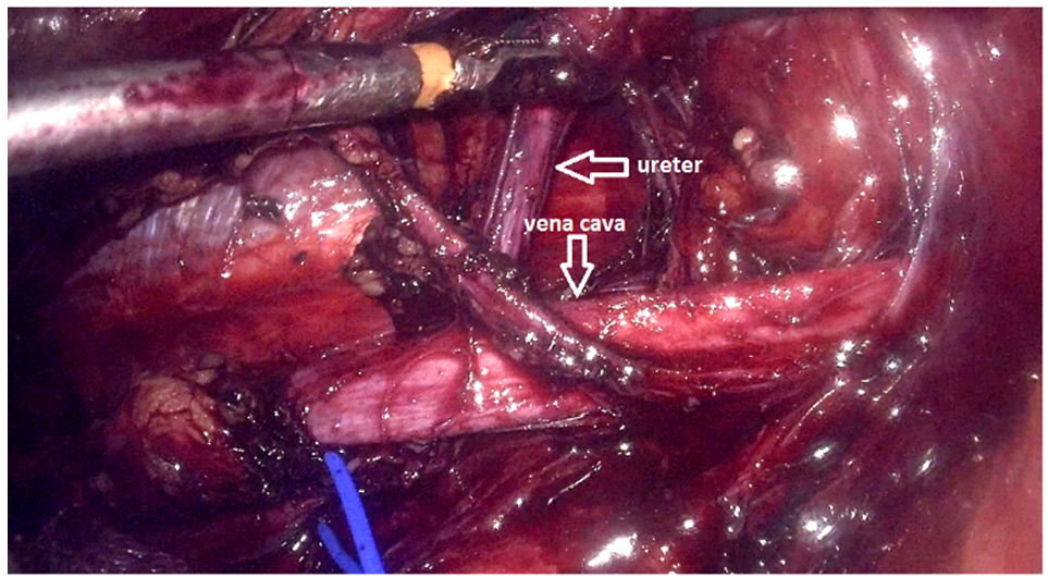
Based on these results, we decided to operate on the patient by transperitoneal laparoscopy. After locating the pelvis and the right ureter, which was dilated, the segment of the retrocaval ureter was located lower than the pyelo-ureteral junction. We sectioned the pathological segment (∼3 cm) of the ureter (Figure 2).
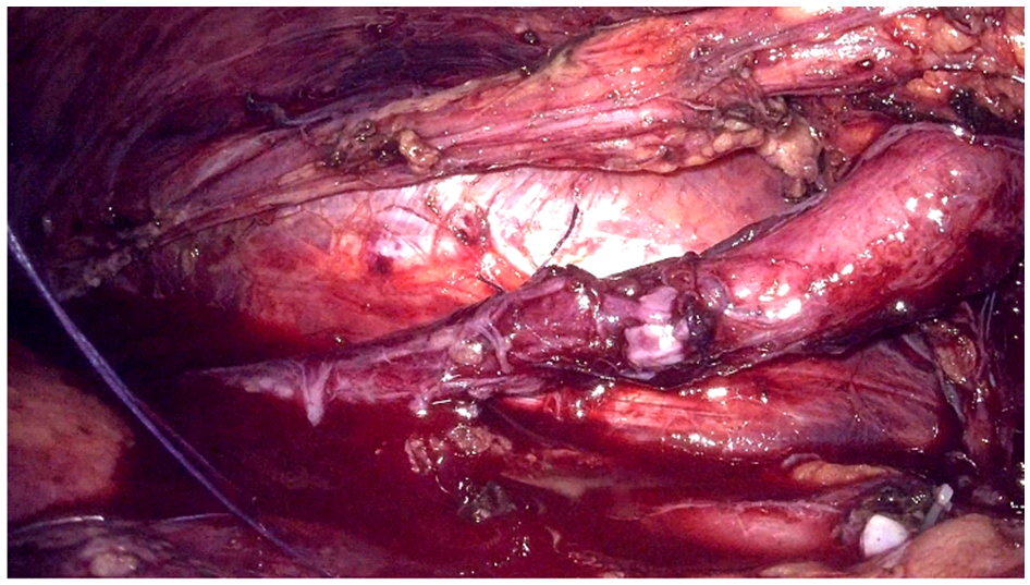
We then performed a uretero-ureteral anastomosis with insertion of a double-j stent (Figure 3).

Figure2: Segment of the retrocaval ureter.

Figure 3: Uretero-ureteral anastomosis after sectioning the stenotic zone.
Discussion
The defect known as retrocaval ureter is an uncommon congenital condition resulting from the aberrant development of infrarenal veins (IVC) from anteriorly positioned subcardinal veins rather than posteriorly located supracardinal veins. A portion of the proximal ureter becomes entrapped, causing the ureter to coil around the IVC. As a result, other names for it include preureteral vena cava and circumcaval ureter [2].
The retrocaval ureter is a rare malformation whose exact frequency is unknown; its incidence is thought to be of the order of 1 per 1,000 births [3]. The anomaly is about three times more common in men, and the age at diagnosis is usually between 20 and 40. Uncertainty about its frequency stems from the fact that it is usually asymptomatic. Currently, imaging tests such as abdominal computed tomography (CT) or magnetic resonance imaging (MRI) are making incidental diagnoses increasingly frequent [1].
The retrocaval ureter has different anatomical types, depending on the position of the crossing of the ureter around the vein vena cava. Radiological examination helps to define the morphology of the malformation, as defined by Bateson and Atkinson [4]. Two types of retrocave ureter have been defined:
- Type 1, the most common form, corresponds to an abrupt hook-shaped trajectory at L3; radiologically, there is an inverted J image on ureterography.
- Type 2 is described as a progressive coiling of the ureter, in which case the initial portion of the ureter is retrocave.
In this case, the abnormality was type 1 and the obstruction was at the right side beside the lateral margin of the IVC at the level of lumbar vertebrae L2–L3.
Patients usually present with symptoms associated with ureteral obstruction and hydronephrosis, such as right lumbar pain, recurrent urinary tract infections and renal lithiasis. In the present case, the patient presented with intermittent low-back pain with no hematuria [5].
Imaging is an essential aspect of the diagnostic and pre-therapeutic work-up of a retrocaval ureter. Each morphological examination (ureteral opacification by Intravenous Urography [IVU], retrograde ureteropyelography, URO-CT or abdominal MRI) helps to determine the level of the anomaly, the extent of dilatation and the local anatomical conditions. Renal scintigraphy is used to assess the functional value of the kidney in cases of obstructive syndrome associated with renal atrophy [1].
Surgical intervention is required in symptomatic patients or those with worsening renal function [5].
Nephrectomy is justified in symptomatic patients whose kidney is destroyed, with a low functional value on scintigraphy, in order to avoid complications [1].
Conservative surgical treatment is indicated for all symptomatic patients with preserved homolateral renal function.
The principle of conservative surgery on the retrocave ureter is to restore the normal anatomical situation, by unhooking the ureter from the inferior vena cava. This may be achieved by sectioning and anastomosing the ureter, or sectioning and anastomosing the inferior vena cava (a technique now abandoned). This may or may not be combined with resection of the retrocaval ureter. Surgery may be performed open, laparoscopic transperitoneally, laparoscopic retroperitoneally, or robot-assisted [1].
Conclusion
Paraclinical imaging is used to diagnose the retrocaval ureter and assess its impact on right kidney function. CT is the imaging test of choice. Treatment is surgical, and can be performed laparoscopically or openly.
After one month, there was no more hydronephrosis, and we removed the double-j stent.
References
- Cornu J.-N., Sèbe P.Uretère rétrocave.EMC Urologie, 2011; 18-158-A-10.
- Qureshi MA, Mulvaney WP. Retrocaval ureter: Report of two cases. The American surgeon, 1965; 31: 50–52.
- Simforoosh N, Nouri-Mahdavi K, Tabibi A. Laparoscopic pyelopyelostomy for retrocaval ureter without excision of the retrocaval segment: first report of 6 cases. J Urol, 2006; 175: 2166-2169.
- Bateson E, Atkinson D. Circumcaval ureter: a new classification. Clin Radiol, 1969; 20: 173-177.
- Maher Al-Hajjaj, Mohamed Tallaa. Retrocaval ureter: a case report Journal of Surgical Case Reports, 2021; 1: 1–3.

