Squamous Cell Carcinoma a Rare Complication of Chronic Intertrigo
Fajri Zineb*
Department of Dermatology, Hassan University Hospital Center II, Morocco
Received Date: 09/03/2024; Published Date: 18/07/2024
*Corresponding author: Fajri Zineb, Department of Medicine, Hassan University Hospital Center II, Morocco
Abstract
Verrucous Carcinoma (VC) is a rare variant of squamous cell carcinoma. These carcinomas are most found in the oral, laryngeal, nasal or genital areas, but can occur anywhere on the integument. It is a locally aggressive tumor. Etiological risk factors are chronic inflammatory skin conditions, trauma, chemical carcinogens, Human Papillomavirus (HPV), the location in intertriginous areas of the foot is exceptional [1]. Here we report a case of verrucous carcinoma in chronic intertrigo.
Keywords: Verrucous carcinoma; Chronic intertrigo; Dermoscopy
Introduction
Verrucous carcinoma is a rare type of Squamous Cell Carcinoma (SCC). These carcinomas are most found in the oral, laryngeal, nasal or genital areas, but can occur anywhere on the integument. It is a locally aggressive tumor. Etiological risk factors are chronic inflammatory skin conditions, trauma, chemical carcinogens, Human Papillomavirus (HPV), It is characterized by a locally aggressive malignancy and a low incidence of metastasis. The localization in intertriginous areas of the foot is rare, only a few cases of intertrigo (SCC) have been reported in the literature, we describe a new case of VC in chronic intertrigo.
Case Report
A ninety-year-old male with no medical history presented with a 4-year history of intertrigo of the 4th intertrigo space initially treated by oral antibiotic and antifungal treatment without improvement.
The evolution was marked by the appearance of an extended, painful, hyperkeratotic tumor gradually increasing in size over a year. The patient reported a notion of naked walking and repeated trauma.
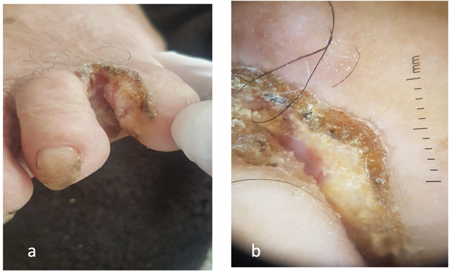
A clinical examination revealed a tumor, approx. 2.5 cm in size, overflowing on the back of the foot with an ulcerated, warty surface with an infiltrated base located in the last intertriginous space of the right foot.
Dermoscopy of the lesion showed papillomatous appearance, keratin deposit, (Figures A: a – b). After performing biopsy, the lesion was diagnosed as verrucous squamous cell carcinoma. Standard radiographs did not show any bony involvement Laboratory test results were normal. No abnormalities on ultrasound of lymph nodes Human papilloma virus viral typing was negative, and no local or distant metastasis was detected.
A wide resection of the lesion was performed with a safety margin of 5 mm, including the fourth toes. Histopathology examination of the excised specimen showed an invasive, differentiated, and keratinizing squamous cell carcinoma of total excision.

Figure A: (a) A tumor, approx 2.5cm in size, overflowing on the back of the foot with an ulcerated, warty surface, (b) Dermoscopy of the lesion showed papillomatous appearance, keratin deposit, hemorrhagic suffusion.
Discussion
Squamous cell carcinoma is a well-differentiated, low-grade malignant carcinoma. first described by Ackermann in 1948, which defined clinical and histological criteria [2]. The preferred sites for this type of carcinoma are the skin, esophagus, oral cavity, larynx, plantar surface, and genitals [2]. intertrigo localization is exceptional. Tumor occurring most frequently at the level of the last two interdigital spaces in benign chronic lesions that do not respond to specific treatment and most predominantly in lesions of mycotic intertrigo.
An etiology of verrucous carcinoma remains less clear, some publications have reported the factors that favour the development of Squamous cell carcinoma at the intertrigo, with maceration being considered the main an etiological factor [3]. The other causes, especially chronic inflammation such as psoriasis, traumatization, Human papilloma virus (HPV) has been associated with this tumor and specifically HPV types 11 and 16 have been described in plantar lesions [4]. remain unexplained.
But in most cases reported in the literature, interdigital squamous cell carcinoma has been considered as an intertrigo mycosis [2,3],
The clinical appearance is that of vegetative, exophytic tumors with a verrucous and ulcerated surface with keratinous debris and an infiltrated base extending beyond the visible limits of the tumor. The tumor then expands in surface and height by overflowing the dorsal side of the feet.
The dermoscopy findings of verrucous carcinoma are the presence of a white background (amorphous keratinous masses, yellowish white to light brown), a verrucous appearance, a polymorphous vascular pattern with more than one type of vessel dominating (consisting of linear, irregular, hairpin, glomerular and, rarely, dotted types), and ulceration.
The most common differential diagnoses are, verrucous leishmaniasis, Bowen's disease, verrucous tuberculosis, verruca vulgaris, deep mycoses, atypical mycobacteriosis, and common warts [5].
Histological features of verrucous carcinoma include a papillomatous surface and broad, blunt tongues of epithelial tissue extending into the dermis, few cytological atypia, mitoses are usually limited to the basal layer and are few, presence of neutrophils [6], which makes difficult the distinction with psedo-epitheliomatous hyperplasia and common wart [7]. The prognosis of Verrucous Carcinoma (VC) is often favorable but local invasiveness and
The progress is mainly local with a rare risk of bone lysis [6]. Metastatic risk is low Surgical excision is the treatment of the choice. Clinical follow-up of these patients is very essential, as recurrences are common even after surgery.
Conclusion
Verrucous carcinoma is a rare subtype of squamous cell carcinoma, the clinical, topographical, and therapeutic characteristics of which should be understood to allow appropriate management. The localization in intertriginous areas of the foot remains unusual and the particularity of the observation is the presentation of squamous cell carcinoma in chronic intertriginous sites. This recent observation reminds us of the importance of regular clinical and dermoscopy monitoring of chronic cutaneous intertrigo, as well as other benign lesions, which should be biopsied in case of doubt in order to allow for an early diagnosis and appropriate treatment.
References
- McKay C, McBride P, Muir J. Plantar verrucous carcinoma masquerading as toe web intertrigo. Australasian Journal of Dermatology, 2012; 53(2): e20-e22.
- Ackerman LV. Verrucous carcinoma of oral cavity. Surgery, 1948; 23: 670–678.
- Terrab Z, Azzouzi S, Benchikhi H, Chlihi A, Lakhdar H. Inter-toes space cutaneous epidermoid carcinoma. Ann Dermatol Venereol, 2006; 133: 456-458.
- Lamchahab EF, Guerrouj B, Bourra B, Marot L, Zouaidia F, Lamzaf O. Aggressive course of intertoe verrucous carcinoma. Ann Dermatol Venereol, 2012; 139: 510-513.
- D Sgouros, M Theofili, V Damaskou, S Theotokoglou, K Theodoropoulos, et al. dermoscopy as a tool in differentiating cutaneous squamous cell carcinoma from its variants. Dermatol Pract Concept, 2021; 11: e2021050.
- Varma R, Asokan N, Sarin A, Rahman N. Unusually localised cutaneous multifocal squamous cell carcinoma. Our Dermatol Online, 2019; 10: 170-172.
- EL Amraoui M, Frikh R, Hjira N, Boui B. Degeneration of chronique intertrigo into squamous cell carcinoma. Our Dermatol Online, 2021; 12: 76.

