Emphysematous Pyelonephritis Complicated by a Perirenal Collection
Tmiri A*, Elbadr M, Kbirou A, Moataz A, Dakir M, Debbagh A and Aboutaieb R
Department of Urology, Ibn Rochd University Hospital Center, Morocco
Received Date: 15/02/2024; Published Date: 04/07/2024
*Corresponding author: Anas Tmiri, Department of Urology, Ibn Rochd University Hospital Center, Casablanca, Morocco
Summary
Emphysematous pyelonephritis constitutes a rare but serious clinical form of acute pyelonephritis, characterized by the presence of gas in the renal parenchyma, excretory cavities or the perirenal space. It is fatal in the absence of early diagnosis and adequate treatment. It constitutes a medical-surgical emergency.
In this present work, we present a case of emphysematous pyelonephritis complicated by a perirenal collection.
Keywords: Acute pyelonephritis; Emphysematous pyelonephritis; Serious urinary tract infection; Surgical treatment; Conservative treatment; Percutaneous drainage
Introduction
Emphysematous pyelonephritis is a rare clinical entity, its incidence is increasing due to better knowledge of the disease, the diffusion of CT scanning, or the increase in the incidence of diabetes, it represents a serious complication of acute pyelonephritis which can be life-threatening, characterized by the presence of gas in the renal parenchyma, the excretory cavities or the perirenal space in connection with non-anaerobic gas-producing bacteria. It is found mainly in diabetics and immunocompromised subjects [1,2].
Treatment must be urgent based on medical treatment and/or surgical treatment, associated with percutaneous drainage [2].
We report a case of emphysematous pyelonephritis associated with a perirenal collection.
Case Report
Patient aged 27, with no particular pathological history, who presented with chronic right lower back pain that had been developing for a year and had been accentuated for a week before her hospitalization, associated with lower urinary tract problems of the irritative type, all evolving in an unquantified febrile context. with chills and night sweats.
The clinical examination noted a conscious patient in fairly good general condition, febrile at 40°C, the urogenital examination revealed right lumbar tenderness with hypogastric tenderness, the rest of the clinical examination was unremarkable.
The biological assessment showed hyperleukocytosis at 34,370/mm3 with predominantly neutrophils (29,902/mm3), CRP at 272 mg/l, with normal renal function and infected ECBU (multi-susceptible Escherichia coli).
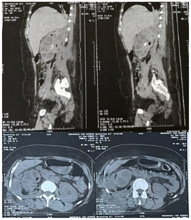
Uro-CT objectified an enlarged right kidney measuring 15.2 cm, a right mid-renal stone of 23 mm with density of 270 HU and dilation of the pyelocalicial cavities with numerous intra-renal air bubbles, infiltration of perirenal fat associated with voluminous collection of the perirenal compartment which extends posteriorly at the level of the external paravertebral soft parts to a height of nearly 6cm, this collection extends widely at the level of the psoas with numerous bubbles at the level of the psoas and in height The extension occurs at the level of a collection of the diaphragmatic pillar and a pulmonary condensation of the right pulmonary base.
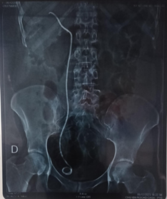
The patient was initially put on triple antibiotic therapy (Ceftriaxone, Amikacin and Metronidazole) with diversion of the urinary tract by raising a JJ catheter and percutaneous drainage of the perirenal collection initially bringing back 700 ml of purulent fluid.
The outcome was favorable with clinical-biological improvement.

Figure 1: Uro-CT reveals emphysematous pyelonephritis with an extensive collection.

Figure 2: Urinary Tract Without Preparation after raising the right JJ probe.
Discussion
Emphysematous Pyelonephritis (EPN) is a severe, necrotizing form of acute bacterial pyelonephritis, first described in1898, resulting in the production of gas within the renal parenchyma. The two main factors contributing to EPN are diabetes and urinary tract obstruction [3,4].
Enterobacteriaceae are incriminated in most cases, E. Coli is the germ most often implicated in 60% of cases, other Gram-negative germs such as Enterobacter aerogenes, Klebsiella sp., and Proteus are incriminated in most other reported cases [3]. It occurs mainly in diabetics, with a female predominance and an average age of 54 years [5].
The clinical presentation is that of severe acute pyelonephritis, with fever, chills, lower back pain, nausea and vomiting [6]. The onset of symptoms can be sudden or more abrupt over two to three weeks. Computed tomography allows the diagnosis to be made, showing non-liquid parenchymal destruction, gas in lumps or streaks extending from the medulla to the cortex, sometimes a crescent of subcapsular or perinephric gas [6].
Huang and Tseng [7] established a scan classification having both prognostic value and an impact on the therapeutic decision:
- Stage 1: gas in the excretory tract only.
- Stage 2: gas in the renal parenchyma without extension into the extrarenal space.
- Stage 3A: extension of gas or abscess of the renal compartment.
- Stage 3B: extension of gas or abscess beyond Gerota's fascia.
- Stage 4: bilateral or single kidney emphysematous pyelonephritis.
EPN has an overall severe prognosis. The overall mortality rate, all therapies combined, is 19%. Four significant prognostic factors were retained by Wan et al.8the prognosis is all the more unfortunate as it is a type I scan, the serum creatinine is greater than 120 mol/l, there is a thrombocytopenia less than 60,000 elements/mm3 and there is hematuria whose importance reflects the severity of renal destruction and/or the presence of venous thrombosis [4,8].
Bilateral forms are uncommon (5 to 20%) but particularly serious since they have a mortality rate approximately 20 times higher than unilateral forms. EPN is a medico surgical emergency. Initial probabilistic antibiotic therapy combines a third-generation cephalosporin or imipenem with a fluoroquinolone or an aminoglycoside. Conservative treatment consisting of percutaneous or surgical drainage can be offered in the absence of poor prognostic factors or when it is a single kidney or a bilateral form. Drainage of the excretory tract by a percutaneous nephrostomy probe or by a ureteral probe will be indicated in forms localized to the excretory tract or in case of obstruction. The use of secondary nephrectomy becomes legitimate in the absence of clinical improvement under drainage. Nephrectomy, first rescue, can be reserved for extensive forms with several organ dysfunctions [4,9].
Conclusion
Emphysematous pyelonephritis is a serious infection that can be life-threatening. It is a condition that particularly affects diabetics. It must be mentioned early in case of urinary infection with serious clinical signs, to allow conservative treatment, the latter is based on antibiotic therapy, resuscitation and drainage of urine and it must not delay a possible salvage nephrectomy.
References
- Bacha MM, Mami I, Gaied H, et al. Pyélonéphrite et cystite emphysémateuses: une complication exceptionnelle chez un transplanté du rein. Néphrologie & Thérapeutique, 2021; 17(6): 458-462. doi: 10.1016/j.nephro.2021.02.003
- Lasri A, Saouli A, Yddoussalah O, et al. La pyélonéphrite emphysémateuse à évolution favorable après traitement médical: à propos de 3 observations. Pan Afr Med J, 2018; 30: 233. doi: 10.11604/pamj.2018.30.223.12086
- Kably MI, Elamraoui F, Chikhaoui N. Pyélonéphrite emphysémateuse : diagnostic radiologique. Annales d’Urologie, 2003; 37(5): 229-232. doi: 10.1016/S0003-4401(03)00093-7
- Derouiche A, Ouni A, Agrebi A, Slama A, Ben Slama MR, Chebil M. La prise en charge des pyélonéphrites emphysémateuses. À propos de 21 cas. Progrès en Urologie, 2008; 18(2): 102-107. doi: 10.1016/j.purol.2007.08.002
- Guillausseau PJ, Farah R, Laloi-Michelin M, Tielmans A, Rymer R, Warnet A. Urinary tract infections and diabetes mellitus. Rev Prat, 2003; 53(16): 1790-1796.
- Ruiz A, Fabre C, Boutault JR, Merzeau C. Pyélonéphrite emphysémateuse. Journal d’imagerie diagnostique et interventionnelle, 2018; 1(3): 172-173. doi: 10.1016/j.jidi.2018.02.010
- Huang JJ, Tseng CC. Emphysematous pyelonephritis: clinicoradiological classification, management, prognosis, and pathogenesis. Arch Intern Med, 2000; 160(6): 797-805. doi: 10.1001/archinte.160.6.797
- Wan YL, Lee TY, Bullard MJ, Tsai CC. Acute gas-producing bacterial renal infection: correlation between imaging findings and clinical outcome. Radiology, 1996; 198(2): 433-438. doi: 10.1148/radiology.198.2.8596845
- Sodqi M, Marih L, Nassib M, Himmich H. Une pyélonéphrite emphysémateuse bilatérale d’évolution favorable après traitement médical seul. Médecine et Maladies Infectieuses, 2006; 36(3): 174-176. doi: 10.1016/j.medmal.2005.12.002

