Large renal cyst infected, About a Case
Tmiri A*, Elbadr M, Kbirou A, Moataz A, Dakir M, Debbagh A and Aboutaieb R
Urology Department, Ibn Rochd University Hospital Center, Casablanca, Morocco
Received Date: 12/02/2024; Published Date: 21/06/2024
*Corresponding author: Anas Tmiri, Urology Department, Ibn Rochd University Hospital Center, Casablanca, Morocco
Summary
Renal cysts represent a very common clinical entity, generally benign and asymptomatic, clinical-radiological monitoring can be proposed in association with analgesic treatment, the surgical indication arises when there is a large and/or symptomatic renal cyst resistant to medical treatment. or complicated, in 5 to 15% of cases renal tumors can be cystic in character with changes defined by the Bosniak classification.
Renal cyst infection constitutes a rare complication which represents 2.5% of complications; emergency diversion may be necessary associated with appropriate medical treatment based on antibiotic therapy.
We report a case of a large renal cyst infected.
Keywords: Renal cyst; Cystic renal masses; Bosniak classification; Renal cyst infected
Introduction
Cystic renal masses describe a spectrum of lesions presenting benign and/or malignant characteristics, they are defined on radiographic and/or pathological findings. Approximately 5–15% of renal tumors have a cystic component, with the Bosniak classification defining a cystic renal mass as containing less than 25% reinforcing tissue [1].
Cyst infections are often caused by an ascending infection of the lower urinary tract; this complication is very rare. only 2.5% of all complications [2].
This work reports a case of a patient with a a large renal cyst infected.
Case Report
A 66-year-old patient, chronic smoker, without particular pathological history, presented with febrile left lower back pain associated with a single episode of pyuria, without hematuria or other accompanying signs, all evolving in a context of febrile sensations and preservation of blood pressure condition.
On admission, the patient was conscious, hemodynamically and respiratory stable, his conjunctivas were discolored, febrile at 38.3°C. The clinical examination revealed left lumbar tenderness, a palpable mass on the left flank, rounded, of firm consistency, with regular boundaries, fixed in relation to the deep plane, painful on palpation. On rectal examination, the prostate is multinodular, firm, non-painful, estimated at 40g, with a soft bladder base.
His biological assessment revealed an inflammatory syndrome with anemia at 5.4 g/dl, associated with an infectious syndrome consisting of leukocytosis at 40,470 with a neutrophil predominance (PNN at 37,430), thrombocytopenia at 1,280,000/mm3, CRP elevated to 172.8 mg/L, acute renal failure with a creatinine level of 56.7 mg/L, a serum potassium level of 5.5 mEq/L and a PSA level of 4.84 ng/ml.
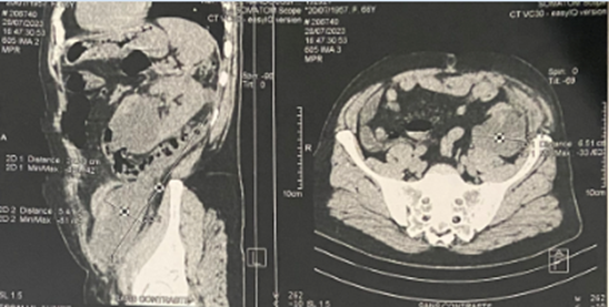
An abdominal and pelvic CT scan carried out before admission revealed a left retroperitoneal infectious collection measuring 22 x 10 cm on a large renal cyst infected with intra-cystic air images within the retroperitoneal and intra-vesical collection with normal sized and contoured kidneys. bumps associated with minimal left UHN and minimal right hydronephrosis.

Figure 1: Abdominopelvic CT revealed a large renal cyst measuring 22 x 10 cm which extends from the kidney to the left inguinal region.
The patient underwent emergency diversion of the infected renal cyst by drain, initially bringing back 500 ml of purulent contents, the cytobacteriological examination revealed an infection of the cystic contents (multi-susceptible Escherichia Coli), he was placed on tri-antibiotic therapy at based on ceftriaxone, aminoglycoside and metronidazole with a good clinico-biological evolution.
Discussion
The definition of renal cysts is purely radiological based on the cystic scan appearance in relation to the renal parenchyma determined by the Bosniak classification which makes it possible to decide between a simple renal cyst (classified 1 and 2) without signs of malignancy, which requires a clinico-radiological monitoring, and renal cysts suspicious for malignancy (classified 3 and 4) which require surgical treatment. Benign renal cysts have been detected incidentally during abdominal evaluations of people over 50 years of age. Cysts can be complicated by bleeding, rupture or infection [1,3].
Infection is a particularly rare cause of complication, responsible for only 2.5% of all complications. The renal cyst may be infected by hematogenous spread of bacteria by means of ascending urinary tract infection with or without vesicoureteral reflux, or surgery, the wall may be thickened, and sometimes calcifications are observed. The infected cyst contains pus and debris. Similar pathological findings are seen in an abscess that has progressed from pyelonephritis to a mature stage of liquefaction and encapsulation [3].
The most efficient imaging test is positron emission tomography coupled with computed tomography using as a marker fluorine 18 incorporated into a glucose molecule forming [18F]-fluorodeoxyglucose ([18F]-FDG-PET/CT) and first-line treatment is 6 weeks of antibiotic therapy with a fluoroquinolone. Aspiration aspiration of the infected cyst is indicated in the initial treatment of very large cysts and for diagnostic purposes in cysts resistant to well-conducted antibiotic treatment [4].
Aspirating the cyst(s) responsible is often a delicate and sometimes dangerous procedure. It only seems indicated to us in very large cysts (mainly therapeutic) or in cases of resistance to well-conducted antibiotic treatment (mainly diagnostic), provided, of course, that the responsible cysts have been formally identified by imagery [4].
Conclusion
Renal cyst infection represents a rare but serious complication which can be life-threatening and which requires adequate management based mainly on good antibiotic therapy associated or not with diversion of the infected contents.
References
- Majed Alrumayyan, Lucshman Raveendran, Keith A Lawson, Antonio Finelli. Cystic Renal Masses: Old and New Paradigms, Urologic Clinics of North America, 2023; 50(2): Pages 227-238. https://doi.org/10.1016/j.ucl.2023.01.003.
- Paul Geertsema, Anna M Leliveld, Niek F Casteleijn. The Presence of Kidney Cyst Infections in Patients with ADPKD After Kidney Transplantation: Need for Urological Analysis? Kidney International Reports, 2022; 7(8): Page 1924. https://doi.org/10.1016/j.ekir.2022.03.039.
- Toprak U, Erdoğan A, Akar E, Karademir AM. Infected renal CYST: Unusual cause of renal vein thrombosis. European Journal of Radiology Extra, 55(3): 97–99. doi: 10.1016/j.ejrex.2005.07.006
- Yves Pirson, Nada Kanaan. Complications infectieuses associées à la polykystose rénale autosomique dominante, Néphrologie & Thérapeutique, 2015 ; 11(2): Pages 73-77. https://doi.org/10.1016/j.nephro.2014.11.008.

