Cutaneous Manifestation in a Patient with Atrial Myxoma: Unconventional Method of Disclosure
Marouane Bouazaze*, Asmaa Bouamoud, Jamila Zarzur and Mohamed Cherti
Department of Medicine, Mohamed V of Rabat, Morocco
Received Date: 18/10/2023; Published Date: 19/03/2024
*Corresponding author: Bouazaze Marouane, Department of Medicine, Mohamed V of Rabat, Morocco
Abstract
Cardiac myxoma is a histologically non-cancerous tumor that can lead to potentially severe systemic complications. Due to the diverse clinical symptoms associated with atrial myxoma, the heart's involvement in the condition can often be unclear. Common clinical indicators include obstructions, embolic events, and constitutional signs and symptoms. Some patients may even be directed to a dermatologist to rule out vasculitis or connective tissue diseases. An accurate diagnosis necessitates a strong suspicion and knowledge of the various ways in which cardiac myxomas can manifest. Non-invasive echocardiography can effectively detect the tumor, and surgical removal can result in a cure.
We present the case of a 53-year-old male who had been suffering from recurring skin rashes and unexplained muscle pain. He was admitted to the emergency room after experiencing a subacute ischemic stroke affecting the left frontal parietal lobes.
Introduction
Cardiac myxomas, the most prevalent primary heart tumors, are primarily found in the left atrium and often remain asymptomatic until they present with embolic manifestations [1].
The diversity of clinical presentations contributes to delays in diagnosis.
Case Report
We present the case of a 53-year-old male who had been experiencing recurrent skin rashes and muscle pain of unknown etiology. He was admitted to the emergency room following a subacute ischemic stroke affecting the left frontal parietal lobes.
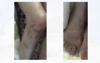
On admission, he displayed incoherent verbal communication and residual weakness on the right side. Cardiovascular examination yielded normal findings, but skin examination revealed maculopapular lesions on the lower limbs (Figure 1).
The ECG displayed sinus rhythm with supraventricular extrasystoles.
Trans-thoracic ultrasound unveiled a mobile, large, and non-obstructive mass with varying echogenicity in the left atrium, measuring 3.5-4.5 cm in diameter and extending from the interatrial septum (Figure 2).
Urgent cardiac surgery was performed, during which the polypoid tumor was successfully removed.
Following the surgery, the patient remained free of cutaneous symptoms.

Figure 1: Skin presentations on the lower limb.

Figure 2: Transthoracic echocardiography revealing a mass of the left atrial.
Discussion
Cardiac myxoma, the most prevalent primary cardiac tumor in adults, occurs at an estimated incidence of 0.5 per million population per year. These tumors typically favor the atrium, predominantly implanting on the inter-atrial septum near the fossa ovalis.
Ventricular myxomas, which constitute just 5% of cases, are primarily situated in the right ventricle [2].
They are typically solitary, with multiple occurrences being rare and linked to familial forms.
The clinical presentation of cardiac myxoma is remarkably diverse, often posing diagnostic challenges for clinicians. Cardiac manifestations can manifest as paroxysmal mitral stenosis or regurgitation, syncope in specific positions, palpitations, dyspnea, chest pain, or heart failure. Concurrently, cutaneous signs can arise due to myxoid emboli, leading to symptoms such as limb ulceration, digital ischemia, erythematous popular eruptions, or splinter hemorrhages in the nails. Autoimmune phenomena, like Raynaud's phenomenon, may also be observed.
Echocardiography is the cornerstone for diagnosis, boasting a sensitivity of 93.3% and specificity of 96.7% [3].
It aids in identifying the myxoma's location, size, mobility, and cardiac implications, effectively distinguishing it from vegetations or thrombi. Its appearance is distinctive: an elongated, translucent mass with a domed and multi-lobed surface composed of gelatinous and fragile tissue. However, echocardiography may not always provide definitive confirmation [4].
Treatment involves surgical excision of the tumor, with an excellent short-term prognosis characterized by nearly negligible operative mortality. The long-term prognosis remains uncertain due to the risk of local or metastatic recurrences, necessitating ongoing monitoring.
Conclusion
Cutaneous manifestations are infrequent in patients with cardiac myxoma, and they may represent the only symptoms.
Dermatologists should be vigilant about these signs as they can indicate underlying cardiac myxomas and prevent further complications.
References
- Glock Y, et al. Le myxome cardiaque: diagnostic, traitement et devenir tardif (à propos d'une série de 15 cas). Coeur (Paris. 1970), 1985; 16(3): p. 253-262.
- Centofanti P, et al. Primary cardiac tumors: early and late results of surgical treatment in 91 patients. The Annals of thoracic surgery, 1999; 68(4): p. 1236-1241.
- Denguir R, et al. Les myxomes cardiaques. Prise en charge chirurgicale. À propos de 20 cas. in Annales de cardiologie et d'angeiologie, 2006.
- Goodwin, J., Symposium of cardiac tumors. Am J Card, 1988; 121: p. 1307-1314.

