Osgood-Schlatter’s Disease: A Little-Known Pathology in Rural Areas
Tamassi Bertrand Essobiyou1,2,*, Sosso Piham Kebalo3, Boris Steffane Zibi Meka II4, Alexandre Palissam Keheou1, Mohamed Issa1 and Anani Abalo4
1General surgery department, Sylvanus Olympio University Hospital Center, Lome, Togo
2General surgery department, Dapaong Regional Hospital Center, Dapaong, Togo
3Pediatric Surgery Department, Sylvanus Olympio University Hospital Center, Lome, Togo
4Traumatology and orthopaedics Department, Sylvanus Olympio University Hospital Center, Lome, Togo
Received Date: 11/09/2023; Published Date: 13/02/2024
*Corresponding author: Dr. Essobiyou Tamassi Bertrand, General surgery department, Sylvanus Olympio University Hospital Center, Lome, Togo; General surgery department, Dapaong Regional Hospital Center, Dapaong, Togo
Abstract
Osgood-Schlatter’s disease is the apophysosis of the anterior tibial tuberosity. It occurs preferentially in adolescent males. Its etiology remains unknown. We report a case of late diagnosis in Togo. A 13-year-old boy with no previous history of the disease presented with bilateral afebrile gonalgia that had been present for 14 months. The examination allowed us to note multiple previous unsuccessful consultations and to make the diagnosis of Osgood-Schlatter disease. Osgood-Schlatter disease is easily diagnosed. Management must be early and adapted to avoid complications.
Keywords: Osgood-Schlatter; Osteochondrosis; Apophysis; Gonalgia; Togo
Introduction
Osgood-Schlatter disease is an osteochondrosis involving the anterior tibial tuberosity [1-2]. Ogden describes Osgood-Schlatter disease as an avulsion of the anterior tibial tuberosity (ATT) [4]. The disease is a disease of the adolescent and is predominantly male [1-4]. The etiopathogenesis of the condition is unknown [3]. It is a condition marked by gonalgia related to heavy physical exertion [1,3]. The diagnosis of the disease is based on clinical presumption [2-3]. Management is relatively easy and is based on rest [2-3]. The prognosis of the condition is usually favourable [2-3]. However, some cases of complications have been reported [2]. In Togo, the lack of documentation on this disease could be the reason for a lack of awareness of this condition on the part of health personnel. We report a case of late-onset Osgood-Schlatter disease in a regional hospital in Togo.
Care Report
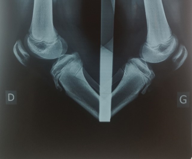
He was a 13 years old adolescent, in the 3rd grade, with no known pathological history, who was seen in a surgical consultation for bilateral gonalgia. The interview revealed knee pain that had been triggered for 2 weeks by sports activities at school, with increasing intensity during exercise. They were calmed by stopping the activity and resting. This is an acute episode on a chronic background of 14 months. The disease evolves in a non-febrile context. Moreover, the parents reported several medical consultations with subsequent treatments based on analgesics (paracetamol, codeine) and anti-inflammatories (ibuprofen, diclofenac) without success. No prolonged exemption from previous sports activity was reported. The patient's examination did not reveal any spontaneous pain. We found a prominence of the anterior tibial tuberosities (ATT) with pain on palpation. Examination of the rest of the knees was normal. There was no limitation of joint range of motion. A comparative radiograph of the knees showed bilateral avulsion with fragmentation of the ossification nuclei of the ATT (Figure 1). This led to the diagnosis of Osgood Schlatter disease. The patient was treated with an exemption from sports activities for 3 weeks. The evolution was favourable with an improvement of the pain after resumption of sports activities. The follow-up over a period of 6 months was satisfactory.

Figure 1: The radiographic appearance of bilateral avulsion with the fragmentation of the ossification nuclei of anterior tibial tuberosities.
Discussion
To our knowledge, no study on Osgood-Schlatter disease has been previously done in Togo. The available national data concern rheumatic diseases in children in general [5-6].
Osteochondrosis is a non-infectious disease that causes abnormal ossification of secondary bone nuclei [7]. 7] Of these, those that affect the apophyses are called apophysoses [7]. Apophysoses are a frequent reason for consultation in children [1,3,7]. Osgood-Schlatter disease is the apophysosis of the anterior tibial tuberosity [1-2,7].
The diagnosis of Osgood-Schlatter disease is relatively easy. The diagnosis is mainly based on clinical findings [1-3,7]. The disease peaks in adolescence during the growth spurt and is most common in males [2-3,7]. Clinically, it is associated with mechanical pain in the anterior aspect of the knee, triggered, maintained and aggravated by exercise and calmed by stopping the exercise and resting [3,7]. Inspection may reveal a prominent ATT and palpation of the ATT is painful [3,7]. This pain may also be caused by the active knee extension manoeuvre [3,7]. Although radiography is the imaging test of choice, it only contributes to a positive diagnosis in rare cases [2-3,7]. It is only used for atypical forms and to eliminate differential diagnoses [1-2,7]. Radiographic abnormalities in Osgood-Schlatter disease include: prominence of the ATT, soft tissue enlargement, fragmentation of the opacification nucleus; the latter may exist physiologically [1,3,7]. Our patient presented with fragmentation of his 2 anterior tibial tuberosities.
The etiopathogenesis of Osgood-Schlatter disease is poorly understood [2-3,7]. A disturbance in endochondral ossification during growth may be the cause [7]. Several authors have reported a multifactorial origin [7]. The microtraumatic component due to overloading remains the most widely described factor [2-3,7].
Conservative treatment is the first line of treatment. It is mainly based on a functional approach [7]. It is based on rest, analgesics and anti-inflammatories [2]. No ideal duration of rest has been reported in the literature. In our case, it was 3 weeks simply for convenience. Surgery has only limited indications [3-4]. It is reserved for complicated forms and hyperalgesic manifestations of the disease [3].
The prognosis is usually favourable, with recovery occurring without sequelae at the end of growth [3,7]. However, this condition is associated with certain complications [3-4,7-8]. We can cite retraction of the hamstrings or the sural triceps, patella alta, genu recurvatum and patellofemoral osteoarthritis [7-8]. Knowledge of this condition is therefore essential for early diagnosis and, above all, for appropriate treatment.
The delay in the diagnosis of our patient was 14 months. The case was reported in the Regional Hospital of Dapaong in Togo. This hospital has an insufficient number of qualified personnel. The whole region has neither a rheumatologist nor a paediatric surgeon. Consultations are carried out by doctors but in most cases by medical assistants and nurses.
Conclusion
Osgood-Schlatter disease is the apophysosis of the anterior tibial tuberosity. It mainly affects adolescent males. Its etiopathogenesis is poorly understood. The diagnosis of the disease is essentially clinical and management is conservative. In Togo, the real problem with this condition is not defined because of the lack of data. An extended study of Osgood-Schlatter disease is therefore essential.
Conflict of interest: The authors declare that they have no known competing financial interests or personal relationships that could have appeared to influence the work reported in this paper.
References
- Rabouin J, Foulquier N, Jousse Joulin S, Devauchelle Pensec V, Garrigues F, Laporte JP, et al. Osgood Schlatter ' s disease: what imaging? Rev Rhum, 2020; 87: A82.
- Nkombo MN, Mulunda DY. Osgood-Schlatter fracture of post-traumatic discovery: a case report. Review of the Congolese Nurse, 2020; 4(2): 23-25.
- Vargas B, Lutz N, Duboit M, Zambelli PY. Osgood-Schlatter's disease. Rev Med Suisse, 2008; 4: 2060-2063.
- Miranda PAM, Trejo LVF, Gutierrez CEB. Osgood-Schlatter's disease. Cuban review of othopaedics and traumatology, 2019; 32(1): 158-168.
- Houzou P, Kakpovi K, Fianyo E, Gbedey G, Koffi-Tessio A, Tagbor KC, et al. Overview of osteoarticular affections in children aged 0 to 15 years in a rheumatological department in Kara (Togo). Rev Rhum, 2021; 88(1): A197.
- Kakpovi K, Fianyo E, Oloude NTA, Sossou KJ, Akolly DAE, Koffi-Tessio VES, et al. Epidemiological profile of rheumatological diseases in children seen in rheumatological consultations in Lomé (TOGO)). Rhum Afr Franc, 2019; 1(1): 7-13.
- Diomande M, Djaha KJM, Eti E, Gbane-Kone M, Gouedji MNS, Ouattara BB, et al. Osteoarticular pathologies of children seen in a rheumatological department in Abidjan: about 70 cases. Rev Cames Santé, 2013; 1(1): 20-23.
- Thepaut M, Master M. Growth apophysoses. The Rheumatologist's Newsletter, 2013; 394: 13-16.

