Dermoscopic Description of Cutaneous Bronchogenic Cyst
Hajar El Bennaye*, Hanane Baybay, Sara Elloudi, Meryem Soughi, Zakia Douhi and Fatima Zahra Mernissi
Department of Dermatology, University Hospital Hassan II, Fes, Morocco
Received Date: 06/09/2023; Published Date: 30/01/2024
*Corresponding author: Dr. El Bennaye Hajar, Department of Dermatology, University Hospital Hassan II, Fes, Morocco
Abstract
Bronchogenic cysts are rare congenital malformations that result from abnormal budding of the tracheobronchial tree.
During embryonic development, the primitive foregut appears during the third week of gestation and divides into a dorsal portion, which elongates to form the esophagus, and a ventral portion, which differentiates to form the tracheobronchial tree. Errors in the development of the ventral foregut will result in bronchogenic cysts [1,2]. The developmental stage at which these errors occur determines the final location of the bronchogenic cysts [3].
Cutaneous presentations of these cysts are rare and are seen shortly after birth or in infancy [3].
They preferentially affect young male children and present clinically as exophytic lesions on the skin with most often the presence of fistulas. The most frequent location is the preseternal area [4].
Dermoscopic description of this entity has never been described to date.
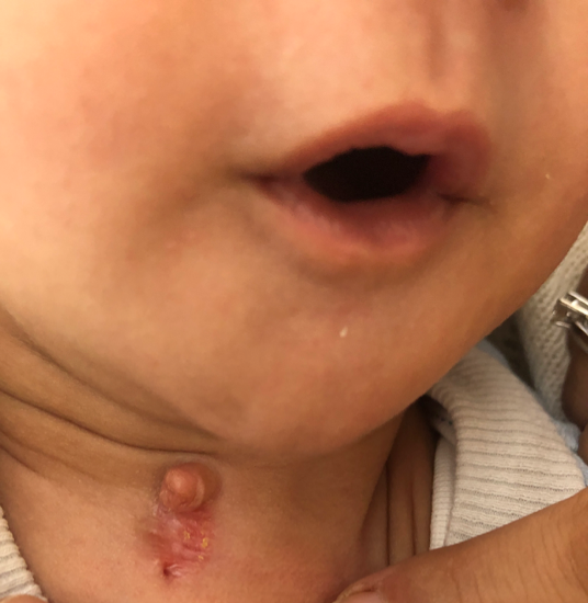
We report the case of a 2-month-old infant with no previous pathological history who presented with a rounded papule of normal skin color measuring about 1 cm in diameter on the right lateral aspect of the neck resting on a slightly atrophic and scaly plaque with a small pertus at its lower part.
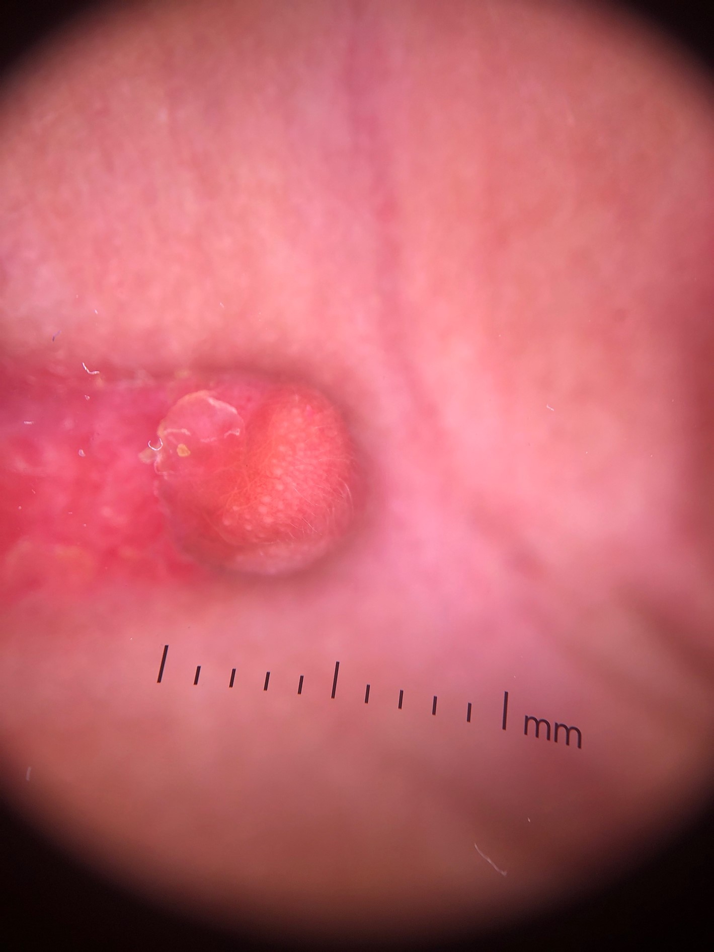
Dermoscopic examination revealed an erythematous background, yellowish scales, a few leucotrichic hairs and coarse round whitish structures as well as a few linear vessels.
The diagnosis of bronchogenic cyst is based on anatomopathological examination of the lesions with standard hematoxylin and eosin staining.
The cyst wall is formed of smooth muscle and sometimes cartilage and shows tracheobronchial differentiation with a respiratory-type epithelium that includes columnar cells, ciliated cells, and mucus-secreting cells [4].
These cysts may be lined with fibrous tissue.
Immunohistochemical study shows CK7 labeling with negativity for TTF-1, CDX-2 and CK20 in favor of a bronchogenic origin [5].
The round whitish structures may correspond histologically to cystic structures lined with fibrous tissue and smooth muscle, the linear vessels may be correlate to inflammation or vasodilatation of dermic vessels.

Figure 1: Clinical picture of a cutaneous bronchogenic cyst.

Figure 2: Dermoscopic examination of the cyst.
References
- Schouten van der Velden AP, Severijnen RS, Wobbes T. A bronchogenic cyst under the scapula with a fistula on the back. Pediatr Surg Int, 2006; 22(10): 857-860. doi: 10.1007/s00383-006-1753-1. Epub 2006 Aug 19. PMID: 16924507.
- Rodgers BM, Harman PK, Johnson AM. Bronchopulmonary foregut malformations. The spectrum of anomalies. Ann Surg, 1998; 203: 517–524.
- Fraga S, Helwig EB, Rossen SH. Bronchogenic cysts in the skin and subcutaneous tissue. Am J Clin Pathol, 1971; 56: 230–238.
- Ramón R, Betlloch I, Guijarro J, Bañuls J, Alfonso R, Silvestre JF. Bronchogenic cyst presenting as a nodular lesion. Pediatr Dermatol, 1999; 16(4): 285-287. doi: 10.1046/j.1525-1470.1999.00075.x. PMID: 10469413.
- Kim PS, Cataletto M, Garnet DJ, Alexeeva V, Selbs E, Katz DS, et al. Unusual presentation of a cutaneous bronchogenic cyst in an asymptomatic neonate. J Pediatr Surg, 2012 ; 47(7): E9-E12. doi: 10.1016/j.jpedsurg.2012.02.024. PMID: 22813830.

