Syndrome of the Pyelo-Ureteral Junction in an Ectopic Kidney, Two Different Surgical Techniques
Mehdi Safieddine, Anas Tmiri*, Amine Moataz, Mohamed Dakir, Adil Debbagh, Rachid Aboutaieb
Urology Department, Ibn Rochd University Hospital Center, Casablanca, Morocco
Received Date: 30/08/2023; Published Date: 18/01/2024
*Corresponding author: Anas Tmiri, Urology Department, Ibn Rochd University Hospital Center, Casablanca, Morocco
Abstract
The Pyelo-Ureteral Junction Syndrome (PUJS) is the most frequent malformative uropathy secondary to a functional or organic obstacle of the pyelo-ureteral junction.
In our work, we report two observations objectifying a syndrome of the pyelo-ureteral junction on ectopic kidney having benefited from a defferent surgical treatment with good clinico-radiological evolution.
The JPU syndrome corresponds to an alteration in the transport of urine from the pyelon to the ureter, the consequence of which is pyelo-calicielle dilation, which, if not treated, entails a risk of progressive alteration of the function of the affected kidney. Renal ectopia is an infrequent urinary malformation.
The reference treatment has been described by Küss, Anderson and Hynes, consisting of resection of the junctional zone and repair by anastomotic suture. The open route has been gradually abandoned in favor of laparoscopic techniques.
Keywords: Syndrome of the pyelo-ureteral junction; Renal ectopia ; Surgical treatment; Pyeloplasty; Laparoscopy
Introduction
The Pyelo-Ureteral Junction Syndrome (PUJS) is the most frequent malformative uropathy secondary to a functional or organic obstacle of the pyelo-ureteral junction with a defect in the flow of urine between the renal pelvis and the proximal urethra, which can occur on an ectopic kidney, this ectopic localization results from an anomaly of migration during the embryonic development, the pelvic localization is the most found at the time of the low ectopies. Its treatment is essentially surgical [1,2].
The incidence of PUJS has been reported in 22-37% of ectopic kidney cases in adults. It is responsible for a urodynamic disorder of the upper excretory tract [3,4].
In our work, we report two observations objectifying a syndrome of pyelo-ureteral junction on ectopic kidney.
Observations
Observation 1:
This is a 30-year-old patient, an active chronic smoker, who presents with chronic pelvic pain of the heaviness type associated with irritative-type lower urinary tract disorders without any notion of hematuria or fever.
The clinical examination found tenderness in the right iliac fossa and the hypogastric region, with no lower back pain or other associated signs.
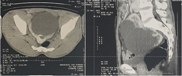
On the radiological level, an abdomino-pelvic ultrasound was performed, objectifying a pelvic cystic mass with no visualization of the right kidney. A URO-CT was performed with visualization of a right kidney in an ectopic pelvic position, the site of major hydronephrosis related to a syndrome of the pyelo-ureteral junction (Figure 1).

Figure 1: URO-CT showing a syndrome of the pyelo-ureteral junction on an ectopic right kidney in the pelvic position.
A dynamic renal scintigraphy with 99mTc-DTPA was performed, showing an asymmetrical relative renal function: 70% on the left and 30% on the right; with absence of spontaneous or provoked emptying on the ectopic right kidney (Figure 2).
The patient underwent open pyeloplasty using the Küss-Anderson-Hynes technique, with drainage by JJ catheter. Postoperative follow-up was good with clinical improvement in pain.

Figure 2: Dynamic renal scintigraphy with 99mTc-DTPA showing an asymmetrical relative renal function with absence of spontaneous or induced emptying in the ectopic right kidney.
Observation 2:
This is a 39-year-old patient, hypertensive, who presents with chronic pain in the right iliac fossa without lower urinary tract disorders or notion of hematuria or fever.
The clinical examination found tenderness in the right iliac fossa without low back pain or other associated signs.
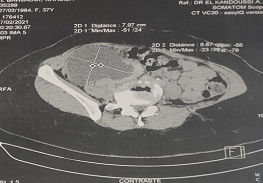
On the radiological level, a uro-scanner objectified an ectopic pelvic right kidney seat of a major hydronephrosis in favor of a syndrome of the pyelo-ureteral junction (Figure 3).

Figure 3: URO-CT showing a syndrome of the pyelo-ureteral junction on an ectopic right kidney in the pelvic position.
The patient underwent transperitoneal laparoscopic pyeloplasty with simple postoperative course. (Figure 4).

Figure 4: Cure of the syndrome of the pyelo-ureteral junction of the ectopic right kidney in the pelvic position by laparoscopy.
Discussion
The SJPU corresponds to an alteration of the transport of urine from the pyelon to the ureter, the consequence of which is a pyelo-calicielle dilation, which, if it is not treated, involves a risk of progressive alteration of the function of the affected kidney. Although this syndrome is mainly of congenital origin, it can manifest itself only from adulthood [5].
Renal ectopia is an infrequent urinary malformation with an incidence of around one in 1000. Lower ectopia is most often pelvic but also lumbar or iliac. Male and left-sided predominance has been reported in the literature [1].
Giant hydronephrosis is defined as the presence of more than 1000 ml of urine in a hydronephrotic sac in an adult, or a kidney that occupies one hemi-abdomen, crosses the midline, and is at least 5 vertebrae [6].
The majority of patients with renal ectopia are often asymptomatic and this ectopia is only detected on the occasion of a complication (hydronephrosis, infection, nephrolithiasis, etc.) or in the context of an X-ray or an ultrasound indicated for other reasons [1]. It is most often discovered in front of an abdominal mass syndrome associated with abdominal pain with signs of digestive, urinary, pulmonary or venous compression. Rupture of giant hydronephrosis is a serious complication [7].
Although the diagnosis is easily revealed by ultrasound in most patients, it can sometimes be confused with other cystic diseases. In such cases, computed tomography and magnetic resonance imaging have been helpful in the differential diagnosis [9]. DMSA kidney scan can be used to measure kidney function and to locate an ectopic kidney, as it can detect kidneys with relative functional value ≥ 5%. A renal scintigraphy with MAG3, sensitized or not to furosemide, makes it possible to calculate the separate function of each kidney and to confirm urinary obstruction [3].
The reference treatment has been described by Küss, Anderson and Hynes, consisting of resection of the junctional zone and repair by anastomotic suture. Open pyeloplasty remains the gold standard in the surgical management of pyelo-ureteral junction syndrome. Kavousi and Schussler [9] first described laparoscopic pyeloplasty by transperitoneal approach in 1993, with the same open surgical principles described by Küss-Anderson-Hynes to reproduce the very satisfactory long-term functional results by reducing postoperative morbidity and convalescence as well as due to cost and accessibility problems encountered during open pyeloplasty, the latter has been gradually abandoned in favor of laparoscopic techniques.Trans-peritoneal laparoscopy is indicated in the cure of SJPU in the case of ectopic kidneys [3,9,10].
The monitoring of patients operated for a syndrome of the pyelo-ureteral junction must most often call upon a dynamic renal scintigraphy which represents the reference examination. This examination must be performed 3 months postoperatively. If there is no abnormality, it is not necessary to continue isotopic monitoring of operated patients [2].
Conclusion
Ectopic pelvic kidney associated with giant hydronephrosis is an extremely rare entity and sometimes difficult to diagnose, hence the importance of antenatal diagnosis and monitoring. In our 2 cases, the conservative attitude, pyeloplasty type, was performed. The evolution was favorable.
References
- Ghfir I, Ben Rais N. Ectopie rénale iliaque explorée par scintigraphie au 99mTc-DTPA et au 99mTc-DMSA. À propos d’un cas. Medecine Nucleaire-imagerie Fonctionnelle Et Metabolique - MED NUCL, 2008; 32: 559-563. doi:10.1016/j.mednuc.2008.06.011
- Bettaieb MA, Fredj MB, Horrigue M, et al. Évaluation du suivi scintigraphique du syndrome de la jonction pyélo-urétérale en postopératoire. Médecine Nucl, 2018; 42(3): 174. doi:10.1016/j.mednuc.2018.03.109
- Hsieh M-Y, Ku M-S, Tsao T-F, Chen S-M, Chao Y-H, Tsai J-D, et al. Rare Case of Atrophic Ectopic Kidney With Giant Hydronephrosis in a 7-Year-Old Girl. Urology, 2013; 81(3): 655‑658.
- Muller CO, Blanc T, Peycelon M, El Ghoneimi A. Laparoscopic treatment of ureteropelvic junction obstruction in five pediatric cases of pelvic kidneys. J Pediatr Urol, 2015; 11(6): 353.e1-353.e5.
- Jacobs JA, Berger BW, Goldman SM, Robbins MA, Young JD. Ureteropelvic obstruction in adults with previously normal pyelograms: a report of 5 cases. J Urol, 1979; 121(2): 242-244. doi:10.1016/s0022-5347(17)56735-x
- Pal BC, Shah SA, Gupta S, Trivedi P. Laparoscopic pyelovesicostomy for giant hydronephrosis in a solitary kidney. Urol Int, 2010; 84(2): 242-244. doi:10.1159/000277607
- Yassine R. Hydronéphrose géante sur rein ectopique pelvien révélée par un syndrome occlusif: Cas rare. Afr J Urol, 2014; 20(4): 211‑4.
- Yapanoğlu T, Alper F, Özbey İ, Aksoy Y, Demi̇rel A. Giant Hydronephrosis Mimicking an Intraabdominal Mass. Turk J Med Sci, 2007; 37(3): 177‑179.
- Papalia R, Simone G, Leonardo C, et al. Retrograde placement of ureteral stent and ureteropelvic anastomosis with two running sutures in transperitoneal laparoscopic pyeloplasty: tips of success in our learning curve. J Endourol, 2009; 23(5): 847-852. doi:10.1089/end.2008.0617
- Bentani N, Moudouni SM, Wakrim B, et al. Cure du syndrome de Jonction Pyelo-Ureterale par voie laparoscopique : Résultats et clés du succès au cours de la courbe d’apprentissage. Afr J Urol, 2012; 18(1): 49-54. doi:10.1016/j.afju.2012.04.011

